PDGF is a major serum mitogen that can exist as a homo or hetero-dimeric protein consisting of disulfide-linked PDGF-A and PDGF‑B chains. The PDGF-AA, PDGF‑BB and PDGF-AB isoforms have been shown to bind to two distinct cell surface PDGF receptors with different affinities. Where as PDGF R alpha binds all three PDGF isoforms with high affinity, PDGF R beta binds PDGF‑BB only with high-affinity. Both PDGF R alpha and PDGF R beta are members of the class III subfamily of receptor tyrosine kinases (RTK) that also includes the receptors for M-CSF, SCF and Flt3 ligand. All class III RTKs are characterized by the presence of five immunoglobulin‑like domains in their extracellular region and a split kinase domain in their intracellular region. PDGF binding induces receptor homo-and hetero‑dimerization and signal transduction. The expression of the alpha and beta receptors is independently regulated in various cell types. Recombinant soluble PDGF R beta binds PDGF with high affinity and is potent PDGF antagonist.
Human phospho-PDGF R beta (Y751) Antibody
R&D Systems | Catalog # AF1767
 by Western Blot.")
Loading...
Key Product Details
Validated by
Biological Validation
Species Reactivity
Validated:
Human
Cited:
Human
Applications
Validated:
Western Blot, Simple Western
Cited:
TaqMan Protein Assay
Label
Unconjugated
Antibody Source
Polyclonal Rabbit IgG
Loading...
Product Specifications
Immunogen
Phosphopeptide containing human PDGF R beta Y751 site
Specificity
Detects human PDGF R beta when phosphorylated at Y751 in Western blots.
Clonality
Polyclonal
Host
Rabbit
Isotype
IgG
Scientific Data Images for Human phospho-PDGF R beta (Y751) Antibody
Detection of Human Phospho-PDGF R beta (Y751) by Western Blot.
Western blot shows lysates of human foreskin fibroblast untreated (-) or treated (+) with 100 ng/mL Recombinant Human PDGF-AA, PDGF-AB, and PDGF-BB (Catalog # 221-AA, 222-AB, and 220-BB, respectively) for 10 minutes. PVDF membrane was probed with 0.5 µg/mL of Rabbit Anti-Human Phospho-PDGF R beta (Y751) Antigen Affinity-purified Polyclonal Antibody (Catalog # AF1767), followed by HRP-conjugated Anti-Rabbit IgG Secondary Antibody (Catalog # HAF008). A specific band was detected for Phospho-PDGF R beta (Y751) at approximately 185 kDa (as indicated). This experiment was conducted under reducing conditions and using Immunoblot Buffer Group 1. by Simple Western<SUP>TM</SUP>.")
Detection of Human Phospho-PDGF R beta (Y751) by Simple WesternTM.
Simple Western lane view shows lysates of CCD-1070Sk human foreskin fibroblast cell line untreated (-) or treated (+) with 100 ng/mL Recombinant Human PDGF-AA (Catalog # 221-AA) for 10 minutes, loaded at 0.2 mg/mL. A specific band was detected for Phospho-PDGF R beta (Y751) at approximately 274 kDa (as indicated) using 5 µg/mL of Rabbit Anti-Human Phospho-PDGF R beta (Y751) Antigen Affinity-purified Polyclonal Antibody (Catalog # AF1767). This experiment was conducted under reducing conditions and using the 66-440 kDa separation system.Applications for Human phospho-PDGF R beta (Y751) Antibody
Application
Recommended Usage
Simple Western
5 µg/mL
Sample: CCD‑1070Sk human foreskin fibroblast cell line treated with Recombinant Human PDGF‑AA (Catalog # 221-AA)
Sample: CCD‑1070Sk human foreskin fibroblast cell line treated with Recombinant Human PDGF‑AA (Catalog # 221-AA)
Western Blot
0.5 µg/mL
Sample: Human foreskin fibroblast treated with Recombinant Human PDGF‑BB (Catalog # 220-BB
Sample: Human foreskin fibroblast treated with Recombinant Human PDGF‑BB (Catalog # 220-BB
Reviewed Applications
Read 1 review rated 5 using AF1767 in the following applications:
Formulation, Preparation, and Storage
Purification
Antigen Affinity-purified
Reconstitution
Reconstitute at 0.2 mg/mL in sterile PBS. For liquid material, refer to CoA for concentration.
Loading...
Formulation
Lyophilized from a 0.2 μm filtered solution in PBS with Trehalose. *Small pack size (SP) is supplied either lyophilized or as a 0.2 µm filtered solution in PBS.
Shipping
Lyophilized product is shipped at ambient temperature. Liquid small pack size (-SP) is shipped with polar packs. Upon receipt, store immediately at the temperature recommended below.
Stability & Storage
Use a manual defrost freezer and avoid repeated freeze-thaw cycles.
- 12 months from date of receipt, -20 to -70 °C as supplied.
- 1 month, 2 to 8 °C under sterile conditions after reconstitution.
- 6 months, -20 to -70 °C under sterile conditions after reconstitution.
Calculators
Background: PDGF R beta
References
- Heldin, C.H. and L. Claesson-Welsh (1994) in Guidebook to Cytokines and Their Receptors, Nicola, N.A. ed. Oxford University Press, New York, p. 202.
Long Name
Platelet-derived Growth Factor Receptor beta
Alternate Names
CD140b, PDGFRB
Gene Symbol
PDGFRB
Additional PDGF R beta Products
Product Documents for Human phospho-PDGF R beta (Y751) Antibody
Certificate of Analysis
To download a Certificate of Analysis, please enter a lot or batch number in the search box below.
Note: Certificate of Analysis not available for kit components.
Product Specific Notices for Human phospho-PDGF R beta (Y751) Antibody
For research use only
Related Research Areas
Citations for Human phospho-PDGF R beta (Y751) Antibody
 Powered by Bioz
Customer Reviews for Human phospho-PDGF R beta (Y751) Antibody (1)
5 out of 5
1 Customer Rating
Have you used Human phospho-PDGF R beta (Y751) Antibody?
Submit a review and receive an Amazon gift card!
$25/€18/£15/$25CAN/¥2500 Yen for a review with an image
$10/€7/£6/$10CAN/¥1110 Yen for a review without an image
Submit a review
Customer Images
Showing
1
-
1 of
1 review
Showing All
Filter By:
-
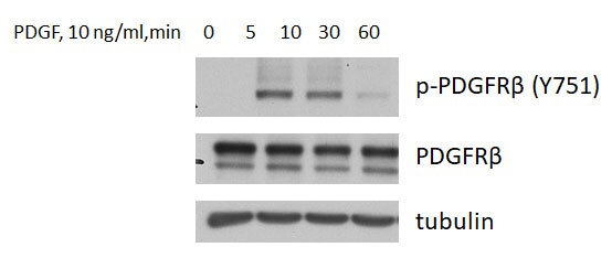
Application: Western BlotSample Tested: G55TL human glioblastoma cell lineSpecies: HumanVerified Customer | Posted 09/29/2017I used this antibody for Western blot in 1:500 dilution; detection with ECL. A strong signal could be detected upon PDGF stimulation of the cells.

There are no reviews that match your criteria.
Protocols
Find general support by application which include: protocols, troubleshooting, illustrated assays, videos and webinars.
- Cellular Response to Hypoxia Protocols
- R&D Systems Quality Control Western Blot Protocol
- Troubleshooting Guide: Western Blot Figures
- Western Blot Conditions
- Western Blot Protocol
- Western Blot Protocol for Cell Lysates
- Western Blot Troubleshooting
- Western Blot Troubleshooting Guide
- View all Protocols, Troubleshooting, Illustrated assays and Webinars


