
Human phospho-PRAS40 (T246) Antibody (1211A)
R&D Systems | Catalog # MAB68901


Scientific images may be resized, cropped, or adjusted for brightness or contrast for presentation purposes. These updates are limited to presentation and do not affect the underlying scientific interpretation of the data. Where available, additional source data may be provided upon request.
Key Product Details
Validated by
Species Reactivity
Applications
Label
Antibody Source
Product Specifications
Immunogen
Accession # Q96B36
Specificity
Clonality
Host
Isotype
Scientific Data Images for Human phospho-PRAS40 (T246) Antibody (1211A)
Detection of Human and Mouse Phospho-PRAS40 (T246) by Western Blot.
Western blot shows lysates of MCF-7 human breast cancer cell line and NIH-3T3 mouse embryonic fibroblast cell line untreated (-) or treated (+) with 100 ng/ml Recombinant Human IGF-I (Catalog # 291-G1) for 60 minutes and 100 ng/ml Recombinant Human PDGF-BB (Catalog # 220-BB) for 20 minutes. PVDF membrane was probed with 1 µg/mL of Rabbit Anti-Human Phospho-PRAS40 (T246) Monoclonal Antibody (Catalog # MAB68901) followed by HRP-conjugated Anti-Rabbit IgG Secondary Antibody (Catalog # HAF008). A specific band was detected for Phospho-PRAS40 (T246) at approximately 40 kDa (as indicated). This experiment was conducted under reducing conditions and using Immunoblot Buffer Group 1.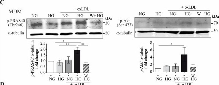
Detection of Phospho-PRAS40 (T246) by Western Blot
The combination of high glucose and oxLDL promotes sCD14 shedding though Akt-PRAS40 pathway activation. MDM were treated with 150 μg/mL of oxLDL for 24 h in culture medium containing 5 mM (normal glucose, NG) or 15 mM (high glucose, HG) of glucose. (A) MDM lysates were exposed to phospho-kinase array spotted membranes. Upper panel: dot images. Lower graph: quantification of dot intensities. (B) Cell lysates were probed with anti-pPRAS40 (Thr246) by Western blotting, and equal loading was determined by probing with alpha -tubulin. Upper panel: representative Western blot images, lower panel: mean ± SEM fold induction levels relative to untreated MDMs (NG) from 2 independent donors. (C–F) Cells were incubated for 45 min with 10 µM of wortmannin (W) for 24 h prior to oxLDL stimulation under NG or HG conditions. (C) PRAS40 (Thr246) and Akt (Ser473) phosphorylation determined by Western blotting. Upper panel: representative images. Lower panel: graph showing fold induction levels relative to untreated cells (NG) from 4 donors. (D) TLR4 and CD14 mRNA expression analyzed by RT-PCR. The data show the mean fold change relative to untreated cells (NG) ± SEM from 4 donors. (E) sCD14 in culture supernatants measured by Western blotting. Upper panel: representative Western blot images. Lower panel: band intensity values/total protein. Fold induction levels relative to untreated cells (NG) from 4 donors are shown. (F) THP1 MΦ were pre-incubated with wortmannin (W) and then treated with NG, HG + oxLDL, and with 2 µg/mL of rsCD14. After 24 h, TNF in culture supernatants was measured by ELISA. Graph showing the mean ± SEM from 3 independent experiments performed in triplicate (* p < 0.05, ** p < 0.01, *** p < 0.001, Mann–Whitney test). MDM: monocyte-derived macrophages. Image collected and cropped by CiteAb from the following open publication (https://pubmed.ncbi.nlm.nih.gov/37237950), licensed under a CC-BY license. Not internally tested by R&D Systems.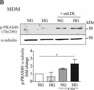
Detection of Phospho-PRAS40 (T246) by Western Blot
The combination of high glucose and oxLDL promotes sCD14 shedding though Akt-PRAS40 pathway activation. MDM were treated with 150 μg/mL of oxLDL for 24 h in culture medium containing 5 mM (normal glucose, NG) or 15 mM (high glucose, HG) of glucose. (A) MDM lysates were exposed to phospho-kinase array spotted membranes. Upper panel: dot images. Lower graph: quantification of dot intensities. (B) Cell lysates were probed with anti-pPRAS40 (Thr246) by Western blotting, and equal loading was determined by probing with alpha -tubulin. Upper panel: representative Western blot images, lower panel: mean ± SEM fold induction levels relative to untreated MDMs (NG) from 2 independent donors. (C–F) Cells were incubated for 45 min with 10 µM of wortmannin (W) for 24 h prior to oxLDL stimulation under NG or HG conditions. (C) PRAS40 (Thr246) and Akt (Ser473) phosphorylation determined by Western blotting. Upper panel: representative images. Lower panel: graph showing fold induction levels relative to untreated cells (NG) from 4 donors. (D) TLR4 and CD14 mRNA expression analyzed by RT-PCR. The data show the mean fold change relative to untreated cells (NG) ± SEM from 4 donors. (E) sCD14 in culture supernatants measured by Western blotting. Upper panel: representative Western blot images. Lower panel: band intensity values/total protein. Fold induction levels relative to untreated cells (NG) from 4 donors are shown. (F) THP1 MΦ were pre-incubated with wortmannin (W) and then treated with NG, HG + oxLDL, and with 2 µg/mL of rsCD14. After 24 h, TNF in culture supernatants was measured by ELISA. Graph showing the mean ± SEM from 3 independent experiments performed in triplicate (* p < 0.05, ** p < 0.01, *** p < 0.001, Mann–Whitney test). MDM: monocyte-derived macrophages. Image collected and cropped by CiteAb from the following open publication (https://pubmed.ncbi.nlm.nih.gov/37237950), licensed under a CC-BY license. Not internally tested by R&D Systems.Applications for Human phospho-PRAS40 (T246) Antibody (1211A)
Western Blot
Sample: MCF‑7 human breast cancer cell line and NIH‑3T3 mouse embryonic fibroblast cell line treated with Recombinant Human IGF‑I (Catalog # 291-G1) and Recombinant Human PDGF‑BB (Catalog # 220-BB)
Formulation, Preparation, and Storage
Purification
Reconstitution
Reconstitute at 0.5 mg/mL in sterile PBS. For liquid material, refer to CoA for concentration.
Formulation
Shipping
Stability & Storage
- 12 months from date of receipt, -20 to -70 °C as supplied.
- 1 month, 2 to 8 °C under sterile conditions after reconstitution.
- 6 months, -20 to -70 °C under sterile conditions after reconstitution.
Calculators
Background: PRAS40
Long Name
Alternate Names
Gene Symbol
UniProt
Additional PRAS40 Products
Product Documents for Human phospho-PRAS40 (T246) Antibody (1211A)
Certificate of Analysis
To download a Certificate of Analysis, please enter a lot or batch number in the search box below.
Note: Certificate of Analysis not available for kit components.
Product Specific Notices for Human phospho-PRAS40 (T246) Antibody (1211A)
For research use only
Related Research Areas
Citations for Human phospho-PRAS40 (T246) Antibody (1211A)
 Powered by Bioz
Customer Reviews for Human phospho-PRAS40 (T246) Antibody (1211A)
There are currently no reviews for this product. Be the first to review Human phospho-PRAS40 (T246) Antibody (1211A) and earn rewards!
Have you used Human phospho-PRAS40 (T246) Antibody (1211A)?
Submit a review and receive an Amazon gift card!
$25/€18/£15/$25CAN/¥2500 for a review with an image
$10/€7/£6/$10CAN/¥1110 for a review without an image
Submit a review
Protocols
Find general support by application which include: protocols, troubleshooting, illustrated assays, videos and webinars.
- Cellular Response to Hypoxia Protocols
- R&D Systems Quality Control Western Blot Protocol
- Troubleshooting Guide: Western Blot Figures
- Western Blot Conditions
- Western Blot Protocol
- Western Blot Protocol for Cell Lysates
- Western Blot Troubleshooting
- Western Blot Troubleshooting Guide
- View all Protocols, Troubleshooting, Illustrated assays and Webinars
Associated Pathways


