
Key Product Details
Species Reactivity
Validated:
Human, Rat
Cited:
Human, Mouse, Rat
Applications
Validated:
Immunohistochemistry, Immunocytochemistry
Cited:
Immunohistochemistry, Western Blot, Flow Cytometry, Immunocytochemistry, Immunoprecipitation
Label
Unconjugated
Antibody Source
Monoclonal Mouse IgG1 Clone # 20B7
Loading...
Product Specifications
Immunogen
E. coli-derived recombinant rat SOX10
aa 1-118
aa 1-118
Specificity
Detects human and rat SOX10. Epitope-mapping experiments indicated that clone 20B7 recognizes a determinant in the first 65 amino acids, which contains sequences unique to SOX10 (1).
Clonality
Monoclonal
Host
Mouse
Isotype
IgG1
Scientific Data Images for SOX10 Antibody (20B7)
SOX10 in BG01V Human Embryonic Stem Cells.
SOX10 was detected in immersion fixed BG01V human embryonic stem cells differentiated to neural crest stem cells using Mouse Anti-Human/Rat SOX10 Monoclonal Antibody (Catalog # MAB2864) at 10 µg/mL for 3 hours at room temperature. Cells were stained using the NorthernLights™ 557-conjugated Anti-Mouse IgG Secondary Antibody (red; Catalog # NL007) and counterstained with DAPI (blue). Specific staining was localized to nuclei. View our protocol for Fluorescent ICC Staining of Cells on Coverslips.
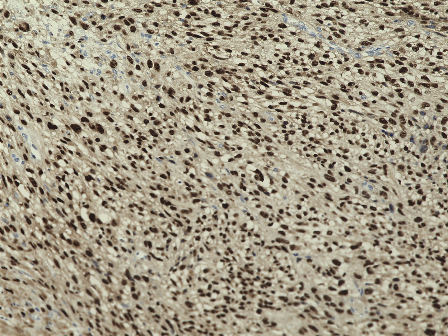
SOX10 in Human Melanoma Tissue.
SOX10 was detected in immersion fixed paraffin-embedded sections of human melanoma tissue using Mouse Anti-Human/Rat SOX10 Monoclonal Antibody (Catalog # MAB2864) at 5 µg/mL for 1 hour at room temperature followed by incubation with the Anti-Mouse IgG VisUCyte™ HRP Polymer Antibody (Catalog # VC001). Tissue was stained using DAB (brown) and counterstained with hematoxylin (blue). Specific staining was localized to nuclei. View our protocol for IHC Staining with VisUCyte HRP Polymer Detection Reagents.
Detection of Rat SOX10 by Immunocytochemistry/Immunofluorescence
Identification of neural crest-derived DPCs. ((a)–(c)) Immunofluorescent cytochemical staining revealed expression of NCSCs-specific markers in DPCs. Primary DPCs were double positive for P75 and Sox10 (a), P75 and Nestin (b), and were positive for Sox9 (c). (d) RT-PCR analysis confirmed the expression of dermal papillae markers ALP and Sox2, and the NCSCs markers Nestin, P75, Sox9, Twist1, and AP2 alpha in DPCs, as well as BMSCs. Scale bar = 20 μm ((a)–(c)). DPCs-P#: DPCs at passage #; BMSCs-P#: BMSCs at passage #. Image collected and cropped by CiteAb from the following publication (https://pubmed.ncbi.nlm.nih.gov/25045659), licensed under a CC-BY license. Not internally tested by R&D Systems.
Detection of Mouse SOX10 by Immunocytochemistry/Immunofluorescence
JCV T-antigen is expressed in neural crest cells.Fluorescent images of bone marrow cells isolated from JCV T-antigen transgenic mice cultured in neural conditions show that these cells are positive for p75 (A,B), nestin (D,E) and SOX-10 (G,H), which are all markers of the neural crest. JCV T-antigen (J,K) was detected in the nucleus of p75 positive cells. Bone marrow cells cultured in mesenchymal media were negative for p75, nestin and SOX-10, and did not express JCV T-antigen (C, F, I, L). Cell nuclei were visualized with DAPI. (Scale bar: 100 µm). (M) Diagram illustrating expression of JCV T-antigen in bone marrow cells. T-antigen expression was associated with neural crest lineage cells of the bone marrow cultured in serum-free neural stem cell media supplemented with bFGF and EFG (left panel), while mesenchymal cells maintained in alpha -MEM in the presence of 20% serum were negative for T-antigen (right panel). (N) FACS analysis of neural crest cells and MSCs demonstrates positivity for JCV T-antigen in 99% of neural crest cells and lack of JCV T-antigen expression in MSCs. Image collected and cropped by CiteAb from the following publication (https://pubmed.ncbi.nlm.nih.gov/23805194), licensed under a CC-BY license. Not internally tested by R&D Systems.Applications for SOX10 Antibody (20B7)
Application
Recommended Usage
Immunocytochemistry
8-25 µg/mL
Sample: Immersion fixed BG01V human embryonic stem cells differentiated to neural crest stem cells
Sample: Immersion fixed BG01V human embryonic stem cells differentiated to neural crest stem cells
Immunohistochemistry
5-25 µg/mL
Sample: Immersion fixed paraffin-embedded sections of human melanoma tissue
Sample: Immersion fixed paraffin-embedded sections of human melanoma tissue
Reviewed Applications
Read 1 review rated 5 using MAB2864 in the following applications:
Formulation, Preparation, and Storage
Purification
Protein A or G purified from hybridoma culture supernatant
Reconstitution
Reconstitute at 0.5 mg/mL in sterile PBS. For liquid material, refer to CoA for concentration.
Loading...
Formulation
Lyophilized from a 0.2 μm filtered solution in PBS with Trehalose. *Small pack size (SP) is supplied either lyophilized or as a 0.2 µm filtered solution in PBS.
Shipping
Lyophilized product is shipped at ambient temperature. Liquid small pack size (-SP) is shipped with polar packs. Upon receipt, store immediately at the temperature recommended below.
Stability & Storage
Use a manual defrost freezer and avoid repeated freeze-thaw cycles.
- 12 months from date of receipt, -20 to -70 °C as supplied.
- 1 month, 2 to 8 °C under sterile conditions after reconstitution.
- 6 months, -20 to -70 °C under sterile conditions after reconstitution.
Calculators
Background: SOX10
References
- Lo, L. et al. (2002) Development 129:1553.
Long Name
SRY-box transcription factor 10
Alternate Names
DOM, PCWH, SOX-10, WS2E, WS4, WS4C
Gene Symbol
SOX10
Additional SOX10 Products
Product Documents for SOX10 Antibody (20B7)
Certificate of Analysis
To download a Certificate of Analysis, please enter a lot or batch number in the search box below.
Note: Certificate of Analysis not available for kit components.
Product Specific Notices for SOX10 Antibody (20B7)
For research use only
Related Research Areas
Citations for SOX10 Antibody (20B7)
 Powered by Bioz
Customer Reviews for SOX10 Antibody (20B7) (1)
5 out of 5
1 Customer Rating
Have you used SOX10 Antibody (20B7)?
Submit a review and receive an Amazon gift card!
$25/€18/£15/$25CAN/¥2500 Yen for a review with an image
$10/€7/£6/$10CAN/¥1110 Yen for a review without an image
Submit a review
Customer Images
Showing
1
-
1 of
1 review
Showing All
Filter By:
-
Application: Immunohistochemistry-ParaffinSample Tested: human melanomaSpecies: HumanVerified Customer | Posted 12/05/2022Human melanoma with SOX10 mAB

There are no reviews that match your criteria.
Protocols
Find general support by application which include: protocols, troubleshooting, illustrated assays, videos and webinars.
- Antigen Retrieval Protocol (PIER)
- Antigen Retrieval for Frozen Sections Protocol
- Appropriate Fixation of IHC/ICC Samples
- Cellular Response to Hypoxia Protocols
- Chromogenic IHC Staining of Formalin-Fixed Paraffin-Embedded (FFPE) Tissue Protocol
- Chromogenic Immunohistochemistry Staining of Frozen Tissue
- ClariTSA™ Fluorophore Kits
- Detection & Visualization of Antibody Binding
- Fluorescent IHC Staining of Frozen Tissue Protocol
- Graphic Protocol for Heat-induced Epitope Retrieval
- Graphic Protocol for the Preparation and Fluorescent IHC Staining of Frozen Tissue Sections
- Graphic Protocol for the Preparation and Fluorescent IHC Staining of Paraffin-embedded Tissue Sections
- Graphic Protocol for the Preparation of Gelatin-coated Slides for Histological Tissue Sections
- ICC Cell Smear Protocol for Suspension Cells
- ICC Immunocytochemistry Protocol Videos
- ICC for Adherent Cells
- IHC Sample Preparation (Frozen sections vs Paraffin)
- Immunocytochemistry (ICC) Protocol
- Immunocytochemistry Troubleshooting
- Immunofluorescence of Organoids Embedded in Cultrex Basement Membrane Extract
- Immunofluorescent IHC Staining of Formalin-Fixed Paraffin-Embedded (FFPE) Tissue Protocol
- Immunohistochemistry (IHC) and Immunocytochemistry (ICC) Protocols
- Immunohistochemistry Frozen Troubleshooting
- Immunohistochemistry Paraffin Troubleshooting
- Preparing Samples for IHC/ICC Experiments
- Preventing Non-Specific Staining (Non-Specific Binding)
- Primary Antibody Selection & Optimization
- Protocol for Heat-Induced Epitope Retrieval (HIER)
- Protocol for Making a 4% Formaldehyde Solution in PBS
- Protocol for VisUCyte™ HRP Polymer Detection Reagent
- Protocol for the Fluorescent ICC Staining of Cell Smears - Graphic
- Protocol for the Fluorescent ICC Staining of Cultured Cells on Coverslips - Graphic
- Protocol for the Preparation & Fixation of Cells on Coverslips
- Protocol for the Preparation and Chromogenic IHC Staining of Frozen Tissue Sections
- Protocol for the Preparation and Chromogenic IHC Staining of Frozen Tissue Sections - Graphic
- Protocol for the Preparation and Chromogenic IHC Staining of Paraffin-embedded Tissue Sections
- Protocol for the Preparation and Chromogenic IHC Staining of Paraffin-embedded Tissue Sections - Graphic
- Protocol for the Preparation and Fluorescent ICC Staining of Cells on Coverslips
- Protocol for the Preparation and Fluorescent ICC Staining of Non-adherent Cells
- Protocol for the Preparation and Fluorescent ICC Staining of Stem Cells on Coverslips
- Protocol for the Preparation and Fluorescent IHC Staining of Frozen Tissue Sections
- Protocol for the Preparation and Fluorescent IHC Staining of Paraffin-embedded Tissue Sections
- Protocol for the Preparation of Gelatin-coated Slides for Histological Tissue Sections
- Protocol for the Preparation of a Cell Smear for Non-adherent Cell ICC - Graphic
- TUNEL and Active Caspase-3 Detection by IHC/ICC Protocol
- The Importance of IHC/ICC Controls
- Troubleshooting Guide: Immunohistochemistry
- View all Protocols, Troubleshooting, Illustrated assays and Webinars
Loading...
Associated Pathways
