
Key Product Details
Validated by
Species Reactivity
Validated:
Cited:
Applications
Validated:
Cited:
Label
Antibody Source
Product Specifications
Immunogen
Pro2-Arg264
Accession # O95863
Specificity
Clonality
Host
Isotype
Scientific Data Images for Human Snail Antibody
Detection of Human Snail by Western Blot.
Western blot shows lysates of A549 human lung carcinoma cell line and JEG-3 human epithelial choriocarcinoma cell line. PVDF membrane was probed with 0.5 µg/mL of Goat Anti-Human Snail Antigen Affinity-purified Polyclonal Antibody (Catalog # AF3639) followed by HRP-conjugated Anti-Goat IgG Secondary Antibody (Catalog # HAF109). A specific band was detected for Snail at approximately 29 kDa (as indicated). This experiment was conducted under reducing conditions and using Immunoblot Buffer Group 1.
Detection of Snail-regulated Genes by Chromatin Immunoprecipitation.
Jurkat human acute T cell leukemia cell line treated with 50 ng/mL PMA and 200 ng/mL calcium ionomycin for 30 minutes was fixed using formaldehyde, resuspended in lysis buffer, and sonicated to shear chromatin. Snail/DNA complexes were immunoprecipitated using 5 µg Goat Anti-Human Snail Antigen Affinity-purified Polyclonal Antibody (Catalog # AF3639) or control antibody (Catalog # AB-108-C) for 15 minutes in an ultrasonic bath, followed by Biotinylated Anti-Goat IgG Secondary Antibody (Catalog # BAF109). Immunocomplexes were captured using 50 µL of MagCellect Streptavidin Ferrofluid (Catalog # MAG999) and DNA was purified using chelating resin solution. TheE-Cadherinpromoter was detected by standard PCR.
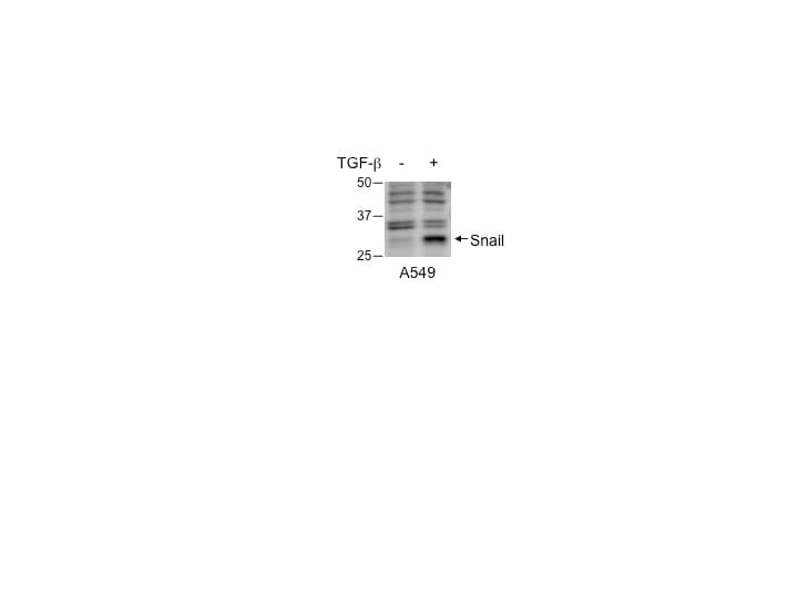
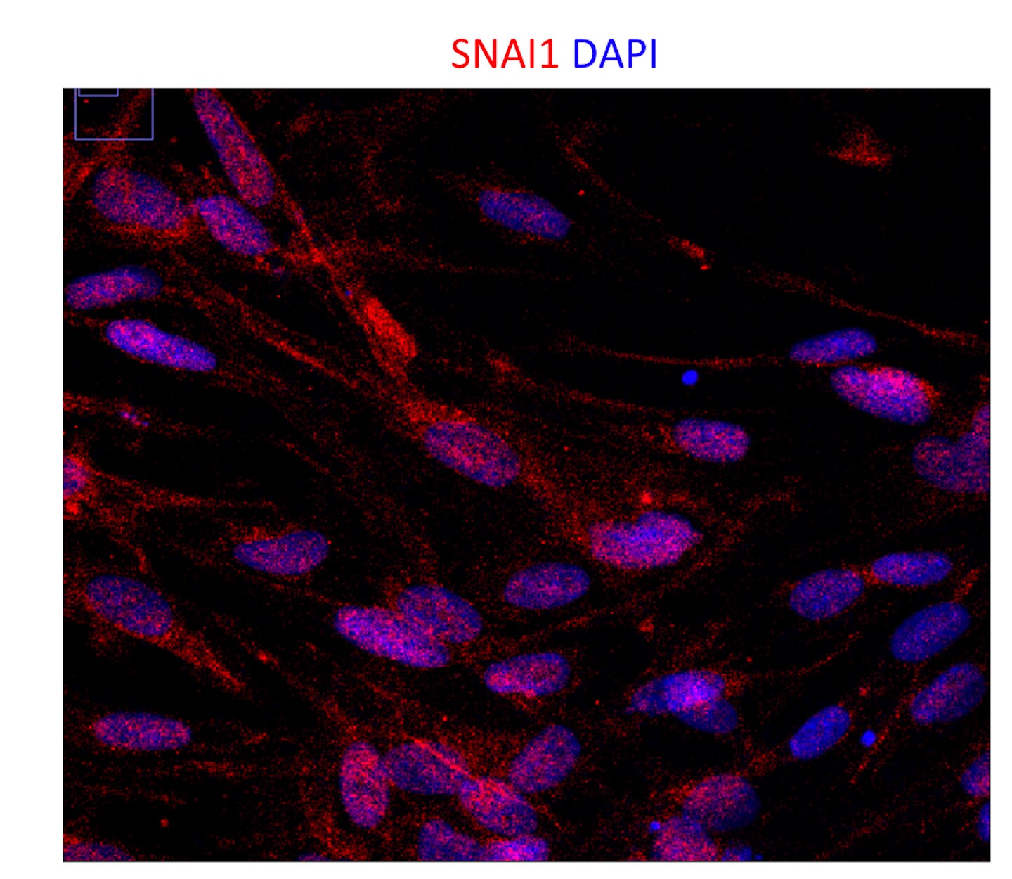
Snail in A549 Human Cell Line.
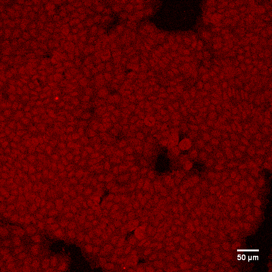
Snail was detected in immersion fixed A549 human lung carcinoma cell line treated with Recombinant Human TGF-beta 1 (left panel, Catalog # 240-B) or untreated (right panel) using Goat Anti-Human Snail Antigen Affinity-purified Polyclonal Antibody (Catalog # AF3639) at 10 µg/mL for 3 hours at room temperature. Cells were stained using the NorthernLights™ 557-conjugated Anti-Goat IgG Secondary Antibody (red; Catalog # NL001) and counterstained with DAPI (blue). Specific staining was localized to nuclei. View our protocol for Fluorescent ICC Staining of Cells on Coverslips.
Detection of Snail in A549 Human Cell Line by Flow Cytometry.
A549 human lung carcinoma cell line was stained with Goat Anti-Human Snail Antigen Affinity-purified Polyclonal Antibody (Catalog # AF3639, filled histogram) or isotype control antibody (Catalog # AB-108-C, open histogram), followed by Fluorescein-conjugated Anti-Goat IgG Secondary Antibody (Catalog # F0109). To facilitate intracellular staining, cells were fixed and permeabilized with FlowX FoxP3 Fixation & Permeabilization Buffer Kit (Catalog # FC012). View our protocol for Staining Intracellular Molecules.
Detection of Human Snail by Western Blot
SerpinA1 was regulated by SnailA. DLD-1 and SW480 cells were transfected with pcDNA-Snail (Snail), control vector pcDNA (vector), Snail siRNA (siSnail), or nontargeting siRNA (siNT), and Snail and serpinA1 protein levels were evaluated by western blot analysis. B. DLD-1 and SW480 cells were transfected with pcDNA-serpinA1 (serpinA1), control vector pcDNA (vector), serpinA1 siRNA (siSerpinA1), or nontargeting siRNA (siNT), and western blot analysis was performed for detection of Snail and SerpinA1 expression. C. DLD-1 and SW480 cells were transfected with pcDNA-Snail (Snail) or control vector pcDNA (vector), and ChIP assays were performed. The presence of the serpinA1 promoter (−516/−4) was verified in immunoprecipitates with either mouse IgG or anti-Snail antibodies, and assay inputs were analyzed using real-time PCR. The samples were loaded on agarose gels. D. Data show promoter enrichment in the anti-Snail immunoprecipitate relative to IgG. Image collected and cropped by CiteAb from the following publication (https://pubmed.ncbi.nlm.nih.gov/26015410), licensed under a CC-BY license. Not internally tested by R&D Systems.
Detection of Human Snail by Western Blot
Snail and serpinA1 promoted tumor progression through fibronectinA. DLD-1 and SW480 cells were transfected with pcDNA-Snail (Snail), control vector pcDNA (vector), Snail siRNA (siSnail), or nontargeting siRNA (siNT), and Snail, serpinA1, and fibronectin protein levels were evaluated by western blot analysis. B. DLD-1 and SW480 cells were transfected with pcDNA-serpinA1 (SerpinA1), control vector pcDNA (vector), serpinA1 siRNA (siSerpinA1), or nontargeting siRNA (siNT), and Snail, serpinA1, and fibronectin protein levels were evaluated by western blot analysis. C. DLD-1 and SW480 cells were transfected with pcDNA-fibronectin (Fibronectin), control vector pcDNA (vector), fibronectin siRNA (siFibronectin), or nontargeting siRNA (siNT), and Snail, serpinA1, and fibronectin protein levels were evaluated by western blot analysis. D., E. Invasion and migration assays were performed using transfected cells. Representative data are shown for cells that invaded (top) and migrated (bottom) in the presence of 1% FBS. *P < 0.05. Image collected and cropped by CiteAb from the following publication (https://pubmed.ncbi.nlm.nih.gov/26015410), licensed under a CC-BY license. Not internally tested by R&D Systems.
Detection of Human Snail by Western Blot
PDGF-AB induces Endo-MT through NF-kB-mediated Snail expression in ECs. a Human brain microvacular ECs were treated with glioma-CM. RNA was isolated and subjected to RNA-seq analysis. Left, heat map for expression of EMT-related transcriptional factors. Right, fold change of these transcriptional factors (n = 3, mean ± SEM). b, c ECs were treated with 100 ng/ml PDGF-AA, PDGF-AB, and PDGF-BB. b Cell lysates were immunoblotted. c RNA was isolated and analyzed by RT-PCR. Results were normalized with GAPDH levels (n = 3, mean ± SEM). d ECs were transfected with siRNAs targeting Snail or control scrambled sequence, and treated with PDGF-AB. Cell lysates were immunoblotted. e ECs were transfected with siRNAs targeting Erg-1, NF-kappa B, or control scrambled sequence, and treated with PDGF-AB. Cell lysates were immunoblotted. f ECs were treated with PDGF-AB for 2 h. Cells were analyzed by immunofluorescence. g, h ECs were treated with PDGF-AB or control medium for 8 h. Nuclear extracts were immunoprecipitated with anti-NF-kappa B antibody or IgG, and subjected to ChIP analysis with primers #1 and #2. g DNA was resolved by agarose electrophoresis, and imaged. Shown are representative results with primer #2. The arrow indicates the amplified DNA in Snail promoter. h Quantitative PCR analysis (n = 3, mean ± SEM). i ECs were transfected with siRNAs targeting Snail or control scrambled sequence, 4 days after treatment with Ki8751, followed by cell viability analysis (n = 3, mean ± SD) Image collected and cropped by CiteAb from the following open publication (https://pubmed.ncbi.nlm.nih.gov/30150753), licensed under a CC-BY license. Not internally tested by R&D Systems.
Detection of Human Snail by Immunohistochemistry
Representative images of positive IHC staining in benign and malignant prostate tissue and a negative control Image collected and cropped by CiteAb from the following open publication (https://pubmed.ncbi.nlm.nih.gov/28877722), licensed under a CC-BY license. Not internally tested by R&D Systems.Applications for Human Snail Antibody
Chromatin Immunoprecipitation (ChIP)
Sample: PMA and calcium ionomycin treated Jurkat human acute T cell leukemia cell line chromatin, E-Cadherin promoter detected by standard PCR.
Immunocytochemistry
Sample: Immersion fixed A549 human lung carcinoma cell line treated with Recombinant Human TGF-beta 1 (Catalog # 240-B)
Intracellular Staining by Flow Cytometry
Sample: A549 human lung carcinoma cell line were fixed and permeabilized with FlowX FoxP3 Fixation & Permeabilization Buffer Kit (Catalog # FC012)
Western Blot
Sample: A549 human lung carcinoma cell line and JEG‑3 human epithelial choriocarcinoma cell line
Reviewed Applications
Read 5 reviews rated 4 using AF3639 in the following applications:
Flow Cytometry Panel Builder
Bio-Techne Knows Flow Cytometry
Save time and reduce costly mistakes by quickly finding compatible reagents using the Panel Builder Tool.
Advanced Features
- Spectra Viewer - Custom analysis of spectra from multiple fluorochromes
- Spillover Popups - Visualize the spectra of individual fluorochromes
- Antigen Density Selector - Match fluorochrome brightness with antigen density

Formulation, Preparation, and Storage
Purification
Reconstitution
Reconstitute at 0.2 mg/mL in sterile PBS. For liquid material, refer to CoA for concentration.
Formulation
Shipping
Stability & Storage
- 12 months from date of receipt, -20 to -70 °C as supplied.
- 1 month, 2 to 8 °C under sterile conditions after reconstitution.
- 6 months, -20 to -70 °C under sterile conditions after reconstitution.
Calculators
Background: Snail
Alternate Names
Gene Symbol
UniProt
Additional Snail Products
Product Documents for Human Snail Antibody
Certificate of Analysis
To download a Certificate of Analysis, please enter a lot or batch number in the search box below.
Note: Certificate of Analysis not available for kit components.
Product Specific Notices for Human Snail Antibody
For research use only
Citations for Human Snail Antibody
 Powered by Bioz
Customer Reviews for Human Snail Antibody (5)
Have you used Human Snail Antibody?
Submit a review and receive an Amazon gift card!
$25/€18/£15/$25CAN/¥2500 Yen for a review with an image
$10/€7/£6/$10CAN/¥1110 Yen for a review without an image
Submit a review
Customer Images
-
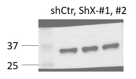
Application: Western BlotSample Tested: Human colorectal cancer cellSpecies: HumanVerified Customer | Posted 10/24/2023SNAIL expression was checked with shCtr or sh-geneX to determine influences on EMT. A clear band was observed at around 30KD.

-
Application: Western BlotSample Tested: A549 human lung carcinoma cell lineSpecies: HumanVerified Customer | Posted 10/17/2019
-
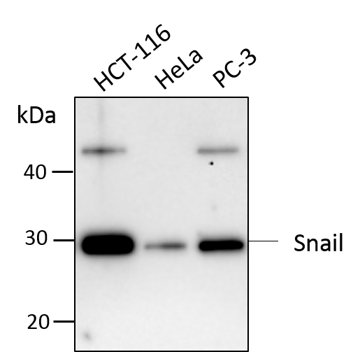
Application: Western BlotSample Tested: HCT-116 human colorectal carcinoma cell line, HeLa human cervical epithelial carcinoma cell line and PC-3 human prostate cancer cell lineSpecies: HumanVerified Customer | Posted 07/23/2018Total cell lysates from HCT-116, HeLa and PC-3 were subjected to western blot. PVDF membrane were probed with 1 um/ml Human Snail Antibody (AF3639). A specific band was detected for Snail at approximately 30 kDa. This experiment was conducted under reducing conditions.
-
Application: Immunocytochemistry/ImmunofluorescenceSample Tested: Bladder cancer tissueSpecies: HumanVerified Customer | Posted 12/10/2017
-
Application: Immunocytochemistry/ImmunofluorescenceSample Tested: mesenchymal cellsSpecies: HumanVerified Customer | Posted 10/18/2016
There are no reviews that match your criteria.
Protocols
Find general support by application which include: protocols, troubleshooting, illustrated assays, videos and webinars.
- 7-Amino Actinomycin D (7-AAD) Cell Viability Flow Cytometry Protocol
- Appropriate Fixation of IHC/ICC Samples
- Cellular Response to Hypoxia Protocols
- ChIP Protocol Video
- Chromatin Immunoprecipitation (ChIP) Protocol
- Chromatin Immunoprecipitation Protocol
- ClariTSA™ Fluorophore Kits
- Detection & Visualization of Antibody Binding
- Extracellular Membrane Flow Cytometry Protocol
- Flow Cytometry Protocol for Cell Surface Markers
- Flow Cytometry Protocol for Staining Membrane Associated Proteins
- Flow Cytometry Staining Protocols
- Flow Cytometry Troubleshooting Guide
- ICC Cell Smear Protocol for Suspension Cells
- ICC Immunocytochemistry Protocol Videos
- ICC for Adherent Cells
- Immunocytochemistry (ICC) Protocol
- Immunocytochemistry Troubleshooting
- Immunofluorescence of Organoids Embedded in Cultrex Basement Membrane Extract
- Immunohistochemistry (IHC) and Immunocytochemistry (ICC) Protocols
- Intracellular Flow Cytometry Protocol Using Alcohol (Methanol)
- Intracellular Flow Cytometry Protocol Using Detergents
- Intracellular Nuclear Staining Flow Cytometry Protocol Using Detergents
- Intracellular Staining Flow Cytometry Protocol Using Alcohol Permeabilization
- Intracellular Staining Flow Cytometry Protocol Using Detergents to Permeabilize Cells
- Preparing Samples for IHC/ICC Experiments
- Preventing Non-Specific Staining (Non-Specific Binding)
- Primary Antibody Selection & Optimization
- Propidium Iodide Cell Viability Flow Cytometry Protocol
- Protocol for Liperfluo
- Protocol for VisUCyte™ HRP Polymer Detection Reagent
- Protocol for the Characterization of Human Th22 Cells
- Protocol for the Characterization of Human Th9 Cells
- Protocol for the Fluorescent ICC Staining of Cell Smears - Graphic
- Protocol for the Fluorescent ICC Staining of Cultured Cells on Coverslips - Graphic
- Protocol for the Preparation and Fluorescent ICC Staining of Cells on Coverslips
- Protocol for the Preparation and Fluorescent ICC Staining of Non-adherent Cells
- Protocol for the Preparation and Fluorescent ICC Staining of Stem Cells on Coverslips
- Protocol for the Preparation of a Cell Smear for Non-adherent Cell ICC - Graphic
- Protocol: Annexin V and PI Staining by Flow Cytometry
- Protocol: Annexin V and PI Staining for Apoptosis by Flow Cytometry
- R&D Systems Quality Control Western Blot Protocol
- TUNEL and Active Caspase-3 Detection by IHC/ICC Protocol
- The Importance of IHC/ICC Controls
- Troubleshooting Guide: Fluorokine Flow Cytometry Kits
- Troubleshooting Guide: Western Blot Figures
- Western Blot Conditions
- Western Blot Protocol
- Western Blot Protocol for Cell Lysates
- Western Blot Troubleshooting
- Western Blot Troubleshooting Guide
- View all Protocols, Troubleshooting, Illustrated assays and Webinars
FAQs for Human Snail Antibody
-
Q: Can snail antibody AF3639 be used for electrophoresis motility shift assays (EMSA) instead of ChIP?
A: For IP/Co-IP and ChIP the antibodies need to provide sufficient binding affinity for the antibodies to stay bound to the protein targets during the precipitation steps, whereas in EMSA the antibodies will only need to bind to the protein targets during the migration through the weak electrophoretic forces. Almost all, if not all, antibodies that bind to their protein targets with reasonable affinity and do not fall off from their protein targets when bound to the targeted, labeled DNA/RNA molecules during PAGE will be rendered in the upper shift in EMSA, provided the binding doesn't block the interaction between the protein targets and the nucleic acids.
Associated Pathways
