STAT5a (Signal Transducer and Activator of Transcription-5a) is one of two closely related genes that belong to the STAT family of transcription factors. It is a 91 kDa cytosolic protein that contains an N-terminal domain (with an NES/Nuclear Export Signal) a coiled-coiled region (with an NLS/Nuclear Localization Signal) a DNA-binding site that recognizes a GAS (Gamma-interferon Activated Site) motif, an SH2 domain that allows for dimerization, and a C-terminal transactivation domain. STAT5a likely exists in a quiescent state as a cytoplasmic anti-parallel homodimer. Following activation of both tyrosine and non-tyrosine kinase membrane-bound receptors, STAT5a is phosphorylated, increasing its MW by some 5-6 kDa. Phosphorylated STAT5a will enter the nucleus as either a homodimer (or heterodimer with STAT5b), or as a complex with the intracellular domain of a tyrosine kinase receptor such as ErbB4. Once in the nucleus, STAT5a will form a homotetramer and bind either GAS or GAS-related sequences in gene regulatory regions. Notably, it is suggested that STAT5a may "cycle" through the nucleus without phosphorylation, a process that would seem to involve Importins alpha 3 and beta 1. STAT5a is related to STAT5b through gene duplication. They show 92% amino acid (aa) sequence identity, with the major differences existing over aa 45-56 and 773-794. The genes are not entirely redundant; STAT5a activates NDGR1 and SH2, while STAT5b regulates the Treg T cell genes, FoxP3 and CD25/IL-2R alpha.

Key Product Details
Species Reactivity
Human
Applications
Intracellular Staining by Flow Cytometry, Immunocytochemistry, CyTOF-ready
Label
Unconjugated
Antibody Source
Monoclonal Mouse IgG1 Clone # 251610
Loading...
Product Specifications
Immunogen
Human STAT5a synthetic peptide
SLDSRLSPPAGLFTSARGSLS
Accession # NP_003143
SLDSRLSPPAGLFTSARGSLS
Accession # NP_003143
Specificity
Detects human STAT5a. This antibody does not cross-react with STAT5b.
Clonality
Monoclonal
Host
Mouse
Isotype
IgG1
Scientific Data Images for Human STAT5a Antibody (251610)
Detection of STAT5a in Jurkat Human Cell Line by Flow Cytometry.
Jurkat human acute T cell leukemia cell line was stained with Mouse Anti-Human STAT5a Monoclonal Antibody (Catalog # MAB21741, filled histogram) or isotype control antibody (Catalog # MAB002, open histogram), followed by Allophycocyanin-conjugated Anti-Mouse IgG F(ab')2Secondary Antibody (Catalog # F0101B). To facilitate intracellular staining, cells were fixed with paraformaldehyde and permeabilized with methanol.
STAT5a in K562 Human Cell Line.
STAT5a was detected in immersion fixed K562 human chronic myelogenous leukemia cell line using Mouse Anti-Human STAT5a Monoclonal Antibody (Catalog # MAB21741) at 25 µg/mL for 3 hours at room temperature. Cells were stained using the NorthernLights™ 557-conjugated Anti-Mouse IgG Secondary Antibody (red; Catalog # NL007) and counterstained with DAPI (blue). Specific staining was localized to cytoplasm. View our protocol for Fluorescent ICC Staining of Cells on Coverslips.Applications for Human STAT5a Antibody (251610)
Application
Recommended Usage
CyTOF-ready
Ready to be labeled using established conjugation methods. No BSA or other carrier proteins that could interfere with conjugation.
Immunocytochemistry
8-25 µg/mL
Sample: Immersion fixed K562 human chronic myelogenous leukemia cell line and human peripheral blood mononuclear cells
Sample: Immersion fixed K562 human chronic myelogenous leukemia cell line and human peripheral blood mononuclear cells
Intracellular Staining by Flow Cytometry
0.25 µg/106 cells
Sample: Jurkat human acute T cell leukemia cell line fixed with paraformaldehyde and permeabilized with methanol
Sample: Jurkat human acute T cell leukemia cell line fixed with paraformaldehyde and permeabilized with methanol
Reviewed Applications
Read 3 reviews rated 4 using MAB21741 in the following applications:
Flow Cytometry Panel Builder
Bio-Techne Knows Flow Cytometry
Save time and reduce costly mistakes by quickly finding compatible reagents using the Panel Builder Tool.
Advanced Features
- Spectra Viewer - Custom analysis of spectra from multiple fluorochromes
- Spillover Popups - Visualize the spectra of individual fluorochromes
- Antigen Density Selector - Match fluorochrome brightness with antigen density

Formulation, Preparation, and Storage
Purification
Protein A or G purified from hybridoma culture supernatant
Reconstitution
Reconstitute at 0.5 mg/mL in sterile PBS. For liquid material, refer to CoA for concentration.
Loading...
Formulation
Lyophilized from a 0.2 μm filtered solution in PBS with Trehalose. See Certificate of Analysis for details.
*Small pack size (-SP) is supplied either lyophilized or as a 0.2 µm filtered solution in PBS.
*Small pack size (-SP) is supplied either lyophilized or as a 0.2 µm filtered solution in PBS.
Shipping
Lyophilized product is shipped at ambient temperature. Liquid small pack size (-SP) is shipped with polar packs. Upon receipt, store immediately at the temperature recommended below.
Stability & Storage
Use a manual defrost freezer and avoid repeated freeze-thaw cycles.
- 12 months from date of receipt, -20 to -70 °C as supplied.
- 1 month, 2 to 8 °C under sterile conditions after reconstitution.
- 6 months, -20 to -70 °C under sterile conditions after reconstitution.
Calculators
Background: STAT5a
Long Name
Signal Transducer and Activator of Transcription 5
Alternate Names
MGF, signal transducer and activator of transcription 5A, STAT5
Gene Symbol
STAT5A
UniProt
Additional STAT5a Products
Product Documents for Human STAT5a Antibody (251610)
Certificate of Analysis
To download a Certificate of Analysis, please enter a lot or batch number in the search box below.
Note: Certificate of Analysis not available for kit components.
Product Specific Notices for Human STAT5a Antibody (251610)
For research use only
Customer Reviews for Human STAT5a Antibody (251610) (3)
4 out of 5
3 Customer Ratings
Have you used Human STAT5a Antibody (251610)?
Submit a review and receive an Amazon gift card!
$25/€18/£15/$25CAN/¥2500 Yen for a review with an image
$10/€7/£6/$10CAN/¥1110 Yen for a review without an image
Submit a review
Customer Images
Showing
1
-
3 of
3 reviews
Showing All
Filter By:
-
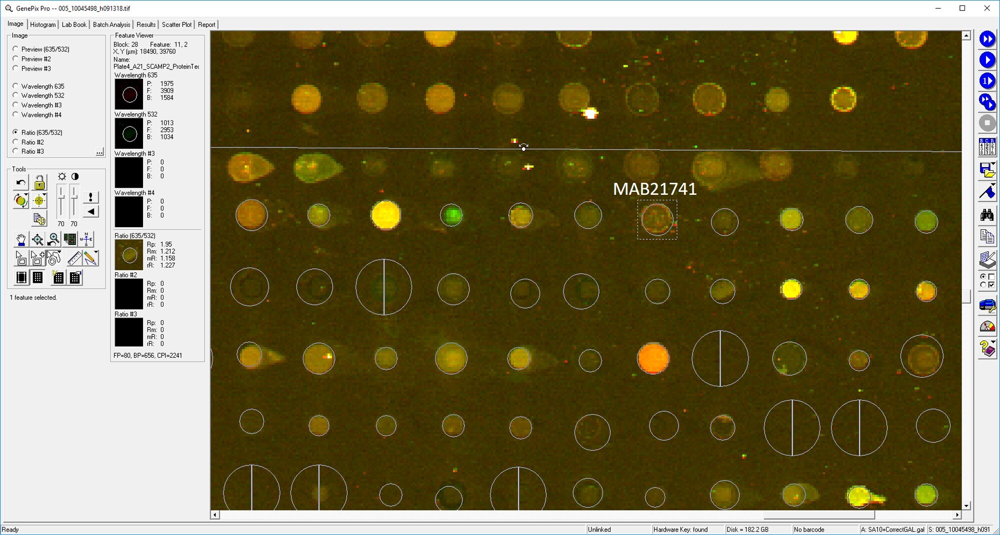
Application: MicroarraysSample Tested: EDTA PlasmaSpecies: HumanVerified Customer | Posted 01/03/2020

-
Application: MicroarraySample Tested: EDTA PlasmaSpecies: HumanVerified Customer | Posted 02/21/2019
-
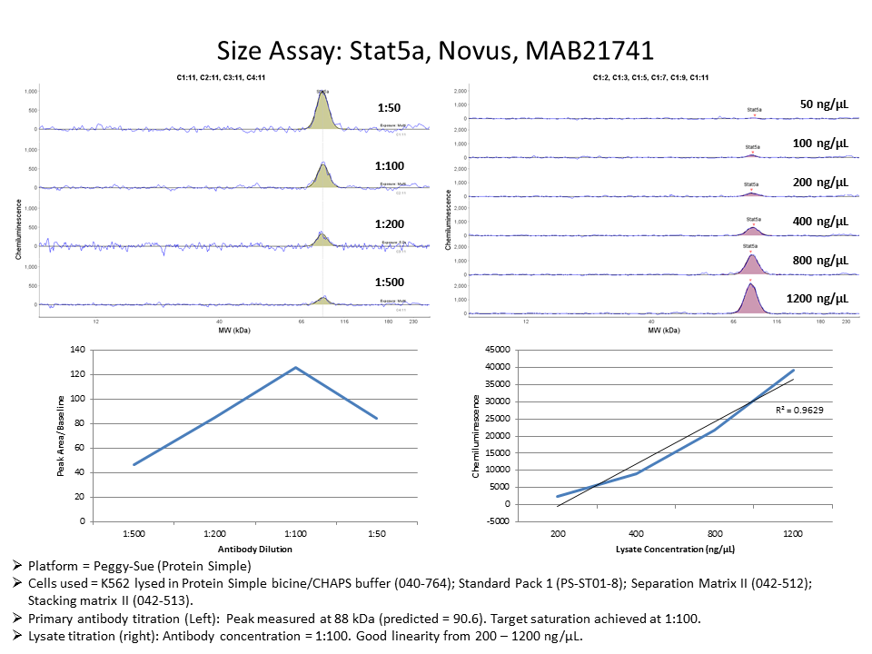
Application: Simple WesternSample Tested: K562 human chronic myelogenous leukemia cell lineSpecies: HumanVerified Customer | Posted 03/22/2017
There are no reviews that match your criteria.
Protocols
Find general support by application which include: protocols, troubleshooting, illustrated assays, videos and webinars.
- 7-Amino Actinomycin D (7-AAD) Cell Viability Flow Cytometry Protocol
- Appropriate Fixation of IHC/ICC Samples
- Cellular Response to Hypoxia Protocols
- ClariTSA™ Fluorophore Kits
- Detection & Visualization of Antibody Binding
- Extracellular Membrane Flow Cytometry Protocol
- Flow Cytometry Protocol for Cell Surface Markers
- Flow Cytometry Protocol for Staining Membrane Associated Proteins
- Flow Cytometry Staining Protocols
- Flow Cytometry Troubleshooting Guide
- ICC Cell Smear Protocol for Suspension Cells
- ICC Immunocytochemistry Protocol Videos
- ICC for Adherent Cells
- Immunocytochemistry (ICC) Protocol
- Immunocytochemistry Troubleshooting
- Immunofluorescence of Organoids Embedded in Cultrex Basement Membrane Extract
- Immunohistochemistry (IHC) and Immunocytochemistry (ICC) Protocols
- Intracellular Flow Cytometry Protocol Using Alcohol (Methanol)
- Intracellular Flow Cytometry Protocol Using Detergents
- Intracellular Nuclear Staining Flow Cytometry Protocol Using Detergents
- Intracellular Staining Flow Cytometry Protocol Using Alcohol Permeabilization
- Intracellular Staining Flow Cytometry Protocol Using Detergents to Permeabilize Cells
- Preparing Samples for IHC/ICC Experiments
- Preventing Non-Specific Staining (Non-Specific Binding)
- Primary Antibody Selection & Optimization
- Propidium Iodide Cell Viability Flow Cytometry Protocol
- Protocol for Liperfluo
- Protocol for VisUCyte™ HRP Polymer Detection Reagent
- Protocol for the Characterization of Human Th22 Cells
- Protocol for the Characterization of Human Th9 Cells
- Protocol for the Fluorescent ICC Staining of Cell Smears - Graphic
- Protocol for the Fluorescent ICC Staining of Cultured Cells on Coverslips - Graphic
- Protocol for the Preparation and Fluorescent ICC Staining of Cells on Coverslips
- Protocol for the Preparation and Fluorescent ICC Staining of Non-adherent Cells
- Protocol for the Preparation and Fluorescent ICC Staining of Stem Cells on Coverslips
- Protocol for the Preparation of a Cell Smear for Non-adherent Cell ICC - Graphic
- Protocol: Annexin V and PI Staining by Flow Cytometry
- Protocol: Annexin V and PI Staining for Apoptosis by Flow Cytometry
- TUNEL and Active Caspase-3 Detection by IHC/ICC Protocol
- The Importance of IHC/ICC Controls
- Troubleshooting Guide: Fluorokine Flow Cytometry Kits
- View all Protocols, Troubleshooting, Illustrated assays and Webinars


