ACE-2, also called ACEH (ACE homologue), is an integral membrane protein and a zinc metalloprotease of the ACE family that also includes somatic and germinal ACE (1). Mouse ACE-2 has about 40% amino acid identity to the N- and C-terminal domains of mouse somatic ACE. The predicted mouse ACE-2 protein sequence consists of 798 amino acids, including a N-terminal signal peptide, a single catalytic domain, a C-terminal membrane anchor, and a short cytoplasmic tail. ACE-2 cleaves angiotensins I and II as a carboxypeptidase. ACE-2 mRNA is found at high levels in testis, kidney and heart and at moderate levels in colon, small intestine and ovary. Classical ACE inhibitors such as captopril and lisinopril do not inhibit ACE-2 activity. Novel peptide inhibitors of ACE-2 do not inhibit ACE activity (2). Genetic data from Drosophila, mice and rats show that ACE-2 is an essential regulator of heart function in vivo (3). In addition, ACE-2 is a key SARS-CoV Spike protein receptor in vivo and has a critical function in acute lung injury (4, 5).

Key Product Details
Species Reactivity
Validated:
Cited:
Applications
Validated:
Cited:
Label
Antibody Source
Product Specifications
Immunogen
Gln18-Thr740
Accession # Q8R0I0
Specificity
Clonality
Host
Isotype
Scientific Data Images for Mouse ACE-2 Antibody
Detection of Mouse ACE‑2 by Western Blot.
Western blot shows lysates of mouse kidney tissue. PVDF membrane was probed with 0.25 µg/mL of Goat Anti-Mouse ACE-2 Antigen Affinity-purified Polyclonal Antibody (Catalog # AF3437) followed by HRP-conjugated Anti-Goat IgG Secondary Antibody (Catalog # HAF019). A specific band was detected for ACE-2 at approximately 95 kDa (as indicated). This experiment was conducted under reducing conditions and using Immunoblot Buffer Group 1.
Detection of ACE-2 in HEK293 Human Cell Line Transfected with Mouse ACE-2 and eGFP by Flow Cytometry.
HEK293 human embryonic kidney cell line transfected with (A) mouse ACE-2 or (B) irrelevant protein, and eGFP was stained with Goat Anti-Mouse ACE-2 Affinity Purified Polyclonal Antibody (Catalog # AF3437) followed by Allophycocyanin-conjugated Anti-Goat IgG Secondary Antibody (F0108). Quadrant markers were set based on Goat IgG control antibody (AB-108-C, data not shown). Staining was performed using our Staining Membrane-associated Proteins protocol.
ACE‑2 in Mouse Kidney.
ACE‑2 was detected in perfusion fixed frozen sections of mouse kidney using 15 µg/mL Goat Anti-Mouse ACE‑2 Antigen Affinity-purified Polyclonal Antibody (Catalog # AF3437) overnight at 4 °C. Tissue was stained (red) and counterstained (green). View our protocol for Fluorescent IHC Staining of Frozen Tissue Sections.
ACE‑2 in Mouse Kidney.
ACE-2 was detected in perfusion fixed frozen sections of mouse kidney using Goat Anti-Mouse ACE-2 Antigen Affinity-purified Polyclonal Antibody (Catalog # AF3437) at 15 µg/mL overnight at 4 °C. Tissue was stained using the Anti-Goat HRP-DAB Cell & Tissue Staining Kit (brown; Catalog # CTS008) and counterstained with hematoxylin (blue). Lower panel shows a lack of labeling if primary antibodies are omitted and tissue is stained only with secondary antibody followed by incubation with detection reagents. View our protocol for Chromogenic IHC Staining of Frozen Tissue Sections.
Detection of Mouse ACE‑2 by Simple WesternTM.
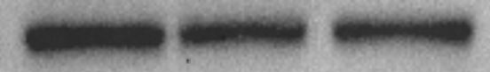
Simple Western lane view shows lysates of mouse kidney tissue, loaded at 0.2 mg/mL. A specific band was detected for ACE-2 at approximately 141 kDa (as indicated) using 2.5 µg/mL of Goat Anti-Mouse ACE-2 Antigen Affinity-purified Polyclonal Antibody (Catalog # AF3437) followed by 1:50 dilution of HRP-conjugated Anti-Goat IgG Secondary Antibody (Catalog # HAF109). This experiment was conducted under reducing conditions and using the 12-230 kDa separation system.
Mouse ACE‑2 ELISA Standard Curve.
Recombinant Mouse ACE‑2 protein was serially diluted 2-fold and captured by Rat Anti-Mouse ACE‑2 Monoclonal Antibody (MAB34371) coated on a Clear Polystyrene Microplate (DY990). Goat Anti-Mouse ACE‑2 Antigen Affinity-purified Polyclonal Antibody (Catalog # AF3437) was biotinylated and incubated with the protein captured on the plate. Detection of the standard curve was achieved by incubating Streptavidin-HRP (DY998) followed by Substrate Solution (DY999) and stopping the enzymatic reaction with Stop Solution (DY994).
Detection of ACE-2 by Western Blot
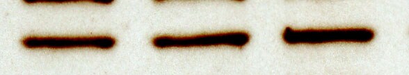
Expression of vasoactive proteins (mRNA and protein levels) in response to different oxygen conditions (mRNA: fold change relative to control condition: 21% O2; protein: % of control condition: 21% O2) as determined by qRT‐PCR using actb as house‐keeping gene and the delta delta Ct method and western blotting from cell lysates. (a) NOS3 mRNA expression, protein expression at 72 hr and Griess reaction (analyzed levels of NO2− and NO3− in the supernatants resulting from NOS activity). (b) ACE1, ACE2 mRNA expression and ACE1, ACE2 intracellular protein expression at 72 hr. (c) ACE1 and ACE2 protein levels in the cell culture supernatant as determined by Western blotting and mRNA expression of sheddases ADAM10 and ADAM17. p‐values result from statistical testing with Student's t‐test (mRNA), one‐way ANOVA (protein) and two‐way ANOVA (Griess) with Tukey's multiple comparison test. *p < .05; **p < .01; ***p < .001, ****p < .0001 Image collected and cropped by CiteAb from the following open publication (https://pubmed.ncbi.nlm.nih.gov/33565273), licensed under a CC-BY license. Not internally tested by R&D Systems.
Detection of ACE-2 by Flow Cytometry
regDCs EXO are taken up by acceptor PBTECs, inhibiting ACE2 expression in vitro. (A) Uptake of Dil labeled EXO (red) by PBTECs, counterstained with nuclear stain DAPI (blue), phalloidin (Alex flour 647) for cell membrane and visualized under confocal microscopy. ACE2 mRNA expression (B) and flow cytometry scattergrams showing ACE2 positive cells percentage (C) in PBTECs treated or not treated with iDCs or regDCs EXO in the presence or absence of TGF beta R1 inhibitor SB431542. (D) representative bar graph of (C). Results shown are representative of three independent experiments (*P < 0.05 by one-way ANOVA followed by Tukey’s multiple comparisons). Image collected and cropped by CiteAb from the following open publication (https://pubmed.ncbi.nlm.nih.gov/33841418), licensed under a CC-BY license. Not internally tested by R&D Systems.
Mouse ACE-2 Standard Curve
Recombinant Mouse ACE‑2 (Catalog # 3437-ZN) was serially diluted and captured by Rat Anti-Mouse ACE‑2 Monoclonal Antibody (Catalog # MAB34371) coated on a Clear Polystyrene Microplate (Catalog # DY990). Goat Anti-Mouse ACE‑2 Antigen Affinity-purified Polyclonal Antibody (Catalog # AF3437) was biotinylated and incubated with the protein captured on the plate. Detection of the standard curve was achieved by incubating Streptavidin-HRP (Catalog # DY998)Applications for Mouse ACE-2 Antibody
ELISA
This antibody functions as an ELISA detection antibody when paired with Rat Anti-Mouse ACE‑2 Monoclonal Antibody (Catalog # MAB34371).
This product is intended for assay development on various assay platforms requiring antibody pairs. We recommend the Mouse ACE-2 DuoSet ELISA Kit (Catalog # DY3437-05) for convenient development of a sandwich ELISA.
Flow Cytometry
Sample: HEK293 human embryonic kidney cell line transfected with mouse ACE-2 and eGFP
Immunohistochemistry
Sample: Perfusion fixed frozen sections of mouse kidney
Immunoprecipitation
Sample: Conditioned cell culture medium spiked with Recombinant Mouse ACE‑2 (Catalog # 3437‑ZN), see our available Western blot detection antibodies
Simple Western
Sample: Mouse kidney tissue
Western Blot
Sample: Mouse kidney tissue
Reviewed Applications
Read 3 reviews rated 4.7 using AF3437 in the following applications:
Flow Cytometry Panel Builder
Bio-Techne Knows Flow Cytometry
Save time and reduce costly mistakes by quickly finding compatible reagents using the Panel Builder Tool.
Advanced Features
- Spectra Viewer - Custom analysis of spectra from multiple fluorochromes
- Spillover Popups - Visualize the spectra of individual fluorochromes
- Antigen Density Selector - Match fluorochrome brightness with antigen density

Formulation, Preparation, and Storage
Purification
Reconstitution
Reconstitute at 0.2 mg/mL in sterile PBS. For liquid material, refer to CoA for concentration.
Formulation
Shipping
Stability & Storage
- 12 months from date of receipt, -20 to -70 °C as supplied.
- 1 month, 2 to 8 °C under sterile conditions after reconstitution.
- 6 months, -20 to -70 °C under sterile conditions after reconstitution.
Calculators
Background: ACE-2
References
- Tipnis, S.R. et al. (2000) J. Biol. Chem. 275:33238.
- Crackower, M.A. et al. (2002) Nature 417:822.
- Huang, L. et al. (2003) J. Biol. Chem. 278:15532.
- Kuba, K. et al. (2005) Nature Med. 11:875.
- Ima, Y. et al. (2005) Nature 436:112.
Long Name
Alternate Names
Entrez Gene IDs
Gene Symbol
UniProt
Additional ACE-2 Products
Product Documents for Mouse ACE-2 Antibody
Certificate of Analysis
To download a Certificate of Analysis, please enter a lot or batch number in the search box below.
Note: Certificate of Analysis not available for kit components.
Product Specific Notices for Mouse ACE-2 Antibody
For research use only
Related Research Areas
Citations for Mouse ACE-2 Antibody
 Powered by Bioz
Customer Reviews for Mouse ACE-2 Antibody (3)
Have you used Mouse ACE-2 Antibody?
Submit a review and receive an Amazon gift card!
$25/€18/£15/$25CAN/¥2500 Yen for a review with an image
$10/€7/£6/$10CAN/¥1110 Yen for a review without an image
Submit a review
Customer Images
-
Application: Western BlotSample Tested: Heart tissueSpecies: MouseVerified Customer | Posted 08/26/2016

-
Application: Western BlotSample Tested: Adult kidneySpecies: MouseVerified Customer | Posted 06/10/2016
-
Application: Western BlotSample Tested: See PMID 22777933Species: MouseVerified Customer | Posted 01/07/2015
There are no reviews that match your criteria.
Protocols
Find general support by application which include: protocols, troubleshooting, illustrated assays, videos and webinars.
- 7-Amino Actinomycin D (7-AAD) Cell Viability Flow Cytometry Protocol
- Antigen Retrieval Protocol (PIER)
- Antigen Retrieval for Frozen Sections Protocol
- Appropriate Fixation of IHC/ICC Samples
- Cellular Response to Hypoxia Protocols
- Chromogenic IHC Staining of Formalin-Fixed Paraffin-Embedded (FFPE) Tissue Protocol
- Chromogenic Immunohistochemistry Staining of Frozen Tissue
- ClariTSA™ Fluorophore Kits
- Detection & Visualization of Antibody Binding
- ELISA Sample Preparation & Collection Guide
- ELISA Troubleshooting Guide
- Extracellular Membrane Flow Cytometry Protocol
- Flow Cytometry Protocol for Cell Surface Markers
- Flow Cytometry Protocol for Staining Membrane Associated Proteins
- Flow Cytometry Staining Protocols
- Flow Cytometry Troubleshooting Guide
- Fluorescent IHC Staining of Frozen Tissue Protocol
- Graphic Protocol for Heat-induced Epitope Retrieval
- Graphic Protocol for the Preparation and Fluorescent IHC Staining of Frozen Tissue Sections
- Graphic Protocol for the Preparation and Fluorescent IHC Staining of Paraffin-embedded Tissue Sections
- Graphic Protocol for the Preparation of Gelatin-coated Slides for Histological Tissue Sections
- How to Run an R&D Systems DuoSet ELISA
- How to Run an R&D Systems Quantikine ELISA
- How to Run an R&D Systems Quantikine™ QuicKit™ ELISA
- IHC Sample Preparation (Frozen sections vs Paraffin)
- Immunofluorescent IHC Staining of Formalin-Fixed Paraffin-Embedded (FFPE) Tissue Protocol
- Immunohistochemistry (IHC) and Immunocytochemistry (ICC) Protocols
- Immunohistochemistry Frozen Troubleshooting
- Immunohistochemistry Paraffin Troubleshooting
- Immunoprecipitation Protocol
- Intracellular Flow Cytometry Protocol Using Alcohol (Methanol)
- Intracellular Flow Cytometry Protocol Using Detergents
- Intracellular Nuclear Staining Flow Cytometry Protocol Using Detergents
- Intracellular Staining Flow Cytometry Protocol Using Alcohol Permeabilization
- Intracellular Staining Flow Cytometry Protocol Using Detergents to Permeabilize Cells
- Preparing Samples for IHC/ICC Experiments
- Preventing Non-Specific Staining (Non-Specific Binding)
- Primary Antibody Selection & Optimization
- Propidium Iodide Cell Viability Flow Cytometry Protocol
- Protocol for Heat-Induced Epitope Retrieval (HIER)
- Protocol for Liperfluo
- Protocol for Making a 4% Formaldehyde Solution in PBS
- Protocol for VisUCyte™ HRP Polymer Detection Reagent
- Protocol for the Characterization of Human Th22 Cells
- Protocol for the Characterization of Human Th9 Cells
- Protocol for the Preparation & Fixation of Cells on Coverslips
- Protocol for the Preparation and Chromogenic IHC Staining of Frozen Tissue Sections
- Protocol for the Preparation and Chromogenic IHC Staining of Frozen Tissue Sections - Graphic
- Protocol for the Preparation and Chromogenic IHC Staining of Paraffin-embedded Tissue Sections
- Protocol for the Preparation and Chromogenic IHC Staining of Paraffin-embedded Tissue Sections - Graphic
- Protocol for the Preparation and Fluorescent IHC Staining of Frozen Tissue Sections
- Protocol for the Preparation and Fluorescent IHC Staining of Paraffin-embedded Tissue Sections
- Protocol for the Preparation of Gelatin-coated Slides for Histological Tissue Sections
- Protocol: Annexin V and PI Staining by Flow Cytometry
- Protocol: Annexin V and PI Staining for Apoptosis by Flow Cytometry
- Quantikine HS ELISA Kit Assay Principle, Alkaline Phosphatase
- Quantikine HS ELISA Kit Principle, Streptavidin-HRP Polymer
- R&D Systems Quality Control Western Blot Protocol
- Sandwich ELISA (Colorimetric) – Biotin/Streptavidin Detection Protocol
- Sandwich ELISA (Colorimetric) – Direct Detection Protocol
- TUNEL and Active Caspase-3 Detection by IHC/ICC Protocol
- The Importance of IHC/ICC Controls
- Troubleshooting Guide: ELISA
- Troubleshooting Guide: Fluorokine Flow Cytometry Kits
- Troubleshooting Guide: Immunohistochemistry
- Troubleshooting Guide: Western Blot Figures
- Western Blot Conditions
- Western Blot Protocol
- Western Blot Protocol for Cell Lysates
- Western Blot Troubleshooting
- Western Blot Troubleshooting Guide
- View all Protocols, Troubleshooting, Illustrated assays and Webinars
Associated Pathways

