BMP-4 is a TGF-beta superfamily ligand that is widely expressed from early embryogenesis through adulthood. It plays an important role in mesenchyme formation, epidermal determination, suppression of neural induction, the development of multiple organs, and tissue repair (1-5). The mouse BMP-4 precursor contains a 273 amino acid (aa) propeptide and a 116 aa mature protein (6). The propeptide is cleaved intracellularly by furin or proprotein convertase 6, enabling the 15 kDa mature BMP-4 monomer to form an active disulfide linked homodimer or heterodimer with BMP-7 (7-9). Mature mouse and human BMP-4 share 98% aa sequence identity. Mouse BMP-4 shares 85% aa sequence identity with mouse BMP-2 and 35%-54% with other mouse BMPs. Compared to BMP-4 homodimers, BMP-4/BMP-7 heterodimers exhibit a greater potency in inducing osteogenic differentiation (9). In Xenopus, the heterodimers can also induce the formation of mesoderm, whereas BMP-4 homodimers only provide ventralizing signals for existing mesoderm (10). BMP-4 signals through tetrameric complexes composed of type I (primarily Activin RIA or BMPR-IA) and type II (primarily Activin RIIA or BMPR-II) receptors (11, 12). The bioavailability of BMP-4 is regulated by its interaction with multiple proteins and glycosaminoglycans (13-15).
Mouse BMP-4 Antibody (1128D)
R&D Systems | Catalog # MAB5020
Recombinant Monoclonal Antibody.

Key Product Details
Species Reactivity
Validated:
Mouse
Cited:
Mouse
Applications
Validated:
Immunohistochemistry, Western Blot
Cited:
Immunohistochemistry-Paraffin
Label
Unconjugated
Antibody Source
Recombinant Monoclonal Rabbit IgG Clone # 1128D
Loading...
Product Specifications
Immunogen
Chinese hamster ovary cell line CHO-derived mature mouse BMP-4
Ser293-Arg408
Accession # P21275
Ser293-Arg408
Accession # P21275
Specificity
Detects mouse BMP-4 in direct ELISAs and Western blots.
Clonality
Monoclonal
Host
Rabbit
Isotype
IgG
Scientific Data Images for Mouse BMP-4 Antibody (1128D)
Detection of Mouse BMP‑4 by Western Blot.
Western blot shows Recombinant Mouse BMP-4 (Catalog # 5020-BP) and lysates of mouse embryo tissue. PVDF membrane was probed with 0.25 µg/mL of Rabbit Anti-Mouse BMP-4 Monoclonal Antibody (Catalog # MAB5020) followed by HRP-conjugated Anti-Rabbit IgG Secondary Antibody (Catalog # HAF008). Specific bands were detected for BMP-4 at approximately 40-55 kDa (as indicated). This experiment was conducted under reducing conditions and using Immunoblot Buffer Group 1.
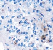
BMP‑4 in Mouse Embryo.
BMP-4 was detected in immersion fixed frozen sections of mouse embryo (15 d.p.c.) using Rabbit Anti-Mouse BMP-4 Monoclonal Antibody (Catalog # MAB5020) at 2 µg/mL overnight at 4 °C. Tissue was stained using the Anti-Rabbit HRP-DAB Cell & Tissue Staining Kit (brown; Catalog # CTS005) and counterstained with hematoxylin (blue). Specific staining was localized to cytoplasm in gastrointestinal tract cells. View our protocol for Chromogenic IHC Staining of Frozen Tissue Sections.Applications for Mouse BMP-4 Antibody (1128D)
Application
Recommended Usage
Immunohistochemistry
1-15 µg/mL
Sample: Immersion fixed frozen sections of mouse embryo (15 d.p.c.)
Sample: Immersion fixed frozen sections of mouse embryo (15 d.p.c.)
Western Blot
0.25 µg/mL
Sample: Mouse embryo tissue
Sample: Mouse embryo tissue
Reviewed Applications
Read 2 reviews rated 4.5 using MAB5020 in the following applications:
Formulation, Preparation, and Storage
Purification
Protein A or G purified from cell culture supernatant
Reconstitution
Reconstitute at 0.5 mg/mL in sterile PBS. For liquid material, refer to CoA for concentration.
Loading...
Formulation
Lyophilized from a 0.2 μm filtered solution in PBS with Trehalose. *Small pack size (SP) is supplied either lyophilized or as a 0.2 µm filtered solution in PBS.
Shipping
Lyophilized product is shipped at ambient temperature. Liquid small pack size (-SP) is shipped with polar packs. Upon receipt, store immediately at the temperature recommended below.
Stability & Storage
Use a manual defrost freezer and avoid repeated freeze-thaw cycles.
- 12 months from date of receipt, -20 to -70 °C as supplied.
- 1 month, 2 to 8 °C under sterile conditions after reconstitution.
- 6 months, -20 to -70 °C under sterile conditions after reconstitution.
Calculators
Background: BMP-4
References
- Zhang, P. et al. (2008) Blood 111:1933.
- Gambaro, K. et al. (2006) Cell Death Differ. 13:1075.
- Simic, P. and S. Vukicevic (2005) Cytokine Growth Factor Rev. 16:299.
- Sadlon, T.J. et al. (2004) Stem Cells 22:457.
- Frank, D.B. et al. (2005) Circ. Res. 97:496.
- Oida, S. et al. (1995) DNA Seq. 5:273.
- Cui, Y. et al. (1998) EMBO J. 17:4735.
- Cui, Y. et al. (2001) Genes Dev. 15:2797.
- Aono, A. et al. (1995) Biochem. Biophys. Res. Commun. 210:670.
- Nishimatsu, S. and G.H. Thomsen (1998) Mech. Dev. 74:75.
- Chen, D. et al. (2004) Growth Factors 22:233.
- Lavery, K. et al. (2008) J. Biol. Chem. April 24 epub.
- Rosen, V. (2006) Ann. N.Y. Acad. Sci. 1068:19.
- Jones, C.M. and J.C. Smith (1998) Dev. Biol. 194:12.
- Takada, T. et al. (2003) J. Biol. Chem. 278:43229.
Long Name
Bone Morphogenetic Protein 4
Alternate Names
BMP2B, BMP2B1, BMP4, MCOPS6, OFC11
Gene Symbol
BMP4
UniProt
Additional BMP-4 Products
Product Documents for Mouse BMP-4 Antibody (1128D)
Certificate of Analysis
To download a Certificate of Analysis, please enter a lot or batch number in the search box below.
Note: Certificate of Analysis not available for kit components.
Product Specific Notices for Mouse BMP-4 Antibody (1128D)
For research use only
Citations for Mouse BMP-4 Antibody (1128D)
 Powered by Bioz
Customer Reviews for Mouse BMP-4 Antibody (1128D) (2)
4.5 out of 5
2 Customer Ratings
Have you used Mouse BMP-4 Antibody (1128D)?
Submit a review and receive an Amazon gift card!
$25/€18/£15/$25CAN/¥2500 Yen for a review with an image
$10/€7/£6/$10CAN/¥1110 Yen for a review without an image
Submit a review
Customer Images
Showing
1
-
2 of
2 reviews
Showing All
Filter By:
-
Application: ImmunohistochemistrySample Tested: Pituitary gland tissueSpecies: MouseVerified Customer | Posted 10/30/2021

-
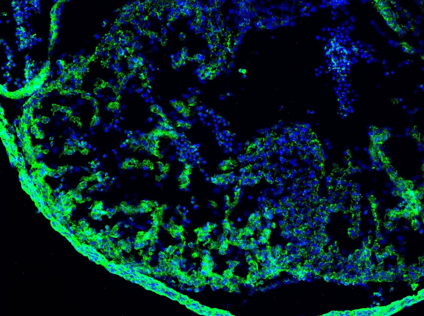
Application: Immunocytochemistry/ImmunofluorescenceSample Tested: E11.5 mouse embryo fixed in 4% PFASpecies: MouseVerified Customer | Posted 12/02/2020Antibody was used to stain heart region on E11.5 mouse embryo.
There are no reviews that match your criteria.
Protocols
Find general support by application which include: protocols, troubleshooting, illustrated assays, videos and webinars.
- Antigen Retrieval Protocol (PIER)
- Antigen Retrieval for Frozen Sections Protocol
- Appropriate Fixation of IHC/ICC Samples
- Cellular Response to Hypoxia Protocols
- Chromogenic IHC Staining of Formalin-Fixed Paraffin-Embedded (FFPE) Tissue Protocol
- Chromogenic Immunohistochemistry Staining of Frozen Tissue
- ClariTSA™ Fluorophore Kits
- Detection & Visualization of Antibody Binding
- Fluorescent IHC Staining of Frozen Tissue Protocol
- Graphic Protocol for Heat-induced Epitope Retrieval
- Graphic Protocol for the Preparation and Fluorescent IHC Staining of Frozen Tissue Sections
- Graphic Protocol for the Preparation and Fluorescent IHC Staining of Paraffin-embedded Tissue Sections
- Graphic Protocol for the Preparation of Gelatin-coated Slides for Histological Tissue Sections
- IHC Sample Preparation (Frozen sections vs Paraffin)
- Immunofluorescent IHC Staining of Formalin-Fixed Paraffin-Embedded (FFPE) Tissue Protocol
- Immunohistochemistry (IHC) and Immunocytochemistry (ICC) Protocols
- Immunohistochemistry Frozen Troubleshooting
- Immunohistochemistry Paraffin Troubleshooting
- Preparing Samples for IHC/ICC Experiments
- Preventing Non-Specific Staining (Non-Specific Binding)
- Primary Antibody Selection & Optimization
- Protocol for Heat-Induced Epitope Retrieval (HIER)
- Protocol for Making a 4% Formaldehyde Solution in PBS
- Protocol for VisUCyte™ HRP Polymer Detection Reagent
- Protocol for the Preparation & Fixation of Cells on Coverslips
- Protocol for the Preparation and Chromogenic IHC Staining of Frozen Tissue Sections
- Protocol for the Preparation and Chromogenic IHC Staining of Frozen Tissue Sections - Graphic
- Protocol for the Preparation and Chromogenic IHC Staining of Paraffin-embedded Tissue Sections
- Protocol for the Preparation and Chromogenic IHC Staining of Paraffin-embedded Tissue Sections - Graphic
- Protocol for the Preparation and Fluorescent IHC Staining of Frozen Tissue Sections
- Protocol for the Preparation and Fluorescent IHC Staining of Paraffin-embedded Tissue Sections
- Protocol for the Preparation of Gelatin-coated Slides for Histological Tissue Sections
- R&D Systems Quality Control Western Blot Protocol
- TUNEL and Active Caspase-3 Detection by IHC/ICC Protocol
- The Importance of IHC/ICC Controls
- Troubleshooting Guide: Immunohistochemistry
- Troubleshooting Guide: Western Blot Figures
- Western Blot Conditions
- Western Blot Protocol
- Western Blot Protocol for Cell Lysates
- Western Blot Troubleshooting
- Western Blot Troubleshooting Guide
- View all Protocols, Troubleshooting, Illustrated assays and Webinars


