FGF-8 is a member of the fibroblast growth factor family that was originally discovered as a growth factor essential for the androgen-dependent growth of mouse mammary carcinoma cells (1‑3). Alternate splicing of mouse FGF-8 mRNA generates eight secreted isoforms, designated a‑h, but only FGF-8a, b, e and f exist in humans (4). FGF-8 contains a 22 amino acid (aa) signal sequence, an N‑terminal domain that varies according to the isoform (30 aa for FGF-8b; 20 aa for the shortest, FGF-8a), a 125 aa FGF domain and a 37 aa proline‑rich C‑terminal sequence. The FGF domain of FGF-8 shares the most aa identity with FGF17 (75%) and FGF-18 (67%), and the three form an FGF subfamily (2). Mouse FGF-8b shares 100% aa identity with human FGF-8b. FGF-8 is widely expressed during embryogenesis, and mediates epithelial-mesenchymal transitions. It plays an organizing and inducing role during gastrulation, and regulates patterning of the midbrain/hindbrain, eye, ear, limbs and heart in the embryo (2, 5‑8). The isoforms may play different roles in development. FGF-8b shows the strongest receptor affinity and oncogenic transforming capacity although FGF-8a and FGF-8e are also transforming and have been found in human prostate, breast or ovarian tumors (1, 5, 9‑12). FGF-8 shows limited expression in the normal adult, but low levels are found in the reproductive and genitourinary tract, peripheral leukocytes and bone marrow hematopoietic cells (3, 9, 13).
Mouse FGF-8 b Isoform Antibody
R&D Systems | Catalog # AF-423-NA

Key Product Details
Species Reactivity
Validated:
Mouse
Cited:
Mouse, Avian - Chicken
Applications
Validated:
Immunohistochemistry, Western Blot, Neutralization
Cited:
Neutralization
Label
Unconjugated
Antibody Source
Polyclonal Goat IgG
Loading...
Product Specifications
Immunogen
E. coli-derived recombinant mouse FGF-8b
Gln23-Arg215
Accession # NP_006110
Gln23-Arg215
Accession # NP_006110
Specificity
Detects mouse FGF‑8b Isoform in direct ELISAs and Western blots. In direct ELISAs and Western blots, less than 1% cross-reactivity with recombinant human (rh) FGF acidic, rhFGF basic, rhFGF-4, rhFGF-5, rhFGF-6, rhFGF-7 and rhFGF-9 is observed. It does, however,cross‑react with recombinant mouse FGF-8c in neutralizing bioassay.
Clonality
Polyclonal
Host
Goat
Isotype
IgG
Endotoxin Level
<0.10 EU per 1 μg of the antibody by the LAL method.
Scientific Data Images for Mouse FGF-8 b Isoform Antibody
Cell Proliferation Induced by FGF‑8 and Neutralization by Mouse FGF‑8 Antibody.
Recombinant Mouse FGF-8b Isoform (Catalog # 423-F8) stimulates proliferation in the the NR6R-3T3 mouse fibroblast cell line in a dose-dependent manner (orange line), as measured by Resazurin (Catalog # AR002). Proliferation elicited by Recombinant Mouse FGF-8b Isoform (60 ng/mL) is neutralized (green line) by increasing concentrations of Goat Anti-Mouse FGF-8b Isoform Antigen Affinity-purified Polyclonal Antibody (Catalog # AF-423-NA). The ND50 is typically 0.4-2.4 µg/mL in the presence of heparin (1 µg/mL).
FGF‑8 in Human Prostate.
FGF-8 was detected in immersion fixed paraffin-embedded sections of human prostate using Goat Anti-Mouse FGF-8 b Isoform Antigen Affinity-purified Polyclonal Antibody (Catalog # AF-423-NA) at 15 µg/mL overnight at 4 °C. Tissue was stained using the Anti-Goat HRP-DAB Cell & Tissue Staining Kit (brown; Catalog # CTS008) and counterstained with hematoxylin (blue). Specific staining was localized to epithelial and endothelial cells. View our protocol for Chromogenic IHC Staining of Paraffin-embedded Tissue Sections.Applications for Mouse FGF-8 b Isoform Antibody
Application
Recommended Usage
Immunohistochemistry
5-15 µg/mL
Sample: Immersion fixed paraffin-embedded sections of human prostate
Sample: Immersion fixed paraffin-embedded sections of human prostate
Western Blot
0.1 µg/mL
Sample: Recombinant Mouse FGF‑8b Isoform (Catalog # 423-F8)
Sample: Recombinant Mouse FGF‑8b Isoform (Catalog # 423-F8)
Neutralization
Measured by its ability to neutralize FGF‑8-induced proliferation in the NR6R‑3T3 mouse fibroblast cell line. Rizzino, A. et al. (1988) Cancer Res. 48:4266. The Neutralization Dose (ND50) is typically 0.4-2.4 µg/mL in the presence of 60 ng/mL Recombinant Mouse FGF‑8b Isoform and 1 µg/mL heparin.
Reviewed Applications
Read 1 review rated 5 using AF-423-NA in the following applications:
Formulation, Preparation, and Storage
Purification
Antigen Affinity-purified
Reconstitution
Reconstitute at 0.2 mg/mL in sterile PBS. For liquid material, refer to CoA for concentration.
Loading...
Formulation
Lyophilized from a 0.2 μm filtered solution in PBS with Trehalose. *Small pack size (SP) is supplied either lyophilized or as a 0.2 µm filtered solution in PBS.
Shipping
Lyophilized product is shipped at ambient temperature. Liquid small pack size (-SP) is shipped with polar packs. Upon receipt, store immediately at the temperature recommended below.
Stability & Storage
Use a manual defrost freezer and avoid repeated freeze-thaw cycles.
- 12 months from date of receipt, -20 to -70 °C as supplied.
- 1 month, 2 to 8 °C under sterile conditions after reconstitution.
- 6 months, -20 to -70 °C under sterile conditions after reconstitution.
Calculators
Background: FGF-8
References
- Mattila, M.M. and P.L. Harkonen (2007) Cytokine Growth Factor Rev. 18:257.
- Reuss, B. and O. von Bohlen und Halbach (2003) Cell Tiss. Res. 313:139.
- Tanaka, A. et al. (1992) Proc. Natl. Acad. Sci. USA 89:8928.
- Gemel, J. et al. (1996) Genomics 35:253.
- Olsen, S.K. et al. (2006) Genes Dev. 20:185.
- Crossley, P.H. et al. (1996) Cell, 84:127.
- Heikinheimo, M. et al. (1994) Mech. Dev. 48:129.
- Sun, X. et al. (1999) Genes Dev. 13:1834.
- Ghosh, A.K. et al. (1996) Cell Growth Differ. 7:1425.
- Mattila, M.M. et al. (2001) Oncogene 20:2791.
- Valve, E. et al. (2000) Int. J. Cancer 88:718.
- Valve, E.M. et al. (2001) Lab. Invest. 81:815.
- Nezu, M. et al. (2005) Biochem. Biophys. Res. Commun. 335:843.
Long Name
Fibroblast Growth Factor 8
Alternate Names
AIGF, FGF8, HBGF-8
Gene Symbol
FGF8
UniProt
Additional FGF-8 Products
Product Documents for Mouse FGF-8 b Isoform Antibody
Certificate of Analysis
To download a Certificate of Analysis, please enter a lot or batch number in the search box below.
Note: Certificate of Analysis not available for kit components.
Product Specific Notices for Mouse FGF-8 b Isoform Antibody
For research use only
Related Research Areas
Citations for Mouse FGF-8 b Isoform Antibody
 Powered by Bioz
Customer Reviews for Mouse FGF-8 b Isoform Antibody (1)
5 out of 5
1 Customer Rating
Have you used Mouse FGF-8 b Isoform Antibody?
Submit a review and receive an Amazon gift card!
$25/€18/£15/$25CAN/¥2500 Yen for a review with an image
$10/€7/£6/$10CAN/¥1110 Yen for a review without an image
Submit a review
Customer Images
Showing
1
-
1 of
1 review
Showing All
Filter By:
-
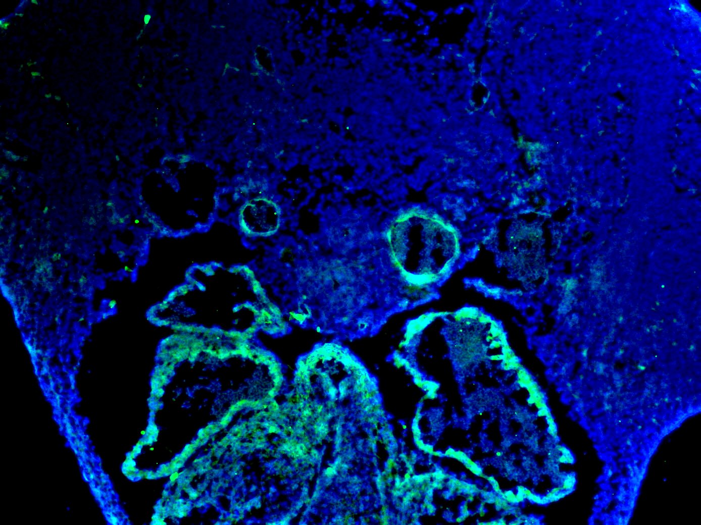
Application: Immunocytochemistry/ImmunofluorescenceSample Tested: E11.5 mouse embryo fixed in 4% PFASpecies: MouseVerified Customer | Posted 02/12/2021Dilution used - 1:50. The staining was done on an E11.5 mouse transverse section (fixed on 4% PFA overnight) and done using standard IF techniques. Blocking - 1 hour with 1% BSA before addition of the primary antibody Secondary antibody - Alexa Fluor Donkey Anti Goat 488

There are no reviews that match your criteria.
Protocols
Find general support by application which include: protocols, troubleshooting, illustrated assays, videos and webinars.
- Antigen Retrieval Protocol (PIER)
- Antigen Retrieval for Frozen Sections Protocol
- Appropriate Fixation of IHC/ICC Samples
- Cellular Response to Hypoxia Protocols
- Chromogenic IHC Staining of Formalin-Fixed Paraffin-Embedded (FFPE) Tissue Protocol
- Chromogenic Immunohistochemistry Staining of Frozen Tissue
- ClariTSA™ Fluorophore Kits
- Detection & Visualization of Antibody Binding
- Fluorescent IHC Staining of Frozen Tissue Protocol
- Graphic Protocol for Heat-induced Epitope Retrieval
- Graphic Protocol for the Preparation and Fluorescent IHC Staining of Frozen Tissue Sections
- Graphic Protocol for the Preparation and Fluorescent IHC Staining of Paraffin-embedded Tissue Sections
- Graphic Protocol for the Preparation of Gelatin-coated Slides for Histological Tissue Sections
- IHC Sample Preparation (Frozen sections vs Paraffin)
- Immunofluorescent IHC Staining of Formalin-Fixed Paraffin-Embedded (FFPE) Tissue Protocol
- Immunohistochemistry (IHC) and Immunocytochemistry (ICC) Protocols
- Immunohistochemistry Frozen Troubleshooting
- Immunohistochemistry Paraffin Troubleshooting
- Preparing Samples for IHC/ICC Experiments
- Preventing Non-Specific Staining (Non-Specific Binding)
- Primary Antibody Selection & Optimization
- Protocol for Heat-Induced Epitope Retrieval (HIER)
- Protocol for Making a 4% Formaldehyde Solution in PBS
- Protocol for VisUCyte™ HRP Polymer Detection Reagent
- Protocol for the Preparation & Fixation of Cells on Coverslips
- Protocol for the Preparation and Chromogenic IHC Staining of Frozen Tissue Sections
- Protocol for the Preparation and Chromogenic IHC Staining of Frozen Tissue Sections - Graphic
- Protocol for the Preparation and Chromogenic IHC Staining of Paraffin-embedded Tissue Sections
- Protocol for the Preparation and Chromogenic IHC Staining of Paraffin-embedded Tissue Sections - Graphic
- Protocol for the Preparation and Fluorescent IHC Staining of Frozen Tissue Sections
- Protocol for the Preparation and Fluorescent IHC Staining of Paraffin-embedded Tissue Sections
- Protocol for the Preparation of Gelatin-coated Slides for Histological Tissue Sections
- R&D Systems Quality Control Western Blot Protocol
- TUNEL and Active Caspase-3 Detection by IHC/ICC Protocol
- The Importance of IHC/ICC Controls
- Troubleshooting Guide: Immunohistochemistry
- Troubleshooting Guide: Western Blot Figures
- Western Blot Conditions
- Western Blot Protocol
- Western Blot Protocol for Cell Lysates
- Western Blot Troubleshooting
- Western Blot Troubleshooting Guide
- View all Protocols, Troubleshooting, Illustrated assays and Webinars

