Frizzled-4, designated CD344, is a 7-transmembrane glycoprotein of the Frizzled family within the G-protein coupled receptor superfamily (1, 2). Frizzled proteins function as receptors for Wnt proteins and can activate canonical Wnt/beta-catenin signaling as well as planar cell polarity and calcium flux pathways (1). Frizzled-4 is particularly important in angiogenic Wnt pathway signaling (1, 5). Frizzleds contain a divergent N-terminal signal peptide, a highly conserved ~120 amino acid (aa) cysteine-rich domain (CRD), a variable length linker region, seven transmembrane domains, and a variable-length C-terminal tail (1). The mouse Frizzled-4 cDNA encodes 537 aa with a 36 aa signal sequence and a 186 aa N-terminal extracellular sequence (4). The portion expressed includes aa 37-180, and shares 93%, 94%, 90%, 89%, and 88% identity with human, rat, equine, bovine and canine Frizzled-4, respectively. This portion competes for Wnt binding with endogenous receptors. In humans, a 122 aa soluble form that diverges at aa 95 is proposed to be a positive regulator of Wnt signaling pathways (5). Frizzled-4 is unusual in binding a non-wnt ligand, Norrin, in addition to binding Wnt ligands (1, 3, 6). Norrin binds the Frizzled-4 CRD, activates Wnt signaling pathways and uses LRP5/6 as co-receptors (3, 6). Deletion of either Frizzled-4 or Norrin in mice results in a similar phenotype including malformation of vasculature in the retina, cerebellar degeneration, and loss of hair cells in the inner ear (1, 3, 7). In humans, blindness due to familial exudative vitreoretinopathy (FEVR) is associated with mutations producing loss of function of Frizzled-4 or Norrin, designated EVR1 and EVR2, respectively (1, 3, 8). Frizzled-4 expression has been found in many tissues, including mouse ovary, where it influences corpus luteum vasculogenesis and is necessary for fertility (4, 9).

Key Product Details
Species Reactivity
Validated:
Mouse
Cited:
Human, Mouse
Applications
Validated:
Immunohistochemistry, Western Blot
Cited:
Immunohistochemistry, Western Blot, ELISA Development
Label
Unconjugated
Antibody Source
Polyclonal Goat IgG
Loading...
Product Specifications
Immunogen
Mouse myeloma cell line NS0-derived recombinant mouse Frizzled-4
Phe37-Glu180
Accession # Q8BLL2
Phe37-Glu180
Accession # Q8BLL2
Specificity
Detects mouse Frizzled-4 in direct ELISAs and Western blots. In direct ELISAs, approximately 5% cross-reactivity with recombinant mouse (rm) Frizzled-8 and less than 2% cross-reactivity with rmFrizzled-3 and rmFrizzled-7 is observed.
Clonality
Polyclonal
Host
Goat
Isotype
IgG
Scientific Data Images for Mouse Frizzled-4 Antibody
Frizzled‑4 in Embryonic Mouse Neuronal Tissue.
Frizzled‑4 was detected in immersion fixed frozen sections of embryonic mouse neuronal tissue (E11) using 15 µg/mL Goat Anti-Mouse Frizzled‑4 Antigen Affinity-purified Polyclonal Antibody (Catalog # AF194) overnight at 4 °C. Tissue was stained with the Anti-Goat HRP-DAB Cell & Tissue Staining Kit (brown; Catalog # CTS008) and counterstained with hematoxylin (blue). Specific labeling was localized to the plasma membrane and cytoplasm of neuronal cells in the midbrain. View our protocol for Chromogenic IHC Staining of Frozen Tissue Sections.Applications for Mouse Frizzled-4 Antibody
Application
Recommended Usage
Immunohistochemistry
5-15 µg/mL
Sample: Immersion fixed frozen sections of embryonic mouse neuronal tissue (E11)
Sample: Immersion fixed frozen sections of embryonic mouse neuronal tissue (E11)
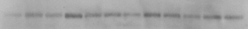
Western Blot
0.1 µg/mL
Sample: Recombinant Mouse Frizzled‑4 Fc Chimera (Catalog # 194-FZ)
Sample: Recombinant Mouse Frizzled‑4 Fc Chimera (Catalog # 194-FZ)
Reviewed Applications
Read 1 review rated 3 using AF194 in the following applications:
Formulation, Preparation, and Storage
Purification
Antigen Affinity-purified
Reconstitution
Reconstitute at 0.2 mg/mL in sterile PBS. For liquid material, refer to CoA for concentration.
Loading...
Formulation
Lyophilized from a 0.2 μm filtered solution in PBS with Trehalose. *Small pack size (SP) is supplied either lyophilized or as a 0.2 µm filtered solution in PBS.
Shipping
Lyophilized product is shipped at ambient temperature. Liquid small pack size (-SP) is shipped with polar packs. Upon receipt, store immediately at the temperature recommended below.
Stability & Storage
Use a manual defrost freezer and avoid repeated freeze-thaw cycles.
- 12 months from date of receipt, -20 to -70 °C as supplied.
- 1 month, 2 to 8 °C under sterile conditions after reconstitution.
- 6 months, -20 to -70 °C under sterile conditions after reconstitution.
Calculators
Background: Frizzled-4
References
- Huang, H-C. and P.S. Klein (2004) Genome Biol. 5:234.
- Parmalee, N.L. and J. Kitajewski (2008) Curr. Drug Targets 9:558.
- Xu, Q. et al. (2004) Cell 116:883.
- Wang, Y. et al. (1996) J. Biol. Chem. 271:4468.
- Sagara, N. et al. (2001) Biochem. Biophys. Res. Commun. 282:750.
- Smallwood, P.M. et al. (2007) J. Biol. Chem. 282:4057.
- Wang, Y. et al. (2001) J. Neurosci. 21:4761.
- Robitaille, J. et al. (2002) Nat. Genet. 32:326.
- Hsieh, M. et al. (2005) Biol. Reprod. 73:1135.
Alternate Names
CD344, Frizzled4, FZD4
Gene Symbol
FZD4
UniProt
Additional Frizzled-4 Products
Product Documents for Mouse Frizzled-4 Antibody
Certificate of Analysis
To download a Certificate of Analysis, please enter a lot or batch number in the search box below.
Note: Certificate of Analysis not available for kit components.
Product Specific Notices for Mouse Frizzled-4 Antibody
For research use only
Related Research Areas
Citations for Mouse Frizzled-4 Antibody
 Powered by Bioz
Customer Reviews for Mouse Frizzled-4 Antibody (1)
3 out of 5
1 Customer Rating
Have you used Mouse Frizzled-4 Antibody?
Submit a review and receive an Amazon gift card!
$25/€18/£15/$25CAN/¥2500 Yen for a review with an image
$10/€7/£6/$10CAN/¥1110 Yen for a review without an image
Submit a review
Customer Images
Showing
1
-
1 of
1 review
Showing All
Filter By:
-
Application: Western BlotSample Tested: primary cortical neuron cultureSpecies: MouseVerified Customer | Posted 07/02/2021used at 0.1ug/ml in 5% Blocking Solution (Biorad) overnight at 4C

There are no reviews that match your criteria.
Protocols
Find general support by application which include: protocols, troubleshooting, illustrated assays, videos and webinars.
- Antigen Retrieval Protocol (PIER)
- Antigen Retrieval for Frozen Sections Protocol
- Appropriate Fixation of IHC/ICC Samples
- Cellular Response to Hypoxia Protocols
- Chromogenic IHC Staining of Formalin-Fixed Paraffin-Embedded (FFPE) Tissue Protocol
- Chromogenic Immunohistochemistry Staining of Frozen Tissue
- ClariTSA™ Fluorophore Kits
- Detection & Visualization of Antibody Binding
- Fluorescent IHC Staining of Frozen Tissue Protocol
- Graphic Protocol for Heat-induced Epitope Retrieval
- Graphic Protocol for the Preparation and Fluorescent IHC Staining of Frozen Tissue Sections
- Graphic Protocol for the Preparation and Fluorescent IHC Staining of Paraffin-embedded Tissue Sections
- Graphic Protocol for the Preparation of Gelatin-coated Slides for Histological Tissue Sections
- IHC Sample Preparation (Frozen sections vs Paraffin)
- Immunofluorescent IHC Staining of Formalin-Fixed Paraffin-Embedded (FFPE) Tissue Protocol
- Immunohistochemistry (IHC) and Immunocytochemistry (ICC) Protocols
- Immunohistochemistry Frozen Troubleshooting
- Immunohistochemistry Paraffin Troubleshooting
- Preparing Samples for IHC/ICC Experiments
- Preventing Non-Specific Staining (Non-Specific Binding)
- Primary Antibody Selection & Optimization
- Protocol for Heat-Induced Epitope Retrieval (HIER)
- Protocol for Making a 4% Formaldehyde Solution in PBS
- Protocol for VisUCyte™ HRP Polymer Detection Reagent
- Protocol for the Preparation & Fixation of Cells on Coverslips
- Protocol for the Preparation and Chromogenic IHC Staining of Frozen Tissue Sections
- Protocol for the Preparation and Chromogenic IHC Staining of Frozen Tissue Sections - Graphic
- Protocol for the Preparation and Chromogenic IHC Staining of Paraffin-embedded Tissue Sections
- Protocol for the Preparation and Chromogenic IHC Staining of Paraffin-embedded Tissue Sections - Graphic
- Protocol for the Preparation and Fluorescent IHC Staining of Frozen Tissue Sections
- Protocol for the Preparation and Fluorescent IHC Staining of Paraffin-embedded Tissue Sections
- Protocol for the Preparation of Gelatin-coated Slides for Histological Tissue Sections
- R&D Systems Quality Control Western Blot Protocol
- TUNEL and Active Caspase-3 Detection by IHC/ICC Protocol
- The Importance of IHC/ICC Controls
- Troubleshooting Guide: Immunohistochemistry
- Troubleshooting Guide: Western Blot Figures
- Western Blot Conditions
- Western Blot Protocol
- Western Blot Protocol for Cell Lysates
- Western Blot Troubleshooting
- Western Blot Troubleshooting Guide
- View all Protocols, Troubleshooting, Illustrated assays and Webinars
Loading...
Associated Pathways

