Complement Component C3d Antibody
R&D Systems | Catalog # AF2655

Key Product Details
Species Reactivity
Validated:
Cited:
Applications
Validated:
Cited:
Label
Antibody Source
Product Specifications
Immunogen
His1002-Arg1303 (Cys1010Ser)
Accession # P01027
Specificity
Clonality
Host
Isotype
Scientific Data Images for Complement Component C3d Antibody
Complement Component C3d in Mouse Kidney.
Complement Component C3d was detected in perfusion fixed frozen sections of mouse kidney using Goat Anti-Mouse Complement Component C3d Antigen Affinity-purified Polyclonal Antibody (Catalog # AF2655) overnight at 4 °C. Tissue was stained using the NorthernLights™ 557-conjugated Anti-Goat IgG Secondary Antibody (red; Catalog # NL001) and counterstained with DAPI (blue). Specific staining was localized to basement membrane. View our protocol for Fluorescent IHC Staining of Frozen Tissue Sections.
Complement Component C3d in Rat Kidney.
Complement Component C3d was detected in perfusion fixed paraffin-embedded sections of rat kidney using Goat Anti-Mouse Complement Component C3d Antigen Affinity-purified Polyclonal Antibody (Catalog # AF2655) at 3 µg/mL for 1 hour at room temperature followed by incubation with the Anti-Goat IgG VisUCyte™ HRP Polymer Antibody (Catalog # VC004). Before incubation with the primary antibody, tissue was subjected to heat-induced epitope retrieval using Antigen Retrieval Reagent-Basic (Catalog # CTS013). Tissue was stained using DAB (brown) and counterstained with hematoxylin (blue). Specific staining was localized to cytoplasm. View our protocol for IHC Staining with VisUCyte HRP Polymer Detection Reagents.
Detection of Mouse Mouse/Rat Complement Component C3d Antibody by Immunohistochemistry
The activation of mTORC1 in KCs enhances complement alternative system.a Expression heatmap of genes of neutrophils chemotaxis or complement activation analyzed by RNA-seq from Tsc1+/+ and Tsc1-/- BMMs (n = 3 each). b Western blotting result was shown the expression of CFB protein in hepatic tissues from mice. c Left, representative co-immunofluorescent staining images for F4/80 with CFB. Scale bar = 50 μm. Right, quantitative determination of F4/80+ and CFB+ cells among groups as indicated, n = 3. d Western blotting assay showing the abundance for TSC1, CFB, and p-S6 in BMMs. e qRT-PCR analysis showing the CFB mRNA abundance in BMMs, n = 3. f Western blotting assay showing the abundance for TSC1, CFB, and p-S6 in KCs. g qRT-PCR analysis showing the CFB mRNA abundance in KCs, n = 3. h Representative immunofluorescent staining images for C3d. Scale bar = 50 μm. i Representative immunostaining images for C5b-9. Scale bar = 50 μm. j Western blotting assay showing the abundance for Rheb and CFB in BMMs. k Western blotting assay showing the abundance for Rheb and CFB in KCs. l Representative co-immunofluorescent staining images for F4/80 with CFB (white arrows). Scale bar = 100 μm. m Quantitative determination of F4/80+ and CFB+ cells among groups as indicated, n = 3. n Representative immunofluorescent staining images for C3d. Scale bar = 100 μm. o Representative immunostaining images for C5b-9 among groups as indicated. Scale bar = 100 μm. *p < 0.05. Image collected and cropped by CiteAb from the following publication (https://pubmed.ncbi.nlm.nih.gov/36494334), licensed under a CC-BY license. Not internally tested by R&D Systems.
Detection of Mouse Mouse/Rat Complement Component C3d Antibody by Immunohistochemistry
Down regulation of CFB in liver protects against Con-A induced liver injury.a Western blotting assay showing CFB expression in mouse livers after shRNA-CFB injection. b Representative co-immunofluorescent staining images for F4/80 with CFB (white arrows). Scale bar = 100 μm. c The strategy for establishing a mouse model of injection of shRNA-CFB and Con-A administration. d The ALT levels in serum of mice exposed to Con-A for 8 h, n = 6. e Representative HE-stained mouse livers. Scale bar = 100 μm. f Liver sections of were immunofluorescent stained with TUNEL. Scale bar = 200 μm. g Left, representative immunofluorescent staining images for ly6b. Scale bar = 100 μm. Right, quantitative determination of ly6b+ cells among groups as indicated, n = 4. h Left, representative immunofluorescent staining images for C3d. Scale bar = 100 μm. Right, quantitative determination of C3d area in a field of vision, n = 4. i Representative immunostaining images for C5b-9. Scale bar = 100 μm. j Left, representative immunofluorescent staining images for F4/80 (white arrows). Scale bar = 100 μm. Right, quantitative determination of F4/80+ cells among groups as indicated, n = 4. *p < 0.05. k Schematic working model on the role of mTORC1 activation in hepatocytes and KCs in the pathogenesis of ALD. Image collected and cropped by CiteAb from the following publication (https://pubmed.ncbi.nlm.nih.gov/36494334), licensed under a CC-BY license. Not internally tested by R&D Systems.Applications for Complement Component C3d Antibody
Immunohistochemistry
Sample: Perfusion fixed frozen sections of mouse kidney and perfusion fixed paraffin-embedded sections of rat kidney
Western Blot
Sample: Recombinant Mouse Complement Component C3d
Reviewed Applications
Read 2 reviews rated 4 using AF2655 in the following applications:
Formulation, Preparation, and Storage
Purification
Reconstitution
Reconstitute at 0.2 mg/mL in sterile PBS. For liquid material, refer to CoA for concentration.
Formulation
Shipping
Stability & Storage
- 12 months from date of receipt, -20 to -70 °C as supplied.
- 1 month, 2 to 8 °C under sterile conditions after reconstitution.
- 6 months, -20 to -70 °C under sterile conditions after reconstitution.
Calculators
Background: Complement Component C3d
Alternate Names
Entrez Gene IDs
Gene Symbol
UniProt
Additional Complement Component C3d Products
Product Documents for Complement Component C3d Antibody
Certificate of Analysis
To download a Certificate of Analysis, please enter a lot or batch number in the search box below.
Note: Certificate of Analysis not available for kit components.
Product Specific Notices for Complement Component C3d Antibody
For research use only
Related Research Areas
Citations for Complement Component C3d Antibody
 Powered by Bioz
Customer Reviews for Complement Component C3d Antibody (2)
Have you used Complement Component C3d Antibody?
Submit a review and receive an Amazon gift card!
$25/€18/£15/$25CAN/¥2500 Yen for a review with an image
$10/€7/£6/$10CAN/¥1110 Yen for a review without an image
Submit a review
Customer Images
-
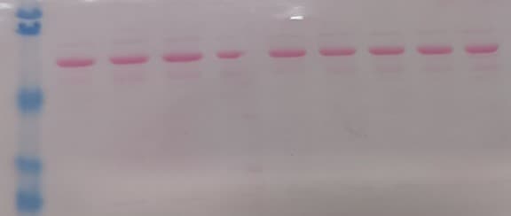
Application: Western BlotSample Tested: SerumSpecies: MouseVerified Customer | Posted 01/27/2024

-
Application: Immunocytochemistry/ImmunofluorescenceSample Tested: A549 human lung carcinoma cell lineSpecies: HumanVerified Customer | Posted 12/19/2016
There are no reviews that match your criteria.
Protocols
Find general support by application which include: protocols, troubleshooting, illustrated assays, videos and webinars.
- Antigen Retrieval Protocol (PIER)
- Antigen Retrieval for Frozen Sections Protocol
- Appropriate Fixation of IHC/ICC Samples
- Cellular Response to Hypoxia Protocols
- Chromogenic IHC Staining of Formalin-Fixed Paraffin-Embedded (FFPE) Tissue Protocol
- Chromogenic Immunohistochemistry Staining of Frozen Tissue
- ClariTSA™ Fluorophore Kits
- Detection & Visualization of Antibody Binding
- Fluorescent IHC Staining of Frozen Tissue Protocol
- Graphic Protocol for Heat-induced Epitope Retrieval
- Graphic Protocol for the Preparation and Fluorescent IHC Staining of Frozen Tissue Sections
- Graphic Protocol for the Preparation and Fluorescent IHC Staining of Paraffin-embedded Tissue Sections
- Graphic Protocol for the Preparation of Gelatin-coated Slides for Histological Tissue Sections
- IHC Sample Preparation (Frozen sections vs Paraffin)
- Immunofluorescent IHC Staining of Formalin-Fixed Paraffin-Embedded (FFPE) Tissue Protocol
- Immunohistochemistry (IHC) and Immunocytochemistry (ICC) Protocols
- Immunohistochemistry Frozen Troubleshooting
- Immunohistochemistry Paraffin Troubleshooting
- Preparing Samples for IHC/ICC Experiments
- Preventing Non-Specific Staining (Non-Specific Binding)
- Primary Antibody Selection & Optimization
- Protocol for Heat-Induced Epitope Retrieval (HIER)
- Protocol for Making a 4% Formaldehyde Solution in PBS
- Protocol for VisUCyte™ HRP Polymer Detection Reagent
- Protocol for the Preparation & Fixation of Cells on Coverslips
- Protocol for the Preparation and Chromogenic IHC Staining of Frozen Tissue Sections
- Protocol for the Preparation and Chromogenic IHC Staining of Frozen Tissue Sections - Graphic
- Protocol for the Preparation and Chromogenic IHC Staining of Paraffin-embedded Tissue Sections
- Protocol for the Preparation and Chromogenic IHC Staining of Paraffin-embedded Tissue Sections - Graphic
- Protocol for the Preparation and Fluorescent IHC Staining of Frozen Tissue Sections
- Protocol for the Preparation and Fluorescent IHC Staining of Paraffin-embedded Tissue Sections
- Protocol for the Preparation of Gelatin-coated Slides for Histological Tissue Sections
- R&D Systems Quality Control Western Blot Protocol
- TUNEL and Active Caspase-3 Detection by IHC/ICC Protocol
- The Importance of IHC/ICC Controls
- Troubleshooting Guide: Immunohistochemistry
- Troubleshooting Guide: Western Blot Figures
- Western Blot Conditions
- Western Blot Protocol
- Western Blot Protocol for Cell Lysates
- Western Blot Troubleshooting
- Western Blot Troubleshooting Guide
- View all Protocols, Troubleshooting, Illustrated assays and Webinars
FAQs for Complement Component C3d Antibody
-
Q: Does Mouse/Rat Complement Component C3d Antibody, Catalog # AF2655, detect/cross-react with C3b or intact C3?
A: Since complement component C3d is derived from C3b, we expect AF2655 to recognize C3b and C3d in western blot. We have not tested AF2655 against intact C3, but there is published literature that shows that intact C3 is not recogniozed by AF2655 in IHC.
-
Q: For Mouse/Rat Complement Component C3d Antibody, Catalog # AF2655, what is the expected molecular weight in Western Blot of the recombinant mouse and natural Complement Component C3d?
A: The mouse Complement Component C3d is expected to have a moleculat weight of ~34 kDa.
-
Q: Does Mouse/Rat Complement Component C3d Antibody, Catalog # AF2655, detect/cross-react with C3b or intact C3?
A: Since complement component C3d is derived from C3b, we expect AF2655 to recognize C3b and C3d in western blot. We have not tested AF2655 against intact C3, but there is published literature that shows that intact C3 is not recogniozed by AF2655 in IHC.
-
Q: For Mouse/Rat Complement Component C3d Antibody, Catalog # AF2655, what is the expected molecular weight in Western Blot of the recombinant mouse and natural Complement Component C3d?
A: The mouse Complement Component C3d is expected to have a moleculat weight of ~34 kDa.