
Key Product Details
Species Reactivity
Validated:
Mouse
Cited:
Human, Mouse, Rat, Transgenic Mouse
Applications
Validated:
Immunohistochemistry, Western Blot, Immunocytochemistry, Simple Western
Cited:
Immunohistochemistry, Immunohistochemistry-Paraffin, Immunohistochemistry-Frozen, Western Blot, Neutralization, Confocal Microscopy, ELISA Development, Functional Assay
Label
Unconjugated
Antibody Source
Polyclonal Goat IgG
Loading...
Product Specifications
Immunogen
E. coli-derived recombinant mouse S100A9
Ala2-Lys113
Accession # P31725
Ala2-Lys113
Accession # P31725
Specificity
Detects mouse S100A9 in direct ELISAs. In direct ELISAs, less than 2% cross‑reactivity with recombinant mouse S100A10 and recombinant human S100B is observed.
Clonality
Polyclonal
Host
Goat
Isotype
IgG
Scientific Data Images for Mouse S100A9 Antibody
Detection of Mouse S100A9 by Western Blot.
Western blot shows lysate of mouse lung tissue. PVDF membrane was probed with 0.2 µg/mL of Goat Anti-Mouse S100A9 Antigen Affinity-purified Polyclonal Antibody (Catalog # AF2065) followed by HRP-conjugated Anti-Goat IgG Secondary Antibody (HAF017). A specific band was detected for S100A9 at approximately 14 kDa (as indicated). This experiment was conducted under reducing conditions and using Immunoblot Buffer Group 1.
S100A9 in XB2 Mouse Cell Line.
S100A9 was detected in immersion fixed XB2 mouse teratoma keratinocyte cell line using 10 µg/mL Goat Anti-Mouse S100A9 Antigen Affinity-purified Polyclonal Antibody (Catalog # AF2065) for 3 hours at room temperature. Cells were stained with the NorthernLights™ 557-conjugated Anti-Goat IgG Secondary Antibody (red; NL001) and counte-rstained with DAPI (blue). View our protocol for Fluorescent ICC Staining of Cells on Coverslips.
S100A9 in Mouse Splenocytes.
S100A9 was detected in immersion fixed mouse splenocytes using Goat Anti-Mouse S100A9 Antigen Affinity-purified Polyclonal Antibody (Catalog # AF2065) at 5 µg/mL for 3 hours at room temperature. Cells were stained using the NorthernLights™ 557-conjugated Anti-Goat IgG Secondary Antibody (red; NL001) and counterstained with DAPI (blue). Specific staining was localized to cell surfaces and cytoplasm. View our protocol for Fluorescent ICC Staining of Non-adherent Cells.
S100A9 in Mouse Spleen.
S100A9 was detected in perfusion fixed frozen sections of mouse spleen using Goat Anti-Mouse S100A9 Antigen Affinity-purified Polyclonal Antibody (Catalog # AF2065) at 0.3 µg/mL for 1 hour at room temperature followed by incubation with the Anti-Goat IgG VisUCyte™ HRP Polymer Antibody (VC004). Tissue was stained using DAB (brown) and counterstained with hematoxylin (blue). Specific staining was localized to cytoplasm in splenocytes. View our protocol for IHC Staining with VisUCyte HRP Polymer Detection Reagents.
Detection of Mouse S100A9 by Simple Western™
Simple Western lane view shows lysates of mouse lung tissue and mouse spleen tissue, loaded at 0.2 mg/mL. A specific band was detected for S100A9 at approximately 20 kDa (as indicated) using 2 µg/mL of Goat Anti-Mouse S100A9 Antigen Affinity-purified Polyclonal Antibody (Catalog # AF2065) followed by 1:50 dilution of HRP-conjugated Anti-Goat IgG Secondary Antibody (HAF109). This experiment was conducted under reducing conditions and using the 12-230 kDa separation system.
Detection of S100A9 by Western Blot
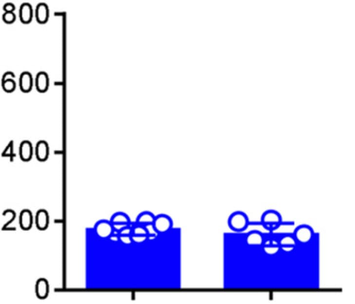
S100A8/A9 proteins augment differentiation and osteoclastic activity derived from iOCPs but not from hOCPs. a Expression of S100A8, S100A9 and RAGE in sorted control and inflamed BM iOCPs and hOCPs. The protein phosphatase 2A catalytic subunit (PP2Ac) is shown as a loading control. b Densitometry measurements of three biological repeats (no statistical analysis presented). c Sorted control and inflamed BM iOCPs and hOCPs (5 × 10⁴) were cultured on the Osteo assay surface with or without anti-RAGE blocking antibodies (bar: 50 µm). d Pit area quantitation. e Sorted iOCPs and hOCPs (5 × 10⁴) from the BM of control mice were cultured on the Osteo assay surface in combination with a recombinant S100A8/A9 heterodimer and anti-RAGE blocking antibodies as indicated (bar: 50 µm). f Pit area quantitation. c–f Depict representative results for two independent experiments, n = 5 for each group. Line: median, box: 25th-75th percentile, whiskers: range. *P < 0.05 (Mann–Whitney test and Holm multiplicity correction) Image collected and cropped by CiteAb from the following open publication (https://pubmed.ncbi.nlm.nih.gov/35396510), licensed under a CC-BY license. Not internally tested by R&D Systems.
Detection of Mouse S100A9 by Immunohistochemistry
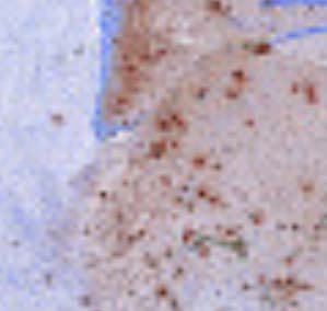
iASPP deficient keratinocytes induce pro-inflammatory gene expression in vitro and attract macrophages in vivo(A) qPCR analysis of mRNA expression levels of inflammatory genes (and iASPP gene Ppp1r13l) in iASPP WT and KO primary keratinocytes at 0, 1, and 6 h after treatment with TNF-alpha. Values are mean + SD. ∗p < 0.05, ∗∗p < 0.01, ∗∗∗p < 0.001, ∗∗∗∗p < 0.0001; n = 3 biological replicates.(B) Left, IHC staining of S100a8 and S100a9 in untreated skin sections from iASPP WT and KO mice (adjacent sections). Scale bar, 50 μm. Right, IB of iASPP and S100a9 expression levels in iASPP WT (−tamoxifen) and KO (+tamoxifen) primary keratinocytes.(C) H&E analysis of acetone- or TPA-treated skin sections from iASPP WT and KO mice. Scale bar, 50 μm. Histograms below show epidermal thickness in the same samples. Values are mean ± SD. n = 3 (WT) and n = 4 (KO) mice in acetone cohort; n = 4 (WT), n = 3 (KO) mice in TPA cohort.(D) IHC staining of F4/80-positive macrophages in acetone- or TPA-treated skin sections from iASPP WT and KO mice. Scale bar, 50 μm. Histograms below show quantification of the same samples. Values are mean + SD. Same cohort as in (C).See STAR Methods for p calculations for (A), (C), and (D). Image collected and cropped by CiteAb from the following open publication (https://pubmed.ncbi.nlm.nih.gov/36261000), licensed under a CC-BY license. Not internally tested by R&D Systems.
Detection of Mouse S100A9 by Immunohistochemistry
iASPP deficient keratinocytes induce pro-inflammatory gene expression in vitro and attract macrophages in vivo(A) qPCR analysis of mRNA expression levels of inflammatory genes (and iASPP gene Ppp1r13l) in iASPP WT and KO primary keratinocytes at 0, 1, and 6 h after treatment with TNF-alpha. Values are mean + SD. ∗p < 0.05, ∗∗p < 0.01, ∗∗∗p < 0.001, ∗∗∗∗p < 0.0001; n = 3 biological replicates.(B) Left, IHC staining of S100a8 and S100a9 in untreated skin sections from iASPP WT and KO mice (adjacent sections). Scale bar, 50 μm. Right, IB of iASPP and S100a9 expression levels in iASPP WT (−tamoxifen) and KO (+tamoxifen) primary keratinocytes.(C) H&E analysis of acetone- or TPA-treated skin sections from iASPP WT and KO mice. Scale bar, 50 μm. Histograms below show epidermal thickness in the same samples. Values are mean ± SD. n = 3 (WT) and n = 4 (KO) mice in acetone cohort; n = 4 (WT), n = 3 (KO) mice in TPA cohort.(D) IHC staining of F4/80-positive macrophages in acetone- or TPA-treated skin sections from iASPP WT and KO mice. Scale bar, 50 μm. Histograms below show quantification of the same samples. Values are mean + SD. Same cohort as in (C).See STAR Methods for p calculations for (A), (C), and (D). Image collected and cropped by CiteAb from the following open publication (https://pubmed.ncbi.nlm.nih.gov/36261000), licensed under a CC-BY license. Not internally tested by R&D Systems.Applications for Mouse S100A9 Antibody
Application
Recommended Usage
Immunocytochemistry
5-15 µg/mL
Sample: Immersion fixed XB2 mouse teratoma keratinocyte cell line and mouse splenocytes
Sample: Immersion fixed XB2 mouse teratoma keratinocyte cell line and mouse splenocytes
Immunohistochemistry
0.3-15 µg/mL
Sample: Perfusion fixed frozen sections of mouse spleen
Sample: Perfusion fixed frozen sections of mouse spleen
Simple Western
2 µg/mL
Sample: Mouse lung tissue and mouse spleen tissue
Sample: Mouse lung tissue and mouse spleen tissue
Western Blot
0.2 µg/mL
Sample: Mouse lung tissue
Sample: Mouse lung tissue
Reviewed Applications
Read 5 reviews rated 4.6 using AF2065 in the following applications:
Formulation, Preparation, and Storage
Purification
Antigen Affinity-purified
Reconstitution
Reconstitute at 0.2 mg/mL in sterile PBS. For liquid material, refer to CoA for concentration.
Loading...
Formulation
Lyophilized from a 0.2 μm filtered solution in PBS with Trehalose. See Certificate of Analysis for details.
*Small pack size (-SP) is supplied either lyophilized or as a 0.2 µm filtered solution in PBS.
*Small pack size (-SP) is supplied either lyophilized or as a 0.2 µm filtered solution in PBS.
Shipping
Lyophilized product is shipped at ambient temperature. Liquid small pack size (-SP) is shipped with polar packs. Upon receipt, store immediately at the temperature recommended below.
Stability & Storage
Use a manual defrost freezer and avoid repeated freeze-thaw cycles.
- 12 months from date of receipt, -20 to -70 °C as supplied.
- 1 month, 2 to 8 °C under sterile conditions after reconstitution.
- 6 months, -20 to -70 °C under sterile conditions after reconstitution.
Calculators
Background: S100A9
Long Name
S100 Calcium Binding Protein A9
Alternate Names
Calgranulin B, MRP-14
Gene Symbol
S100A9
UniProt
Additional S100A9 Products
Product Documents for Mouse S100A9 Antibody
Certificate of Analysis
To download a Certificate of Analysis, please enter a lot or batch number in the search box below.
Note: Certificate of Analysis not available for kit components.
Product Specific Notices for Mouse S100A9 Antibody
For research use only
Citations for Mouse S100A9 Antibody
 Powered by Bioz
Customer Reviews for Mouse S100A9 Antibody (5)
4.6 out of 5
5 Customer Ratings
Have you used Mouse S100A9 Antibody?
Submit a review and receive an Amazon gift card!
$25/€18/£15/$25CAN/¥2500 Yen for a review with an image
$10/€7/£6/$10CAN/¥1110 Yen for a review without an image
Submit a review
Customer Images
Showing
1
-
5 of
5 reviews
Showing All
Filter By:
-
Application: ELISASample Tested: A549 human lung carcinoma cell lineSpecies: HumanVerified Customer | Posted 05/24/2022

-
Application: ImmunohistochemistrySample Tested: Adult brainSpecies: HumanVerified Customer | Posted 04/04/2022
-
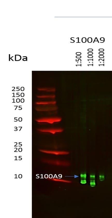
Application: Western BlotSample Tested: mouse fecesSpecies: MouseVerified Customer | Posted 05/03/2017S100A9 detection in mouse feces at 1:500, 1:1000 and 1:2000 dilution. Incubation time was 2 hrs at RT. Antibody in 1% milk TBST
-
Application: Immunohistochemistry-FrozenSample Tested: See PMID 21228116Species: MouseVerified Customer | Posted 01/06/2015
-
Application: ImmunofluorescenceSample Tested: See PMID 23481567Species: MouseVerified Customer | Posted 01/06/2015
There are no reviews that match your criteria.
Protocols
Find general support by application which include: protocols, troubleshooting, illustrated assays, videos and webinars.
- Antigen Retrieval Protocol (PIER)
- Antigen Retrieval for Frozen Sections Protocol
- Appropriate Fixation of IHC/ICC Samples
- Cellular Response to Hypoxia Protocols
- Chromogenic IHC Staining of Formalin-Fixed Paraffin-Embedded (FFPE) Tissue Protocol
- Chromogenic Immunohistochemistry Staining of Frozen Tissue
- ClariTSA™ Fluorophore Kits
- Detection & Visualization of Antibody Binding
- Fluorescent IHC Staining of Frozen Tissue Protocol
- Graphic Protocol for Heat-induced Epitope Retrieval
- Graphic Protocol for the Preparation and Fluorescent IHC Staining of Frozen Tissue Sections
- Graphic Protocol for the Preparation and Fluorescent IHC Staining of Paraffin-embedded Tissue Sections
- Graphic Protocol for the Preparation of Gelatin-coated Slides for Histological Tissue Sections
- ICC Cell Smear Protocol for Suspension Cells
- ICC Immunocytochemistry Protocol Videos
- ICC for Adherent Cells
- IHC Sample Preparation (Frozen sections vs Paraffin)
- Immunocytochemistry (ICC) Protocol
- Immunocytochemistry Troubleshooting
- Immunofluorescence of Organoids Embedded in Cultrex Basement Membrane Extract
- Immunofluorescent IHC Staining of Formalin-Fixed Paraffin-Embedded (FFPE) Tissue Protocol
- Immunohistochemistry (IHC) and Immunocytochemistry (ICC) Protocols
- Immunohistochemistry Frozen Troubleshooting
- Immunohistochemistry Paraffin Troubleshooting
- Preparing Samples for IHC/ICC Experiments
- Preventing Non-Specific Staining (Non-Specific Binding)
- Primary Antibody Selection & Optimization
- Protocol for Heat-Induced Epitope Retrieval (HIER)
- Protocol for Making a 4% Formaldehyde Solution in PBS
- Protocol for VisUCyte™ HRP Polymer Detection Reagent
- Protocol for the Fluorescent ICC Staining of Cell Smears - Graphic
- Protocol for the Fluorescent ICC Staining of Cultured Cells on Coverslips - Graphic
- Protocol for the Preparation & Fixation of Cells on Coverslips
- Protocol for the Preparation and Chromogenic IHC Staining of Frozen Tissue Sections
- Protocol for the Preparation and Chromogenic IHC Staining of Frozen Tissue Sections - Graphic
- Protocol for the Preparation and Chromogenic IHC Staining of Paraffin-embedded Tissue Sections
- Protocol for the Preparation and Chromogenic IHC Staining of Paraffin-embedded Tissue Sections - Graphic
- Protocol for the Preparation and Fluorescent ICC Staining of Cells on Coverslips
- Protocol for the Preparation and Fluorescent ICC Staining of Non-adherent Cells
- Protocol for the Preparation and Fluorescent ICC Staining of Stem Cells on Coverslips
- Protocol for the Preparation and Fluorescent IHC Staining of Frozen Tissue Sections
- Protocol for the Preparation and Fluorescent IHC Staining of Paraffin-embedded Tissue Sections
- Protocol for the Preparation of Gelatin-coated Slides for Histological Tissue Sections
- Protocol for the Preparation of a Cell Smear for Non-adherent Cell ICC - Graphic
- R&D Systems Quality Control Western Blot Protocol
- TUNEL and Active Caspase-3 Detection by IHC/ICC Protocol
- The Importance of IHC/ICC Controls
- Troubleshooting Guide: Immunohistochemistry
- Troubleshooting Guide: Western Blot Figures
- Western Blot Conditions
- Western Blot Protocol
- Western Blot Protocol for Cell Lysates
- Western Blot Troubleshooting
- Western Blot Troubleshooting Guide
- View all Protocols, Troubleshooting, Illustrated assays and Webinars
Loading...