Key Product Details
Species Reactivity
Validated:
Porcine
Cited:
Human
Applications
Validated:
Western Blot
Cited:
Flow Cytometry
Label
Unconjugated
Antibody Source
Polyclonal Goat IgG
Loading...
Product Specifications
Immunogen
Mouse myeloma cell line NS0-derived recombinant porcine CD34
Ala20-Thr294 (Thr32Ala, Ala52Thr, Pro71Ser, Val126Ala, His278Asn)
Accession # NP_999251
Ala20-Thr294 (Thr32Ala, Ala52Thr, Pro71Ser, Val126Ala, His278Asn)
Accession # NP_999251
Specificity
Detects porcine CD34 in direct ELISAs and Western blots. In direct ELISAs, less than 20% cross‑reactivity with recombinant rat CD34 is observed, and less than 5% cross-reactivity with recombinant mouse CD34 and recombinant human CD34 is observed.
Clonality
Polyclonal
Host
Goat
Isotype
IgG
Applications for Porcine CD34 Antibody
Application
Recommended Usage
Western Blot
0.1 µg/mL
Sample: Recombinant Porcine CD34
Sample: Recombinant Porcine CD34
Reviewed Applications
Read 1 review rated 5 using AF3890 in the following applications:
Formulation, Preparation, and Storage
Purification
Antigen Affinity-purified
Reconstitution
Reconstitute at 0.2 mg/mL in sterile PBS. For liquid material, refer to CoA for concentration.
Loading...
Formulation
Lyophilized from a 0.2 μm filtered solution in PBS with Trehalose. *Small pack size (SP) is supplied either lyophilized or as a 0.2 µm filtered solution in PBS.
Shipping
Lyophilized product is shipped at ambient temperature. Liquid small pack size (-SP) is shipped with polar packs. Upon receipt, store immediately at the temperature recommended below.
Stability & Storage
Use a manual defrost freezer and avoid repeated freeze-thaw cycles.
- 12 months from date of receipt, -20 to -70 °C as supplied.
- 1 month, 2 to 8 °C under sterile conditions after reconstitution.
- 6 months, -20 to -70 °C under sterile conditions after reconstitution.
Calculators
Background: CD34
Alternate Names
CD34, HPCA1
Gene Symbol
CD34
UniProt
Additional CD34 Products
Product Documents for Porcine CD34 Antibody
Certificate of Analysis
To download a Certificate of Analysis, please enter a lot or batch number in the search box below.
Note: Certificate of Analysis not available for kit components.
Product Specific Notices for Porcine CD34 Antibody
For research use only
Related Research Areas
Citations for Porcine CD34 Antibody
 Powered by Bioz
Customer Reviews for Porcine CD34 Antibody (1)
5 out of 5
1 Customer Rating
Have you used Porcine CD34 Antibody?
Submit a review and receive an Amazon gift card!
$25/€18/£15/$25CAN/¥2500 Yen for a review with an image
$10/€7/£6/$10CAN/¥1110 Yen for a review without an image
Submit a review
Customer Images
Showing
1
-
1 of
1 review
Showing All
Filter By:
-
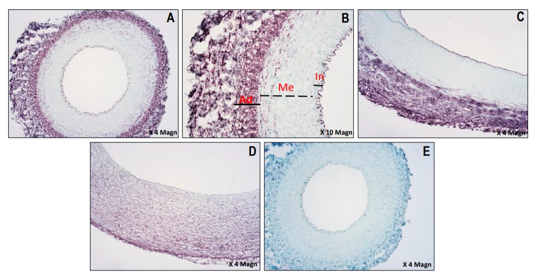
Application: Immunohistochemistry-FrozenSample Tested: porcine arteries and ArteriesSpecies: PorcineVerified Customer | Posted 12/03/2020Expression of CD34 (purple stain) in the healthy pig carotid (A, B), abdominal aorta (C) and coronary artery (D). Control with nuclei staining only (E). Intima: In, Media: Me, Adventitia: AdPig healthy arterial sections of 7µm were fixed in ice cold acetone for 7min. Once dry, blocking buffer 4% FCS/TBS was added to all tissues for 30min to minimize non-specific binding, followed by 10 min incubation with Bloxall, endogenous peroxidase blocking solution at room temperature. Tissues were washed in Tris buffered saline (TBS) for 5 min and incubated for 20min with normal horse serum (Vector laboratories # S-2012-50). Polyclonal goat anti-CD34 (R&D systems # AF3890) was diluted at 1:80 in 0.5% FCS/TBS and added to the arterial tissues for 1 hour incubation at room temperature. Following 5 min wash in TBS, tissues were incubated for 30min with ImmPRESS®HRP horse anti-goat reagent (Vector laboratories # MP-7405). To detect CD34 antigen, tissues were washed in TBS and incubated with VIP HRP substrate (Vector®VIP Substrate kit, Peroxidase (HRP) # SK-4600) for 1 min 30sec then placed immediately in MilliQ water to stop the reaction. Nuclei were counter-stained with methyl green (Sigma-Aldrich # M8884-25G). Control tissues were stained similarly in the absence of primary antibodies. Tissue sections were immediately dehydrated and mounted with ENTELLAN mounting medium (Proscitech, Cat#IA013). Images were captured on Nikon eclipse ME600 microscope and DS-Fi2 camera.

There are no reviews that match your criteria.
Protocols
Find general support by application which include: protocols, troubleshooting, illustrated assays, videos and webinars.
- Cellular Response to Hypoxia Protocols
- R&D Systems Quality Control Western Blot Protocol
- Troubleshooting Guide: Western Blot Figures
- Western Blot Conditions
- Western Blot Protocol
- Western Blot Protocol for Cell Lysates
- Western Blot Troubleshooting
- Western Blot Troubleshooting Guide
- View all Protocols, Troubleshooting, Illustrated assays and Webinars


