IL-1 is a name that designates two pleiotropic cytokines, IL-1 alpha (IL-1F1) and IL-1 beta (IL-1F2), which are the products of distinct genes. IL-1 alpha and IL-1 beta are structurally related polypeptides that share approximately 27% amino acid (aa) identity in porcine. Both proteins are produced by a wide variety of cells in response to inflammatory agents, infections, or microbial endotoxins. While IL-1 alpha and IL-1 beta are regulated independently, they bind to the same receptor and exert identical biological effects. IL-1 RI binds directly to IL-1 alpha or IL-1 beta and then associates with IL-1 R accessory protein (IL-1 R3/IL-1 R AcP) to form a high-affinity receptor complex that is competent for signal transduction. IL-1 RII has high affinity for IL-1 beta but functions as a decoy receptor and negative regulator of IL-1 beta activity. IL-1ra functions as a competitive antagonist by preventing IL-1 alpha and IL-1 beta from interacting with IL-1 RI (1-4). The porcine IL-1 beta cDNA encodes a 267 aa precursor. A 114 aa propeptide is cleaved intracellularly by the cysteine protease IL-1 beta -converting enzyme (Caspase-1/ICE) to generate the active cytokine (5, 6). The 17 kDa mature porcine IL-1 beta shares 63%-70% aa sequence identity with canine, cotton rat, equine, feline, human, mouse, rat, and rhesus IL-1 beta.
Porcine IL‑1 beta /IL‑1F2 Antibody
R&D Systems | Catalog # AF681

Key Product Details
Species Reactivity
Validated:
Porcine
Cited:
Porcine
Applications
Validated:
Western Blot, Neutralization
Cited:
Western Blot, Neutralization, Immunocytochemistry, Co-Immunoprecipitation
Label
Unconjugated
Antibody Source
Polyclonal Goat IgG
Loading...
Product Specifications
Immunogen
E. coli-derived recombinant porcine IL‑1 beta /IL‑1F2
Ala115-Pro267
Accession # P26889
Ala115-Pro267
Accession # P26889
Specificity
Detects porcine IL‑1 beta /IL‑1F2 in direct ELISAs. In direct ELISAs, less than 15% cross‑reactivity with recombinant human IL‑1 beta, recombinant mouse IL‑1 beta, recombinant rat IL‑1 beta, recombinant feline IL‑1 beta, recombinant canine IL‑1 beta, recombinant equine IL‑1 beta, recombinant guinea pig IL‑1 beta, recombinant rabbit IL‑1 beta, recombinant rhesus monkey IL‑1 beta and recombinant cotton rat IL-1 beta is observed.
Clonality
Polyclonal
Host
Goat
Isotype
IgG
Endotoxin Level
<0.10 EU per 1 μg of the antibody by the LAL method.
Scientific Data Images for Porcine IL‑1 beta /IL‑1F2 Antibody
Cell Proliferation Induced by IL‑1 beta /IL‑1F2 and Neutrali-zation by Porcine IL‑1 beta /IL‑1F2 Antibody.
Recombinant Porcine IL-1 beta /IL-1F2 (Catalog # 681-PI) stimulates proliferation in the D10.G4.1 mouse helper T cell line in a dose-dependent manner (orange line). Proliferation elicited by Recombinant Porcine IL-1 beta /IL-1F2 (20 ng/mL) is neutralized (green line) by increasing concentrations of Goat Anti-Porcine IL-1 beta / IL-1F2 Antigen Affinity-purified Polyclonal Antibody (Catalog # AF681). The ND50 is typically 4-20 µg/mL.
Detection of Porcine IL-1 beta /IL-1F2 by Western Blot
Silencing of TREM2 increases the production of inflammatory cytokines and type I interferons.(A) PAMs were transfected with siNC or siTREM2-1 for 24 h, gene expression of IL-1 beta, IL-6, IL-8, TNF-alpha, IFN-alpha, and IFN-beta are shown using qRT-PCR. (B) Protein expression of IL-1 beta, IL-10, IL-4, and TNF-alpha are shown, as detected by western blot analysis in the TREM2 knockdown assay. GAPDH is shown as an internal control. (C) PAMs were transfected with siNC or siTREM2-1 for 24 h and infected with PPRSV (MOI = 1). Cells were collected at the indicated periods. Proinflammatory cytokine (IL-1 beta, IL-6, and IL-8) and type I interferons (IFN-alpha and IFN-beta ) mRNA expression is shown, as detected by qRT-PCR. (D) PAMs were treated as described in A and infected with PRRSV at different MOIs (0.5 or 1), transcription of IL-1 beta, TNF-alpha, and IFN-beta is shown, as measured by qRT-PCR. Data are representative of the results of three independent experiments (mean ± SE). Significant differences compared to the control group are denoted by * (P <.05), ** (P <.01) and *** (P <.001). Image collected and cropped by CiteAb from the following open publication (https://pubmed.ncbi.nlm.nih.gov/32401783), licensed under a CC-BY license. Not internally tested by R&D Systems.Applications for Porcine IL‑1 beta /IL‑1F2 Antibody
Application
Recommended Usage
Western Blot
0.1 µg/mL
Sample: Recombinant Porcine IL‑1 beta /IL‑1F2 (Catalog # 681-PI)
Sample: Recombinant Porcine IL‑1 beta /IL‑1F2 (Catalog # 681-PI)
Neutralization
Measured by its ability to neutralize IL‑1 beta /IL‑1F2-induced proliferation in the D10.G4.1 mouse helper T cell line. Symons, J. A. et al. (1987) in Lymphokines and Interferons, a Practical Approach. Clemens, M. J. et al. (eds): IRL Press. 272. The Neutralization Dose (ND50) is typically 4-20 µg/mL in the presence of 20 ng/mL Recombinant Porcine IL‑1 beta /IL‑1F2.
Reviewed Applications
Read 1 review rated 5 using AF681 in the following applications:
Formulation, Preparation, and Storage
Purification
Antigen Affinity-purified
Reconstitution
Reconstitute at 0.2 mg/mL in sterile PBS. For liquid material, refer to CoA for concentration.
Loading...
Formulation
Lyophilized from a 0.2 μm filtered solution in PBS with Trehalose. See Certificate of Analysis for details.
*Small pack size (-SP) is supplied either lyophilized or as a 0.2 µm filtered solution in PBS.
*Small pack size (-SP) is supplied either lyophilized or as a 0.2 µm filtered solution in PBS.
Shipping
Lyophilized product is shipped at ambient temperature. Liquid small pack size (-SP) is shipped with polar packs. Upon receipt, store immediately at the temperature recommended below.
Stability & Storage
Use a manual defrost freezer and avoid repeated freeze-thaw cycles.
- 12 months from date of receipt, -20 to -70 °C as supplied.
- 1 month, 2 to 8 °C under sterile conditions after reconstitution.
- 6 months, -20 to -70 °C under sterile conditions after reconstitution.
Calculators
Background: IL-1 beta/IL-1F2
References
- Allan, S.M. et al. (2005) Nat. Rev. Immunol. 5:629.
- Boraschi, D. and A. Tagliabue (2006) Vitam. Horm. 74:229.
- Kornman, K.S. (2006) Am. J. Clin. Nutr. 83:475S.
- Isoda, K. and F. Ohsuzu (2006) J. Atheroscler. Thromb. 13:21.
- Huether, M. et al. (1993) Gene 129:285.
- Martinon, F. and J. Tschopp (2007) Cell Death Differ. 14:10.
Long Name
Interleukin 1 beta
Alternate Names
IL-1b, IL-1F2, IL1 beta, IL1B
Entrez Gene IDs
Gene Symbol
IL1B
UniProt
Additional IL-1 beta/IL-1F2 Products
Product Documents for Porcine IL‑1 beta /IL‑1F2 Antibody
Certificate of Analysis
To download a Certificate of Analysis, please enter a lot or batch number in the search box below.
Note: Certificate of Analysis not available for kit components.
Product Specific Notices for Porcine IL‑1 beta /IL‑1F2 Antibody
For research use only
Related Research Areas
Citations for Porcine IL‑1 beta /IL‑1F2 Antibody
 Powered by Bioz
Customer Reviews for Porcine IL‑1 beta /IL‑1F2 Antibody (1)
5 out of 5
1 Customer Rating
Have you used Porcine IL‑1 beta /IL‑1F2 Antibody?
Submit a review and receive an Amazon gift card!
$25/€18/£15/$25CAN/¥2500 Yen for a review with an image
$10/€7/£6/$10CAN/¥1110 Yen for a review without an image
Submit a review
Customer Images
Showing
1
-
1 of
1 review
Showing All
Filter By:
-
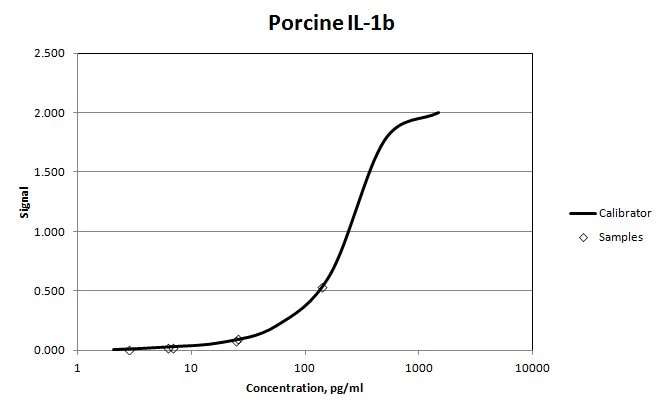
Application: ELISASample Tested: Serum and PlasmaSpecies: PorcineVerified Customer | Posted 11/07/2017We used MAB6911 as the capture antibody for Porcine IL-1b in a sandwich ELISA. Biotinylated AF681 was used as the detection antibody. The antigen standard was 681-PI. We were able to quantify serum and plasma samples.

There are no reviews that match your criteria.
Protocols
Find general support by application which include: protocols, troubleshooting, illustrated assays, videos and webinars.
- Cellular Response to Hypoxia Protocols
- R&D Systems Quality Control Western Blot Protocol
- Troubleshooting Guide: Western Blot Figures
- Western Blot Conditions
- Western Blot Protocol
- Western Blot Protocol for Cell Lysates
- Western Blot Troubleshooting
- Western Blot Troubleshooting Guide
- View all Protocols, Troubleshooting, Illustrated assays and Webinars
Loading...
Associated Pathways


Innate Lymphoid Cell Differentiation Pathways
 NOD-like Receptor Signaling Pathways
 Th17 Differentiation Pathway
 Toll-Like Receptor Signaling Pathways
