
Loading...
Key Product Details
Species Reactivity
Validated:
Multi-Species
Cited:
Human, Porcine, Avian - Chicken
Applications
Validated:
Western Blot, Neutralization, Simple Western
Cited:
Immunohistochemistry, Western Blot, Neutralization, Flow Cytometry, ELISA Detection
Label
Unconjugated
Antibody Source
Polyclonal Rabbit IgG
Loading...
Product Specifications
Immunogen
Porcine platelet-derived TGF-beta 2
Specificity
Detects TGF-beta 2 and TGF-beta 1.2 in direct ELISAs and Western blots. In direct ELISAs and Western blots, less than 2% cross-reactivity with TGF-beta 3 and TGF-beta 1 is observed.
Clonality
Polyclonal
Host
Rabbit
Isotype
IgG
Endotoxin Level
<0.10 EU per 1 μg of the antibody by the LAL method.
Scientific Data Images for TGF-beta 2 Antibody
Detection of Human TGF‑ beta 2 by Western Blot.
Western blot shows lysates of human heart tissue and human breast cancer tissue. PVDF membrane was probed with 0.1 µg/mL of Rabbit Anti-TGF-beta 2 Polyclonal Antibody (Catalog # AB-12-NA) followed by HRP-conjugated Anti-Rabbit IgG Secondary Antibody (Catalog # HAF008). A specific band was detected for TGF-beta 2 at approximately 70 kDa (as indicated). This experiment was conducted under reducing conditions and using Immunoblot Buffer Group 1.
Detection of Human TGF‑ beta 2 by Simple WesternTM.
Simple Western lane view shows lysates of human heart tissue and human breast cancer tissue, loaded at 0.2 mg/mL. A specific band was detected for TGF‑ beta 2 at approximately 64 kDa (as indicated) using 5 µg/mL of Rabbit Anti-TGF‑ beta 2 Polyclonal Antibody (Catalog # AB-12-NA). This experiment was conducted under reducing conditions and using the 12-230 kDa separation system.
TGF‑ beta 2 Inhibition of IL‑4-dependent Cell Proliferation and Neutralization by TGF‑ beta 2 Antibody.
Porcine TGF-beta 2 (Catalog # 102-B2) inhibits Recombinant Mouse IL-4 (Catalog # 404-ML) induced proliferation in the HT-2 mouse T cell line in a dose-dependent manner (orange line). Inhibition of Recombinant Mouse IL-4 (7.5 ng/mL) activity elicited by Porcine TGF-beta 2 (1 ng/mL) is neutralized (green line) by increasing concentrations of Rabbit Anti-TGF-beta 2 Polyclonal Antibody (Catalog # AB-12-NA). The ND50 is typically 0.25-1.25 µg/mL.
Detection of Human TGF-beta 2 Antibody by Western Blot
MiR-30b induces expression of TGF beta 2.(A) HUVECs were transfected with either control mimic (con) or miR-30b mimic (30b) and levels of TGF beta 1 and TGF beta 2 mRNA were assessed by qRT-PCR. Expression levels relative to control mimic transfected cells and normalized to beta -actin expression are presented as the mean ± SEM (n = 2). Overexpression of miR-30b significantly increases TGF beta 2 expression. * P < 0.05, ** P < 0.01 as determined by unpaired Student’s t-test. (B) Cells were transfected with 20 nM of either control mimic (control) or miR-30b mimic (miR-30b) and protein lysates were collected after 48 hours for assessment of TGF beta 2 protein levels by western blot. beta -actin was used as endogenous control. (C) ELISAs for TGF beta 1 and TGF beta 2 were performed with 24 hour conditioned supernates from HUVECs transfected with 20 nM of either control or miR-30b mimic. Data represents the mean ± SEM (n = 2). Overexpression of miR-30b significantly increases TGF beta 2 secretion into cell culture supernate. * P = 0.044 as determined by unpaired Student’s t-test. (D) HUVECs were transfected with 20 nM of either control mimic (control) or miR-30b mimic (miR-30b) and protein lysates were collected after 48 hours for assessment of Smad2 phosphorylation by western blot. Image collected and cropped by CiteAb from the following publication (https://pubmed.ncbi.nlm.nih.gov/28977001), licensed under a CC-BY license. Not internally tested by R&D Systems.
Detection of Human TGF-beta 2 Antibody by Western Blot
MiR-30b induces expression of TGF beta 2.(A) HUVECs were transfected with either control mimic (con) or miR-30b mimic (30b) and levels of TGF beta 1 and TGF beta 2 mRNA were assessed by qRT-PCR. Expression levels relative to control mimic transfected cells and normalized to beta -actin expression are presented as the mean ± SEM (n = 2). Overexpression of miR-30b significantly increases TGF beta 2 expression. * P < 0.05, ** P < 0.01 as determined by unpaired Student’s t-test. (B) Cells were transfected with 20 nM of either control mimic (control) or miR-30b mimic (miR-30b) and protein lysates were collected after 48 hours for assessment of TGF beta 2 protein levels by western blot. beta -actin was used as endogenous control. (C) ELISAs for TGF beta 1 and TGF beta 2 were performed with 24 hour conditioned supernates from HUVECs transfected with 20 nM of either control or miR-30b mimic. Data represents the mean ± SEM (n = 2). Overexpression of miR-30b significantly increases TGF beta 2 secretion into cell culture supernate. * P = 0.044 as determined by unpaired Student’s t-test. (D) HUVECs were transfected with 20 nM of either control mimic (control) or miR-30b mimic (miR-30b) and protein lysates were collected after 48 hours for assessment of Smad2 phosphorylation by western blot. Image collected and cropped by CiteAb from the following publication (https://pubmed.ncbi.nlm.nih.gov/28977001), licensed under a CC-BY license. Not internally tested by R&D Systems.Applications for TGF-beta 2 Antibody
Application
Recommended Usage
Simple Western
5 µg/mL
Sample: Human heart tissue and human breast cancer tissue
Sample: Human heart tissue and human breast cancer tissue
Western Blot
0.1 µg/mL
Sample: Human heart tissue and human breast cancer tissue
Sample: Human heart tissue and human breast cancer tissue
Neutralization
Measured by its ability to neutralize TGF‑ beta 2 inhibition of IL‑4-dependent proliferation in the HT‑2 mouse T cell line [Tsang, M. et al. (1995) Cytokine 7:389]. The Neutralization Dose (ND50) is typically 0.25-1.25 µg/mL in the presence of 1 ng/mL Porcine TGF‑ beta 2 and 7.5 ng/mL Recombinant Mouse IL‑4.
Reviewed Applications
Read 1 review rated 3 using AB-12-NA in the following applications:
Formulation, Preparation, and Storage
Purification
Protein A or G purified
Reconstitution
Reconstitute at 1 mg/mL in sterile PBS.
Loading...
Formulation
Lyophilized from a 0.2 μm filtered solution in PBS with Trehalose.
Shipping
The product is shipped at ambient temperature. Upon receipt, store it immediately at the temperature recommended below.
Stability & Storage
Use a manual defrost freezer and avoid repeated freeze-thaw cycles.
- 12 months from date of receipt, -20 to -70 °C as supplied.
- 1 month, 2 to 8 °C under sterile conditions after reconstitution.
- 6 months, -20 to -70 °C under sterile conditions after reconstitution.
Calculators
Background: TGF-beta 2
References
- Sporn, M.B. (2006) Cytokine Growth Factor Rev. 17:3.
- Dunker, N. and K. Krieglstein, 2000, Eur. J. Biochem. 267:6982.
- Wahl, S.M. (2006) Immunol. Rev. 213:213.
- Chang, H. et al. (2002) Endocr. Rev. 23:787.
- Lin, J.S. et al. (2006) Reproduction 132:179.
- Hinck, A.P. et al. (1996) Biochemistry 35:8517.
- Mittl, P.R.E. et al. (1996) Protein Sci. 5:1261.
- deMartin, R. et al. (1987) EMBO J. 6:3673.
- Miyazono, K. et al. (1988) J. Biol. Chem. 263:6407.
- Oklu, R. and R. Hesketh (2000) Biochem. J. 352:601.
- de Caestecker, M. et al. (2004) Cytokine Growth Factor Rev. 15:1.
- Zuniga, J.E. et al. (2005) J. Mol. Biol. 354:1052.
Long Name
Transforming Growth Factor beta 2
Alternate Names
TGFB2, TGFbeta 2
Gene Symbol
TGFB2
Additional TGF-beta 2 Products
Product Documents for TGF-beta 2 Antibody
Certificate of Analysis
To download a Certificate of Analysis, please enter a lot or batch number in the search box below.
Note: Certificate of Analysis not available for kit components.
Product Specific Notices for TGF-beta 2 Antibody
For research use only
Citations for TGF-beta 2 Antibody
 Powered by Bioz
Customer Reviews for TGF-beta 2 Antibody (1)
3 out of 5
1 Customer Rating
Have you used TGF-beta 2 Antibody?
Submit a review and receive an Amazon gift card!
$25/€18/£15/$25CAN/¥2500 Yen for a review with an image
$10/€7/£6/$10CAN/¥1110 Yen for a review without an image
Submit a review
Customer Images
Showing
1
-
1 of
1 review
Showing All
Filter By:
-
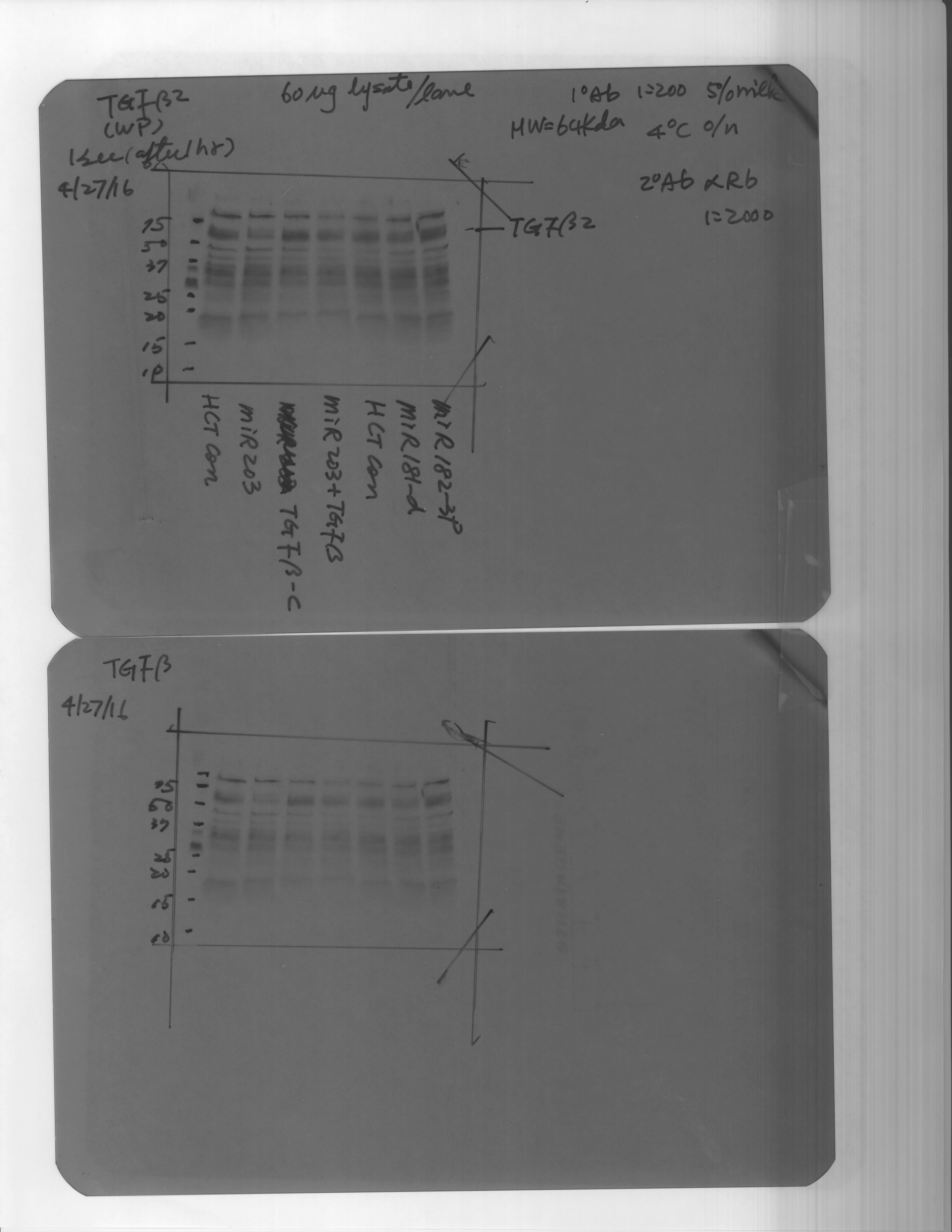
Application: Western BlotSample Tested: colon cancer cell lysateSpecies: HumanVerified Customer | Posted 05/02/2016

There are no reviews that match your criteria.
Protocols
Find general support by application which include: protocols, troubleshooting, illustrated assays, videos and webinars.
- Cellular Response to Hypoxia Protocols
- R&D Systems Quality Control Western Blot Protocol
- Troubleshooting Guide: Western Blot Figures
- Western Blot Conditions
- Western Blot Protocol
- Western Blot Protocol for Cell Lysates
- Western Blot Troubleshooting
- Western Blot Troubleshooting Guide
- View all Protocols, Troubleshooting, Illustrated assays and Webinars
Loading...
Associated Pathways


