![Western Blot: TNF-alpha AntibodyAzide Free [NB600-587]](https://resources.rndsystems.com/images/products/TNF-alpha-Antibody---Azide-Free-Western-Blot-NB600-587-img0012.jpg "Western Blot: TNF-alpha AntibodyAzide Free [NB600-587]")
Loading...
Key Product Details
Validated by
Knockout/Knockdown
Species Reactivity
Validated:
Human, Mouse, Rat, Chicken, Primate
Cited:
Human, Mouse, Rat, Avian - Chicken
Applications
Validated:
Immunohistochemistry, Immunohistochemistry-Paraffin, Western Blot, ELISA, Immunocytochemistry/ Immunofluorescence
Cited:
Immunohistochemistry, Immunohistochemistry-Paraffin, Western Blot, IF/IHC
Label
Unconjugated
Antibody Source
Polyclonal Rabbit Serum
Loading...
Product Specifications
Immunogen
The whole rabbit serum was prepared by repeated immunizations with recombinant human TNF-alpha produced in E.coli. (Uniprot: P01375)
Reactivity Notes
Rat reactivity reported in scientific literature (PMID: 25894537), Mouse reactivity reported in scientific literature (PMID:22567116). Chicken reactivity reported in scientific literature (PMID: 30298003).
Localization
Type 2 membrane protein or soluble form derived from the membrane by proteolytic cleavage.
Specificity
The antiserum is directed against mature 17,000 MW human TNFa and is useful in determining its presence in various assays. In general, this antibody also detects primate TNFa in the same formats using similar dilutions. The antibody does not recognize human TNFb (lymphotoxin). This antiserum will recognize the cell-bound precursor of TNFa as a 26,000 protein in immunoblots, particularly in denatured samples. This antibody is also useful for neutralization of human and primate TNFa activity in bioassays. It does not neutralize the biological activity of lymphotoxin. For neutralization, it is recommended to incubate the sample with a 1:200 dilution of the antibody for at least 4 hours before being tested. A control of similarly diluted normal rabbit IgG is recommended.
Clonality
Polyclonal
Host
Rabbit
Isotype
Serum
Description
Store vial at -20C prior to opening. Aliquot contents and freeze at -20C or below for extended storage. Avoid cycles of freezing and thawing. Centrifuge product if not completely clear after standing at room temperature. This product is stable for several weeks at 4C as an undiluted liquid. Dilute only prior to immediate use.
Scientific Data Images for TNF-alpha Antibody
Western Blot: TNF-alpha AntibodyAzide Free [NB600-587]
Western Blot: TNF-alpha Antibody - Azide Free [NB600-587] - Analysis using Azide/BSA FREE version of NB600-587. Membrane blocked in 1% BSA-TBS-T 30 min RT, 1:1000 dilution in 1% BSA-TBS-T 4C.![Western Blot: TNF-alpha AntibodyAzide Free [NB600-587]](https://resources.rndsystems.com/images/products/TNF-alpha-Antibody---Azide-Free-Western-Blot-NB600-587-img0014.jpg "Western Blot: TNF-alpha AntibodyAzide Free [NB600-587]")
Western Blot: TNF-alpha AntibodyAzide Free [NB600-587]
TNF-alpha-Antibody---Azide-Free-Western-Blot-NB600-587-img0014.jpg![Immunocytochemistry/ Immunofluorescence: TNF-alpha Antibody - Azide Free [NB600-587]](https://resources.rndsystems.com/images/products/TNF-alpha-Antibody---Azide-Free-Immunocytochemistry-Immunofluorescence-NB600-587-img0015.jpg "Immunocytochemistry/ Immunofluorescence: TNF-alpha Antibody - Azide Free [NB600-587]")
![Immunohistochemistry: TNF-alpha Antibody - Azide Free [NB600-587]](https://resources.rndsystems.com/images/products/TNF-alpha-Antibody---Azide-Free-Immunohistochemistry-NB600-587-img0008.jpg "Immunohistochemistry: TNF-alpha Antibody - Azide Free [NB600-587]")
Immunohistochemistry: TNF-alpha Antibody - Azide Free [NB600-587]
Immunohistochemistry: TNF-alpha Antibody - Azide Free [NB600-587] - Staining of formalin/PFA-fixed paraffin-embedded sections of human artery tissue sections. Sections were fixed in formaldehyde and subjected to heat mediated antigen retrieval in citrate buffer (pH 6.0). Slides were blocked for ten minutes with 1.5% serum. Primary antibody was diluted 1:100 and incubated with samples for 24 hours at 4C. HRP-conjugated goat anti-rabbit antibody was used as the secondary antibody.![Western Blot: TNF-alpha AntibodyAzide Free [NB600-587]](https://resources.rndsystems.com/images/products/TNF-alpha-Antibody---Azide-Free-Western-Blot-NB600-587-img0013.jpg "Western Blot: TNF-alpha AntibodyAzide Free [NB600-587]")
Western Blot: TNF-alpha AntibodyAzide Free [NB600-587]
TNF-alpha-Antibody---Azide-Free-Western-Blot-NB600-587-img0013.jpg
TNF-alpha Antibody - Azide Free
Fluorescent immunohistochemistry showing staining of human colon by anti-TNF alpha (formalin/PFA-fixed paraffin-embedded sections). Samples were formaldehyde-fixed, then blocked in 10% serum for 2 hours at 20C. The primary antibody was diluted 1:100 and incubated with the sample for 2 hours at 20C. Alexa Fluor(R) 680 goat polyclonal secondary antibody was used diluted 1:5000.
TNF-alpha Antibody - Azide Free
Immunohistochemistry using polyclonal TNFa antibody showing staining of formalin/PFA-fixed paraffin-embedded sections of human artery tissue sections. Sections were fixed in formaldehyde and subjected to heat mediated antigen retrieval in citrate buffer (pH 6.0). Slides were blocked for ten minutes with 1.5% serum. Primary antibody was diluted 1:100 and incubated with samples for 24 hours at 4C. HRP-conjugated goat anti-rabbit antibody was used as the secondary antibody.
TNF-alpha Antibody - Azide Free
Western blot of TNF-alpha Antibody - Azide Free. Lane 1: 250ng human recombinant TNF. Lane 2: 500ng human recombinant TNF. Lane 3: 1000ng human recombinant TNF. Membrane blocked in Blotto
Knockdown Validated: TNF-alpha Antibody [NB600-587] -
LMP2 knockdown by shRNA reduced the expression of TNF-alpha and IL-1 beta after MCAO. (a) Immunofluorescence labeling revealed that both TNF-alpha and IL-1 beta immunoreactivity was significantly upregulated in the ipsilateral ischemic hemisphere compared with sham-operated animals. In contrast, LMP2 knockdown by shRNA significantly reduced the expression of TNF-alpha and IL-1 beta (A). Five rats in each group were used for immunofluorescent assays (n=5). (b) Western blot confirmed that TNF-alpha and IL-1 beta protein levels were reduced to 37.4 and 44.8% compared with the Cont-shRNA group, respectively (*P<0.001, compared with the sham-operated controls; #P<0.001 compared with the cont-shRNA group). Scale bars=50 μm. Five rats in each group were used for western blot (n=5) Image collected and cropped by CiteAb from the following open publication (https://pubmed.ncbi.nlm.nih.gov/25633295), licensed under a CC-BY license. Not internally tested by Novus Biologicals.Applications for TNF-alpha Antibody
Application
Recommended Usage
ELISA
1:1000-1:5000
Immunocytochemistry/ Immunofluorescence
1:100
Immunohistochemistry
1:100-1:500
Immunohistochemistry-Paraffin
1:100-1:500
Western Blot
1:500-1:2000
Application Notes
This product has been tested for use in immunohistochemistry, immunofluorescence, and immunoblotting. It recognizes the 17,000 MW TNFalpha. Reactivity in other immunoassays is unknown.
Reviewed Applications
Read 1 review rated 5 using NB600-587 in the following applications:
Formulation, Preparation, and Storage
Purification
Whole Antiserum
Formulation
Antiserum
Preservative
No Preservative
Concentration
Please see the vial label for concentration. If unlisted please contact technical services.
Shipping
The product is shipped with polar packs. Upon receipt, store it immediately at the temperature recommended below.
Stability & Storage
Store at -20C short term. Aliquot and store at -80C long term. Avoid freeze-thaw cycles.
Background: TNF-alpha
TNF-alpha is critical for normal immune response; however, dysregulation of TNF-alpha production can result in various pathologies (2,4,5). Excessive production of pro-inflammatory cytokines including interleukin 1 (IL-1), IL-6, and TNF-alpha has been implicated in an array of autoimmune diseases like rheumatoid arthritis (RA), inflammatory bowel disease (IBD), and psoriasis (2,4,5). Anti-TNF monoclonal antibodies, including Infliximab, and soluble TNFR have been approved for the treatment of autoimmune and TNF-mediated diseases (5). Additionally, data suggests that TNF inhibitors can be beneficial for treating patients experiencing immune-related adverse events associated with immune checkpoint inhibitor cancer treatment (6).
References
1. Holbrook J, Lara-Reyna S, Jarosz-Griffiths H, McDermott M. Tumour necrosis factor signalling in health and disease. F1000Res. 2019;8:F1000 Faculty Rev-111. https://doi.org/10.12688/f1000research.17023.1
2. Jang DI, Lee AH, Shin HY, et al. The Role of Tumor Necrosis Factor Alpha (TNF-alpha) in Autoimmune Disease and Current TNF-alpha Inhibitors in Therapeutics. Int J Mol Sci. 2021;22(5):2719. https://doi.org/10.3390/ijms22052719
3. Horiuchi T, Mitoma H, Harashima S, Tsukamoto H, Shimoda T. Transmembrane TNF-alpha: structure, function and interaction with anti-TNF agents. Rheumatology (Oxford). 2010;49(7):1215-1228. https://doi.org/10.1093/rheumatology/keq031
4. Webster JD, Vucic D. The Balance of TNF Mediated Pathways Regulates Inflammatory Cell Death Signaling in Healthy and Diseased Tissues. Front Cell Dev Biol. 2020;8:365. https://doi.org/10.3389/fcell.2020.00365
5. Kalliolias GD, Ivashkiv LB. TNF biology, pathogenic mechanisms and emerging therapeutic strategies. Nat Rev Rheumatol. 2016; 12(1):49-62. https://doi.org/10.1038/nrrheum.2015.169
6. Chen AY, Wolchok JD, Bass AR. TNF in the era of immune checkpoint inhibitors: friend or foe?. Nat Rev Rheumatol. 2021;17(4):213-223. doi:10.1038/s41584-021-00584-4
Long Name
Tumor Necrosis Factor alpha
Alternate Names
Cachetin, DIF, TNF, TNF-A, TNFA, TNFalpha, TNFG1F, TNFSF1A, TNFSF2
Entrez Gene IDs
7124 (Human)
Gene Symbol
TNF
UniProt
Additional TNF-alpha Products
Product Documents for TNF-alpha Antibody
Certificate of Analysis
To download a Certificate of Analysis, please enter a lot or batch number in the search box below.
Product Specific Notices for TNF-alpha Antibody
This product is for research use only and is not approved for use in humans or in clinical diagnosis. Primary Antibodies are guaranteed for 1 year from date of receipt.
Related Research Areas
Citations for TNF-alpha Antibody
 Powered by Bioz
Customer Reviews for TNF-alpha Antibody (1)
5 out of 5
1 Customer Rating
Have you used TNF-alpha Antibody?
Submit a review and receive an Amazon gift card!
$25/€18/£15/$25CAN/¥2500 Yen for a review with an image
$10/€7/£6/$10CAN/¥1110 Yen for a review without an image
Submit a review
Customer Images
Showing
1
-
1 of
1 review
Showing All
Filter By:
-
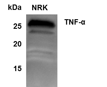
Application: Western BlotSample Tested: NRK whole cell lysateSpecies: RatVerified Customer | Posted 05/22/2015Western Blot of TNF-alpha in NRK cells

There are no reviews that match your criteria.
Protocols
Find general support by application which include: protocols, troubleshooting, illustrated assays, videos and webinars.
- Antigen Retrieval Protocol (PIER)
- Antigen Retrieval for Frozen Sections Protocol
- Appropriate Fixation of IHC/ICC Samples
- Cellular Response to Hypoxia Protocols
- Chromogenic IHC Staining of Formalin-Fixed Paraffin-Embedded (FFPE) Tissue Protocol
- Chromogenic Immunohistochemistry Staining of Frozen Tissue
- ClariTSA™ Fluorophore Kits
- Detection & Visualization of Antibody Binding
- ELISA Sample Preparation & Collection Guide
- ELISA Troubleshooting Guide
- Fluorescent IHC Staining of Frozen Tissue Protocol
- Graphic Protocol for Heat-induced Epitope Retrieval
- Graphic Protocol for the Preparation and Fluorescent IHC Staining of Frozen Tissue Sections
- Graphic Protocol for the Preparation and Fluorescent IHC Staining of Paraffin-embedded Tissue Sections
- Graphic Protocol for the Preparation of Gelatin-coated Slides for Histological Tissue Sections
- How to Run an R&D Systems DuoSet ELISA
- How to Run an R&D Systems Quantikine ELISA
- How to Run an R&D Systems Quantikine™ QuicKit™ ELISA
- ICC Cell Smear Protocol for Suspension Cells
- ICC Immunocytochemistry Protocol Videos
- ICC for Adherent Cells
- IHC Sample Preparation (Frozen sections vs Paraffin)
- Immunocytochemistry (ICC) Protocol
- Immunocytochemistry Troubleshooting
- Immunofluorescence of Organoids Embedded in Cultrex Basement Membrane Extract
- Immunofluorescent IHC Staining of Formalin-Fixed Paraffin-Embedded (FFPE) Tissue Protocol
- Immunohistochemistry (IHC) and Immunocytochemistry (ICC) Protocols
- Immunohistochemistry Frozen Troubleshooting
- Immunohistochemistry Paraffin Troubleshooting
- Preparing Samples for IHC/ICC Experiments
- Preventing Non-Specific Staining (Non-Specific Binding)
- Primary Antibody Selection & Optimization
- Protocol for Heat-Induced Epitope Retrieval (HIER)
- Protocol for Making a 4% Formaldehyde Solution in PBS
- Protocol for VisUCyte™ HRP Polymer Detection Reagent
- Protocol for the Fluorescent ICC Staining of Cell Smears - Graphic
- Protocol for the Fluorescent ICC Staining of Cultured Cells on Coverslips - Graphic
- Protocol for the Preparation & Fixation of Cells on Coverslips
- Protocol for the Preparation and Chromogenic IHC Staining of Frozen Tissue Sections
- Protocol for the Preparation and Chromogenic IHC Staining of Frozen Tissue Sections - Graphic
- Protocol for the Preparation and Chromogenic IHC Staining of Paraffin-embedded Tissue Sections
- Protocol for the Preparation and Chromogenic IHC Staining of Paraffin-embedded Tissue Sections - Graphic
- Protocol for the Preparation and Fluorescent ICC Staining of Cells on Coverslips
- Protocol for the Preparation and Fluorescent ICC Staining of Non-adherent Cells
- Protocol for the Preparation and Fluorescent ICC Staining of Stem Cells on Coverslips
- Protocol for the Preparation and Fluorescent IHC Staining of Frozen Tissue Sections
- Protocol for the Preparation and Fluorescent IHC Staining of Paraffin-embedded Tissue Sections
- Protocol for the Preparation of Gelatin-coated Slides for Histological Tissue Sections
- Protocol for the Preparation of a Cell Smear for Non-adherent Cell ICC - Graphic
- Quantikine HS ELISA Kit Assay Principle, Alkaline Phosphatase
- Quantikine HS ELISA Kit Principle, Streptavidin-HRP Polymer
- R&D Systems Quality Control Western Blot Protocol
- Sandwich ELISA (Colorimetric) – Biotin/Streptavidin Detection Protocol
- Sandwich ELISA (Colorimetric) – Direct Detection Protocol
- TUNEL and Active Caspase-3 Detection by IHC/ICC Protocol
- The Importance of IHC/ICC Controls
- Troubleshooting Guide: ELISA
- Troubleshooting Guide: Immunohistochemistry
- Troubleshooting Guide: Western Blot Figures
- Western Blot Conditions
- Western Blot Protocol
- Western Blot Protocol for Cell Lysates
- Western Blot Troubleshooting
- Western Blot Troubleshooting Guide
- View all Protocols, Troubleshooting, Illustrated assays and Webinars
FAQs for TNF-alpha Antibody
Showing
1
-
4 of
4 FAQs
Showing All
-
Q: Can your TNF-alpha products be used to treat TBI victims and therefore avoid the perispinal injections with Enbrel?
A: I am very sorry, but all of our products are for scientific research use only, and none are intended or approved for use in humans.
-
Q: I am interested in a TNF alpha antibody, cross reactive for human, rat and mouse (host species: rabbit). Could your product NBP1-19532 be used in western blotting applications? Or do you have a similar product in your catalog which could fit with my request?
A:
The antibody you mention, NBP1-19532, has not yet been validated in Western blot. I would instead recommend either NBP1-67821 or NB600-587. These are both rabbit polyclonal antibodies that cross-reacts with human, mouse and rat and have been used in Western blotting.
-
Q: I need to know about TNF-alpha antibody kits for mice.
A:
We currently have two TNFalpha ELISA kits specific for mouse: catalog numbers KA0257 and NBP1-92670.
-
Q: I would like to ask you for help. I need an antibody for Elisa to detect human TNF alpha in a supernatant from a cell culture (meaning supernatant after centrifugation of collected cell suspension from a plate well). Which of your antibodies against human tnf alpha would be suitable? I would like to buy only the primary antibody.
A:
I would recommend catalog number NBP1-67821 or NB600-587; however, a full list of our anti-human TNF alpha antibodies suitable for use in ELISA can be found using this link.
-
Q: Can your TNF-alpha products be used to treat TBI victims and therefore avoid the perispinal injections with Enbrel?
A: I am very sorry, but all of our products are for scientific research use only, and none are intended or approved for use in humans.
-
Q: I am interested in a TNF alpha antibody, cross reactive for human, rat and mouse (host species: rabbit). Could your product NBP1-19532 be used in western blotting applications? Or do you have a similar product in your catalog which could fit with my request?
A:
The antibody you mention, NBP1-19532, has not yet been validated in Western blot. I would instead recommend either NBP1-67821 or NB600-587. These are both rabbit polyclonal antibodies that cross-reacts with human, mouse and rat and have been used in Western blotting.
-
Q: I need to know about TNF-alpha antibody kits for mice.
A:
We currently have two TNFalpha ELISA kits specific for mouse: catalog numbers KA0257 and NBP1-92670.
-
Q: I would like to ask you for help. I need an antibody for Elisa to detect human TNF alpha in a supernatant from a cell culture (meaning supernatant after centrifugation of collected cell suspension from a plate well). Which of your antibodies against human tnf alpha would be suitable? I would like to buy only the primary antibody.
A:
I would recommend catalog number NBP1-67821 or NB600-587; however, a full list of our anti-human TNF alpha antibodies suitable for use in ELISA can be found using this link.
Loading...
Associated Pathways


IL-15 Signaling Pathways and their Primary Biological Effects in Different Immune Cell Types
 Innate Lymphoid Cell Differentiation Pathways
 mTOR Signaling Pathway
 NOD-like Receptor Signaling Pathways
 Th1 Differentiation Pathway
 TNF Superfamily Pathway: Human Ligand-Receptor Interactions & their Associated Functions
 Toll-Like Receptor Signaling Pathways
