Tumor Microenvironment: Immunosuppressive Cells
Many cell types are thought to contribute to the generation of an immunosuppressive tumor microenvironment (TME) including tumor-associated M2 macrophages (TAMs), regulatory T cells (Tregs), myeloid-derived suppressor cells (MDSCs), tolerogenic dendritic cells (tolDCs), and cancer-associated fibroblasts (CAFs). Read below for a brief description of each cell type and the mechanisms by which they contribute to immune suppression. Click the links to browse our offering of relevant proteins, antibodies, ELISAs, multiplex kits, and small molecules to support tumor microenvironment research.
Tumor-associated Macrophages
Tumor-associated M2 macrophages (TAMs) are a heterogeneous population of cells that display a range of phenotypes depending on the type of tumor and their locations in the tumor microenvironment (TME). TAMs are commonly the most abundant infiltrating leukocyte in most tumors and are predominantly thought to have pro-tumor effects. These include both immunosuppressive effects in addition to pro-angiogenic and metastatic effects. The mechanisms by which TAMs mediate immunosuppression are detailed below.
Mechanisms of TAM-Mediated Immunosuppression
TAMs express indoleamine 2,3-dioxygenase (IDO) and tryptophan 2,3-dioxygenase (TDO) in some contexts, which deplete L-tryptophan through kynurenine production. This metabolic pathway inhibits the activity of CD4+ and CD8+ T cells and natural killer (NK) cells, while promoting the differentiation of regulatory T cells (Tregs), thereby supporting tumor immune evasion.
TAMs express HLA-E and HLA-G, which engage inhibitory receptors on T cells and NK cells, suppressing their cytotoxicity, proliferation, and cytokine production. HLA-G additionally induces tolerogenic dendritic cells, which drive Treg development, further promoting an immunosuppressive microenvironment.
TAMs contribute to immunosuppression by secreting IL-10 and TGF-β1, which inhibit effector T cell functions, promote regulatory T cell expansion, impair NK cell activity, and suppress dendritic cell maturation. They also produce prostaglandin E2 (PGE2) that further inhibits NK and T cell cytotoxicity, promotes MDSC expansion, and skews immune responses toward immunosuppressive phenotypes. Factors secreted by TAMs not only inhibit the anti-tumor immune response, but also drive tumor cell proliferation, angiogenesis, invasion, and metastasis.
TAMs indirectly suppress anti-tumor immunity by secreting chemokines that recruit regulatory T cells, which then inhibit effector T cells, NK cells, and antigen-presenting cells (APCs) through diverse immunosuppressive mechanisms.
Select Products for Tumor-Associated Macrophage Research - Listed by Target
Featured Resources for Tumor-Associated Macrophages
Tumor-Associated Macrophage Markers
M2 macrophages play a critical role in tumor progression by promoting immunosuppression, tissue remodeling, and tumor growth. Explore the cell surface markers and secreted factors commonly used to identify M2 macrophages with our interactive cell markers tool.
M2 Macrophage Flow Cytometry Panel
Easily identify M2 macrophages with our M2 Macrophage Flow Cytometry Panel. This panel features fluorochrome-conjugated antibodies against CD3, CD20, and CD56 to exclude other immune cell types, plus antibodies for key M2 macrophage markers, including CD163, CD206, CD204, and VEGF.
Macrophage-Mediated Immunosuppression
Visualize how tumor-associated macrophages (TAMs) suppress anti-tumor immunity with our detailed interactive pathway. This pathway graphically highlights key TAM-mediated mechanisms of immunosuppression, with molecules in the pathway linked directly to products for quick access to reagents to advance macrophage research.
Regulatory T Cells
Regulatory T cells (Tregs) are a heterogeneous subset of CD4+ T cells with suppressive properties that play a central role in maintaining immune homeostasis and self-tolerance, dampening inflammation, and preventing autoimmunity. They also constitute a major suppressive cell population in the tumor microenvironment, contributing to tumor immune evasion. Tregs function by inhibiting the activities of CD4+ and CD8+ effector T cells, natural killer cells, NKT cells, and antigen-presenting cells (APCs) through multiple mechanisms outlined below.
Mechanisms of Treg-Mediated Suppression
Tregs suppress T cell responses by secreting immunosuppressive cytokines such as TGF-β1, IL-10, and IL-35, which inhibit Teff cell differentiation, proliferation, activation, and cytokine production, while promoting conversion to immunosuppressive phenotypes. Additionally, membrane-bound LAP-TGF-β1 and Galectin-1 induce Teff cell growth arrest and apoptosis through both soluble and contact-dependent mechanisms.
Cytokines secreted by Tregs including TGF-β1, IL-10, and IL-35 induce infectious tolerance by converting activated Tconv cells into regulatory cell phenotypes. TGF-β promotes FoxP3+ Tregs, IL-10 drives Tr1 cells, and IL-35 induces iTregs, thereby amplifying immunosuppression within the tumor microenvironment and facilitating tumor immune evasion.
Tregs suppress effector T cells by depleting local IL-2 via high CD25/IL-2 Rα expression, causing IL-2 deprivation-induced apoptosis.
Tregs also inhibit Teff cell proliferation and IL-2 synthesis by directly transferring inhibitory cAMP through the gap junctions of Teff cells.
Tregs express CD39 and CD73 to degrade ATP/ADP into adenosine, which inhibits dendritic cell maturation, suppresses Teff and NK cell functions via A2a/A2b receptor signaling, and promotes both a tolerogenic dendritic cell phenotype and enhanced Treg numbers and suppressive activity.
Tregs contribute to immunosuppression by secreting granzymes, particularly granzyme B, which mediates Teff cell apoptosis primarily through perforin-dependent pathways, with some evidence for perforin-independent mechanisms. Tregs also induce cytolysis of B cells, NK cells, and CD8+ T cells through granzyme B and perforin-dependent pathways.
Tregs indirectly suppress Teff cells by using CTLA-4 to downregulate dendritic cell expression of the co-stimulatory molecules, B7-1/CD80 and B7-2/CD86. Binding of CTLA-4 and CD80 or CD86 also triggers the production of indoleamine 2,3-dioxygenase (IDO) by dendritic cells, generating pro-apoptotic metabolites that suppress Teff cell activity. LAG-3 expressed by Tregs binds to MHC class II on immature dendritic cells, thereby blocking their maturation and limiting T cell activation.
Select Products for Regulatory T Cell Research - Listed by Target
Featured Resources for Regulatory T Cells
Regulatory T Cells Product Guide
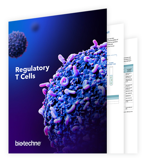
Regulatory T cells (Tregs) play a pivotal role in maintaining immune balance but can hinder effective anti-tumor responses. Use this guide to explore our comprehensive range of antibodies, recombinant proteins, small molecules, and ELISA kits designed for studying Treg biology, tracking their suppressive mechanisms, and modulating their function in disease contexts.
Identification of Novel Treg Surface Markers
The discovery of blocking and neutralizing antibodies that target Treg cell surface markers holds promise for enhancing anti-tumor therapies. In this study, our scientists screened 1,800 antibodies using flow cytometry to systematically uncover approximately 30 potential novel Treg markers.
Treg-Mediated Suppression Mechanisms
Explore how regulatory T cells (Tregs) modulate the tumor microenvironment using our interactive pathway. This graphic shows the key mechanisms Tregs use to suppress immune activity and promote tumor progression, with molecules in the pathway linked to relevant products to support Treg research.
Myeloid-derived Suppressor Cells
Myeloid-derived suppressor cells (MDSCs) are a heterogenous population of immature myeloid progenitor cells that fail to differentiate into granulocytes, macrophages, and dendritic cells. In tumor biology, immature myeloid cell accumulation is of significant interest as these cells display immunosuppressive properties that are thought to contribute to the inhibition of anti-tumor immune responses. MDSC-mediated mechanisms of immunosuppression are detailed below.
Mechanisms of MDSC-Mediated Immunosuppression
MDSCs suppress anti-tumor immunity by depleting key amino acids necessary for T cell proliferation and functions. Increased uptake and metabolism of L-arginine via high levels of ARG1 activity in MDSCs leads to low levels present in the microenvironment, resulting in reduced expression of TCR-CD3 zeta and T cell proliferative arrest. Cystine is also sequestered by MDSCs, limiting its ability to be taken up by APCs and subsequently reduced and exported as cysteine to be provided to T cells during antigen presentation. This compromises T cell activation, proliferation, and differentiation. In the presence of MDSCs, L-tryptophan levels are also reduced through indoleamine 2,3-dioxygenase (IDO)-mediated degradation by the kynurenine pathway, which inhibits the proliferation and activity of CD4+ and CD8+ T cells and natural killer (NK) cells and promotes the differentiation of regulatory T cells (Tregs).
MDSCs inhibit anti-tumor immunity by producing reactive oxygen species (ROS) and reactive nitrogen species (RNS). Hydrogen peroxide, peroxynitrite, and nitric oxide are produced by MDSCs via NADPH oxidase, ARG1, and iNOS. Collectively, these molecules impair T cell activation, proliferation, and migration, and induce apoptosis, and nitration or nitrosylation of multiple target molecules including TCR, CD3, CD8, and CCL2, which inhibits T cell activation and intra-tumoral migration.
MDSCs suppress T cell functions by expressing PD-L1, Fas Ligand, and Galectin-9, which engage T cell receptors PD-1, Fas/CD95, and Tim-3, respectively, leading to T cell exhaustion or apoptosis depending on the ligand-receptor interaction.
MDSCs produce IL-10 and TGF-β1 which suppress effector T cell activation, differentiation, proliferation, and cytokine production while promoting suppressive T cell expansion. TGF-β1 induces FoxP3+ regulatory T cells, while IL-10 promotes the conversion of activated conventional T cells to IL-10-, TGF-β1-secreting Tr1 cells. Additionally, TGF-β1 down-regulates the expression of NKG2D and NKp30 on NK cells and CD8+ T cells, reduces NK cell proliferation and cytotoxicity, skews macrophages toward an M2 phenotype, and inhibits dendritic cell maturation, migration, co-stimulation, and IL-12 production.
MDSCs inhibit T cell activation by expressing ADAM17, a protease that cleaves L-Selectin/CD62L on naïve T cells, preventing their homing from the blood and lymphatics to the lymph nodes where activation occurs.
MDSCs express CD39 and CD73 to generate immunosuppressive extracellular adenosine, which inhibits effector T, NK, and NKT cell functions, promotes Treg expansion and suppressive activity, and prevents dendritic cell maturation and pro-inflammatory cytokine production via A2a/A2b receptor signaling. MDSCs also produce PGE2 through the up-regulated expression of COX-2 and PGES1. PGE2 signals through receptors expressed on the surface of MDSCs, enhancing their own suppressive activity and expansion. PGE2 also inhibits NK cell cytotoxicity, dendritic cell differentiation, IL-2 signaling, and Th1/CD8+ T cell responses.
Select Products for Myeloid-Derived Suppressor Cell Research - Listed by Target
Featured Resources for Myeloid-Derived Suppressor Cells
Myeloid-derived Suppressor Cells Guide

MDSCs promote immunosuppression in the tumor microenvironment and are a major obstacle for cancer immunotherapies. Use this guide to explore our wide selection of antibodies, ELISA Kits, and small molecule inhibitors for studying MDSC biology, uncovering pathways driving their accumulation, and identifying targets to inhibit their immunosuppressive activities.
Blog: Targeting MDSCs for Cancer Immunotherapy
High levels of circulating MDSCs in cancer patients correlate with clinical stage, metastatic burden, and resistance to both chemotherapy and immunotherapy. Read this blog post summarizing the strategies currently being investigated to target MDSCs in the tumor microenvironment and improve the efficacy of anti-cancer treatments.
MDSC-Mediated Immunosuppression
See how MDSCs help shape the tumor microenvironment with our interactive pathway. This pathway graphically outlines the key mechanisms by which MDSCs suppress immune responses and promote tumor progression. Molecules featured in the pathway are linked directly to products, making it easy to find the tools needed for investigating MDSCs.
Tolerogenic Dendritic Cells
Tolerogenic dendritic cells (tolDCs) are a specialized subset of dendritic cells, characterized by a stable, immature or semi-mature phenotype, that promote immune tolerance rather than activation. Unlike conventional dendritic cells that initiate immune responses against pathogens or abnormal cells, tolDCs actively suppress immune activation and contribute to immune homeostasis. In the tumor microenvironment, factors such as chronic inflammation, hypoxia, and tumor-derived signals induce the differentiation of dendritic cells into this tolerogenic state. These tolDCs then play a pivotal role in dampening anti-tumor immunity by modulating T cell responses and fostering an immunosuppressive environment that allows tumor cells to evade immune surveillance. Key mechanisms by which tolerogenic DCs mediate immunosuppression in the tumor microenvironment (TME) are outlined below.
Mechanisms of TolDC-Mediated Immunosuppression
Tolerogenic DCs promote the differentiation and expansion of Tregs, which in turn inhibit the activation and function of effector immune cells, creating an environment that favors tumor immune evasion. This process is often supported by high expression of immunosuppressive enzymes like indoleamine 2,3-dioxygenase (IDO), which contributes to Treg induction and function.
Tolerogenic DCs express checkpoint ligands like PD-L1 that engage inhibitory receptors on T cells, leading to reduced T cell activation, proliferation, and cytokine production within the tumor microenvironment.
Through multiple signals, including antigen presentation with low levels of co-stimulatory molecule expression, tolerogenic DCs induce a state of T cell unresponsiveness or tolerance to tumor antigens, thereby limiting effective immune targeting of cancer cells.
Tolerogenic DCs reduce pro-inflammatory signals that would normally promote anti-tumor responses, further shifting the tumor microenvironment towards immunosuppression.
Tolerogenic DCs facilitate the accumulation and function of MDSCs, which synergize to suppress T cell activity and sustain an immunosuppressive niche within tumors.
Select Products for Tolerogenic Dendritic Cell Research - Listed by Target
Featured Resources for Dendritic Cells
Dendritic Cells Guide

Dendritic cells are central regulators of immune responses, playing vital roles in both immune activation and tolerance. This guide provides access to our comprehensive collection of proteins, antibodies, ELISAs, and multiplex assays designed to support dendritic cell research, from culture and characterization to in-depth analysis of dendritic cell functions.
Dendritic Cells Wall Poster
Dendritic cells bridge innate and adaptive immunity by capturing and presenting antigens to T cells. This poster illustrates distinct dendritic cell subsets in humans and mice, highlighting their development, tissue-specific distribution, and unique markers.
Dendritic Cell Markers
Dendritic cells are a heterogeneous cell population consisting of multiple subsets specialized for distinct tissues and functions. Identify key cell surface markers and secreted factors unique to each dendritic cell subset with our interactive cell markers tool.
Cancer-associated Fibroblasts
Cancer-associated fibroblasts (CAFs) are activated fibroblasts abundant in the tumor stroma. CAFs remodel the extracellular matrix (ECM) and secrete signaling molecules such as growth factors, cytokines, and chemokines that influence the tumor microenvironment and promote tumor progression. They can be identified by expression of the membrane protein Fibroblast Activation Protein alpha/FAP. CAFs inhibit anti-tumor immune responses by at least three mechanisms, which are detailed below.
Mechanisms of CAF-Mediated Immunosuppression
CAFs produce dense ECM proteins like collagen and fibronectin, which increase stromal stiffness and create a mechanical barrier preventing T cell migration into the tumor core.
CAF-derived factors shift the composition of immune cell infiltration in the tumor microenvironment toward immunosuppressive phenotypes, which indirectly limits the presence and function of effector T cells within the tumor.
Select Products for Cancer-Associated Fibroblast Research - Listed by Target
A Look Inside a Tumor Wall Poster
The tumor microenvironment plays a central role in inhibiting anti-tumor immune responses. Request this poster as a visual reference showing the key mechanisms used by Tregs, MDSCs, TAMs, and tumor-derived exosomes that drive immunosuppression in the tumor microenvironment.
Immune Checkpoint Targets eBook
Immune checkpoint proteins play a central role in regulating the activities of different immune cell types and represent some of the most promising targets for cancer immunotherapy. Learn about the latest research on current and emerging immune checkpoint targets in this eBook.
T Cell-Based Therapies eBook
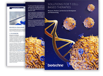
Gain essential insights into overcoming therapeutic challenges posed by the tumor microenvironment. This eBook provides a look at the biological obstacles and manufacturing complexities shaping the future of T cell therapies and solutions to accelerate advancements in the field.