2B4, also known as CD244 and SLAMF4, is a 66 kDa type I transmembrane glycoprotein in the SLAM subgroup of the CD2 protein family. SLAM family proteins have an extracellular domain (ECD) with two or four Ig-like domains and at least two cytoplasmic immunoreceptor tyrosine-based switch motifs (ITSMs). 2B4 interacts with CD48, while other SLAM family proteins interact homophilically (1‑4). Mature human 2B4 consists of a 208 amino acid (aa) ECD with two Ig-like domains, a 21 aa transmembrane segment, and a 120 aa cytoplasmic domain with four ITSMs (5, 6). Three additional splice variants of human 2B4 have deletions of the short region between the Ig-like domains, the second Ig-like domain, or a portion of the cytoplasmic tail. Within the ECD, human 2B4 shares 46% and 40% aa sequence identity with mouse and rat 2B4, respectively. The ECD of human 2B4 shares 17%‑24% aa sequence identity with comparable regions of human CD2 family members BLAME, CD2F-10, CD84, CD229, CRACC, NTB-A, and SLAM. 2B4 is expressed on all NK cells, gamma delta T cells, monocytes, some CD4+ and CD8+ T cells, and some dendritic cells (7). CD48 mediates 2B4+ cell interactions with nearly all hematopoietic cell types, including cells of the same type (8‑10). 2B4/CD48 signaling cooperates with other receptor systems to either promote or inhibit NK and CD8+ T cell activation (7‑13). The inhibitory activities are distinct from those of MHC I restricted inhibitory NK cell receptors (12, 13). Ligation of 2B4 with antibodies or CD48 constructs can either directly trigger inhibitory signaling or disrupt an inhibitory interaction, leading to cellular activation (9, 12). The inhibitory effect is associated with the long form of 2B4, while the activation is associated with the short form (9, 14). 2B4 can also induce signaling through CD48 (10, 15).
Human 2B4/CD244/SLAMF4 Antibody (146510)
R&D Systems | Catalog # MAB1039

Key Product Details
Species Reactivity
Validated:
Human
Cited:
Human
Applications
Validated:
Immunohistochemistry, Western Blot
Cited:
Neutralization
Label
Unconjugated
Antibody Source
Monoclonal Mouse IgG1 Clone # 146510
Loading...
Product Specifications
Immunogen
Mouse myeloma cell line NS0-derived recombinant human 2B4/CD244/SLAMF4
Cys22-Arg221
Accession # NP_057466
Cys22-Arg221
Accession # NP_057466
Specificity
Detects human 2B4/CD244/SLAMF4 in direct ELISAs and Western blots. In Western blots, no cross-reactivity with recombinant mouse 2B4/CD244/SLAMF4 is observed.
Clonality
Monoclonal
Host
Mouse
Isotype
IgG1
Scientific Data Images for Human 2B4/CD244/SLAMF4 Antibody (146510)
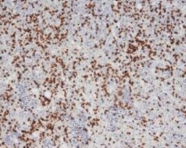
2B4/CD244/SLAMF4 in Human Spleen.
2B4/CD244/SLAMF4 was detected in immersion fixed paraffin-embedded sections of human spleen using Mouse Anti-Human 2B4/CD244/SLAMF4 Monoclonal Antibody (Catalog # MAB1039) at 1 µg/mL for 1 hour at room temperature followed by incubation with the Anti-Mouse IgG VisUCyte™ HRP Polymer Antibody (Catalog # VC001). Tissue was stained using DAB (brown) and counterstained with hematoxylin (blue). Specific staining was localized to splenocytes. View our protocol for IHC Staining with VisUCyte HRP Polymer Detection Reagents.
Detection of Human 2B4/CD244/SLAMF4 by Flow Cytometry
NK cell surface molecules involved in the cell-cell interaction that promotes TNF production by monocytes. (a) The effect of IL-15 on the expression of cell surface molecules by natural killer (NK) cells. NK cells were cultured with IL-15 at 50 ng/ml or medium alone for 24 hours, and the expression of different surface molecules was then assessed by flow cytometry. Grey-filled histograms represent the expression of each molecule on resting NK cells, the grey-line histograms represent the expression on IL-15 activated NK cells, and the dotted-line histograms represent the negative control. One representative experiment is shown. (b) Effect of different antibodies against NK cell surface molecules on TNF production in co-cultures of NK and THP-1 cells. IL-15-stimulated NK cells were co-cultured with THP-1 cells at a 10:1 cell ratio for 24 hours in the presence of different monoclonal antibodies (see the Materials and methods section for further information). The TNF concentration in cell-free supernatants was quantified with an enzyme immunoassay. The results show the percentage TNF production and are expressed as means ± SEM for eight independent experiments (see the Materials and methods section for definition). Staistical significance:*p < 0.01; §p < 0.05; analysis-of-variance test. (c) Expression of CD244 and CD48 on monocytes and THP-1 cells. The solid-line histogram represents the expression of each molecule and the grey histogram the negative control. Image collected and cropped by CiteAb from the following open publication (https://arthritis-research.biomedcentral.com/articles/10.1186/ar1955), licensed under a CC-BY license. Not internally tested by R&D Systems.Applications for Human 2B4/CD244/SLAMF4 Antibody (146510)
Application
Recommended Usage
Immunohistochemistry
1-25 µg/mL
Sample: Immersion fixed paraffin-embedded sections of human spleen
Sample: Immersion fixed paraffin-embedded sections of human spleen
Western Blot
1 µg/mL
Sample: Recombinant Human 2B4/CD244/SLAMF4 Fc Chimera (Catalog # 1039-2B)
Sample: Recombinant Human 2B4/CD244/SLAMF4 Fc Chimera (Catalog # 1039-2B)
Reviewed Applications
Read 1 review rated 5 using MAB1039 in the following applications:
Formulation, Preparation, and Storage
Purification
Protein A or G purified from hybridoma culture supernatant
Reconstitution
Reconstitute at 0.5 mg/mL in sterile PBS. For liquid material, refer to CoA for concentration.
Loading...
Formulation
Lyophilized from a 0.2 μm filtered solution in PBS with Trehalose. *Small pack size (SP) is supplied either lyophilized or as a 0.2 µm filtered solution in PBS.
Shipping
Lyophilized product is shipped at ambient temperature. Liquid small pack size (-SP) is shipped with polar packs. Upon receipt, store immediately at the temperature recommended below.
Stability & Storage
Use a manual defrost freezer and avoid repeated freeze-thaw cycles.
- 12 months from date of receipt, -20 to -70 °C as supplied.
- 1 month, 2 to 8 °C under sterile conditions after reconstitution.
- 6 months, -20 to -70 °C under sterile conditions after reconstitution.
Calculators
Background: 2B4/CD244/SLAMF4
References
- Bhat, R. et al. (2006) J. Leukoc. Biol. 79:417.
- Veillette, A. (2006) Nat. Rev. Immunol. 6:56.
- McNerney, M.E. et al. (2005) Mol. Immunol. 42:489.
- Assarsson, E. et al. (2005) J. Immunol. 175:2045.
- Boles, K.S. et al. (1999) Tissue Antigens 54:27.
- Kubin, M.Z. et al. (1999) Eur. J. Immunol. 29:3466.
- Nakajima, H. et al. (1999) Eur. J. Immunol. 29:1676.
- Lee, K.M. et al. (2006) Blood 107:3181.
- Mooney, J.M. et al. (2004) J. Immunol. 173:3953.
- Assarsson, E. et al. (2004) J. Immunol. 173:174.
- Bryceson, Y.T. et al. (2006) Blood 107:159.
- Lee, K-M. et al. (2004) J. Exp. Med. 199:1245.
- McNerney, M.E. et al. (2005) Blood 106:1337.
- Schatzle, J.D. et al. (1999) Proc. Natl. Acad. Sci. USA 96:3870.
- Messmer, B. et al. (2006) J. Immunol. 176:4646.
Alternate Names
CD244, NAIL, NKR2B4, Nmrk, SLAMF4
Gene Symbol
CD244
UniProt
Additional 2B4/CD244/SLAMF4 Products
Product Documents for Human 2B4/CD244/SLAMF4 Antibody (146510)
Certificate of Analysis
To download a Certificate of Analysis, please enter a lot or batch number in the search box below.
Note: Certificate of Analysis not available for kit components.
Product Specific Notices for Human 2B4/CD244/SLAMF4 Antibody (146510)
For research use only
Citations for Human 2B4/CD244/SLAMF4 Antibody (146510)
 Powered by Bioz
Customer Reviews for Human 2B4/CD244/SLAMF4 Antibody (146510) (1)
5 out of 5
1 Customer Rating
Have you used Human 2B4/CD244/SLAMF4 Antibody (146510)?
Submit a review and receive an Amazon gift card!
$25/€18/£15/$25CAN/¥2500 Yen for a review with an image
$10/€7/£6/$10CAN/¥1110 Yen for a review without an image
Submit a review
Customer Images
Showing
1
-
1 of
1 review
Showing All
Filter By:
-
Application: ImmunohistochemistrySample Tested: Spleen tissueSpecies: HumanVerified Customer | Posted 06/16/2022

There are no reviews that match your criteria.
Protocols
Find general support by application which include: protocols, troubleshooting, illustrated assays, videos and webinars.
- Antigen Retrieval Protocol (PIER)
- Antigen Retrieval for Frozen Sections Protocol
- Appropriate Fixation of IHC/ICC Samples
- Cellular Response to Hypoxia Protocols
- Chromogenic IHC Staining of Formalin-Fixed Paraffin-Embedded (FFPE) Tissue Protocol
- Chromogenic Immunohistochemistry Staining of Frozen Tissue
- ClariTSA™ Fluorophore Kits
- Detection & Visualization of Antibody Binding
- Fluorescent IHC Staining of Frozen Tissue Protocol
- Graphic Protocol for Heat-induced Epitope Retrieval
- Graphic Protocol for the Preparation and Fluorescent IHC Staining of Frozen Tissue Sections
- Graphic Protocol for the Preparation and Fluorescent IHC Staining of Paraffin-embedded Tissue Sections
- Graphic Protocol for the Preparation of Gelatin-coated Slides for Histological Tissue Sections
- IHC Sample Preparation (Frozen sections vs Paraffin)
- Immunofluorescent IHC Staining of Formalin-Fixed Paraffin-Embedded (FFPE) Tissue Protocol
- Immunohistochemistry (IHC) and Immunocytochemistry (ICC) Protocols
- Immunohistochemistry Frozen Troubleshooting
- Immunohistochemistry Paraffin Troubleshooting
- Preparing Samples for IHC/ICC Experiments
- Preventing Non-Specific Staining (Non-Specific Binding)
- Primary Antibody Selection & Optimization
- Protocol for Heat-Induced Epitope Retrieval (HIER)
- Protocol for Making a 4% Formaldehyde Solution in PBS
- Protocol for VisUCyte™ HRP Polymer Detection Reagent
- Protocol for the Preparation & Fixation of Cells on Coverslips
- Protocol for the Preparation and Chromogenic IHC Staining of Frozen Tissue Sections
- Protocol for the Preparation and Chromogenic IHC Staining of Frozen Tissue Sections - Graphic
- Protocol for the Preparation and Chromogenic IHC Staining of Paraffin-embedded Tissue Sections
- Protocol for the Preparation and Chromogenic IHC Staining of Paraffin-embedded Tissue Sections - Graphic
- Protocol for the Preparation and Fluorescent IHC Staining of Frozen Tissue Sections
- Protocol for the Preparation and Fluorescent IHC Staining of Paraffin-embedded Tissue Sections
- Protocol for the Preparation of Gelatin-coated Slides for Histological Tissue Sections
- R&D Systems Quality Control Western Blot Protocol
- TUNEL and Active Caspase-3 Detection by IHC/ICC Protocol
- The Importance of IHC/ICC Controls
- Troubleshooting Guide: Immunohistochemistry
- Troubleshooting Guide: Western Blot Figures
- Western Blot Conditions
- Western Blot Protocol
- Western Blot Protocol for Cell Lysates
- Western Blot Troubleshooting
- Western Blot Troubleshooting Guide
- View all Protocols, Troubleshooting, Illustrated assays and Webinars


