BMI-1 (B cell-specific Moloney-MLV integration site #1) is a 45 kDa protooncogene that is a class II member of the Polycomb group of genes. It participates in the formation of a large multimeric complex termed PRC1 that inhibits target gene transcription. Loss of BMI-1 function precludes stem cells from self-replicating. Human BMI-1 contains an N-terminal RING-finger domain (aa 17-56), an NLS (aa 81-95) and a C-terminal Pro/Ser-rich region (aa 251-326). Human BMI-1 shares 99%, 97%, 99% and 99% aa sequence identity with bovine, mouse, feline and canine BMI-1, respectively.

Key Product Details
Species Reactivity
Validated:
Human
Cited:
Human
Applications
Validated:
Western Blot, Intracellular Staining by Flow Cytometry, Chromatin Immunoprecipitation (ChIP), CyTOF-ready
Cited:
Western Blot
Label
Unconjugated
Antibody Source
Monoclonal Mouse IgG2A Clone # 384515
Loading...
Product Specifications
Immunogen
E. coli-derived recombinant human BMI-1
Asp96-Gly326
Accession # P35226
Asp96-Gly326
Accession # P35226
Specificity
Detects human BMI-1 in direct ELISAs and Western blots.
Clonality
Monoclonal
Host
Mouse
Isotype
IgG2A
Scientific Data Images for Human BMI-1 Antibody (384515)
Detection of Human BMI‑1 by Western Blot.
Western blot shows lysates of HeLa human cervical epithelial carcinoma cell line. Gels were loaded with 30 µg of whole cell lysate (WCL), 20 µg of cytoplasmic (Cyto), and 10 µg of nuclear extracts (Nuc). PVDF membrane was probed with 0.1 µg/mL Mouse Anti-Human BMI-1 Monoclonal Antibody (Catalog # MAB33341) followed by HRP-conjugated Anti-Mouse IgG Secondary Antibody (Catalog # HAF007). A specific band for BMI-1 was detected at approximately 45 kDa (as indicated). This experiment was conducted under reducing conditions and using Immunoblot Buffer Group 4.
Detection of BMI‑1-regulated Genes by Chromatin Immunoprecipitation.
HeLa human cervical epithelial carcinoma cell line was fixed using formaldehyde, resuspended in lysis buffer, and sonicated to shear chromatin. BMI-1/DNA complexes were immunoprecipitated using 5 µg Mouse Anti-Human BMI-1 Monoclonal Antibody (Catalog # MAB33341) or control antibody (Catalog # MAB003) for 15 minutes in an ultrasonic bath, followed by Biotinylated Anti-Mouse IgG Secondary Antibody (Catalog # BAF007). Immunocomplexes were captured using 50 µL of MagCellect Streptavidin Ferrofluid (Catalog # MAG999) and DNA was purified using chelating resin solution. Thehoxc13promoter was detected by standard PCR.
Detection of BMI‑1 in HeLa Human Cell Line by Flow Cytometry.
HeLa human cervical epithelial carcinoma cell line was stained with Mouse Anti-Human BMI-1 Monoclonal Antibody (Catalog # MAB33341, filled histogram) or isotype control antibody (Catalog # MAB003, open histogram), followed by Phycoerythrin-conjugated Anti-Mouse IgG F(ab')2Secondary Antibody (Catalog # F0102B). To facilitate intracellular staining, cells were fixed with paraformaldehyde and permeabilized with saponin.Applications for Human BMI-1 Antibody (384515)
Application
Recommended Usage
Chromatin Immunoprecipitation (ChIP)
5 µg/5 x 106 cells
Sample: HeLa human cervical epithelial carcinoma cell line chromatin, hoxc13 promoter detected by standard PCR
Sample: HeLa human cervical epithelial carcinoma cell line chromatin, hoxc13 promoter detected by standard PCR
CyTOF-ready
Ready to be labeled using established conjugation methods. No BSA or other carrier proteins that could interfere with conjugation.
Intracellular Staining by Flow Cytometry
2.5 µg/106 cells
Sample: HeLa human cervical epithelial carcinoma cell line fixed with paraformaldehyde and permeabilized with saponin
Sample: HeLa human cervical epithelial carcinoma cell line fixed with paraformaldehyde and permeabilized with saponin
Western Blot
0.1 µg/mL
Sample: HeLa human cervical epithelial carcinoma cell line
Sample: HeLa human cervical epithelial carcinoma cell line
Reviewed Applications
Read 1 review rated 5 using MAB33341 in the following applications:
Flow Cytometry Panel Builder
Bio-Techne Knows Flow Cytometry
Save time and reduce costly mistakes by quickly finding compatible reagents using the Panel Builder Tool.
Advanced Features
- Spectra Viewer - Custom analysis of spectra from multiple fluorochromes
- Spillover Popups - Visualize the spectra of individual fluorochromes
- Antigen Density Selector - Match fluorochrome brightness with antigen density

Formulation, Preparation, and Storage
Purification
Protein A or G purified from hybridoma culture supernatant
Reconstitution
Reconstitute at 0.5 mg/mL in sterile PBS. For liquid material, refer to CoA for concentration.
Loading...
Formulation
Lyophilized from a 0.2 μm filtered solution in PBS with Trehalose. *Small pack size (SP) is supplied either lyophilized or as a 0.2 µm filtered solution in PBS.
Shipping
Lyophilized product is shipped at ambient temperature. Liquid small pack size (-SP) is shipped with polar packs. Upon receipt, store immediately at the temperature recommended below.
Stability & Storage
Use a manual defrost freezer and avoid repeated freeze-thaw cycles.
- 12 months from date of receipt, -20 to -70 °C as supplied.
- 1 month, 2 to 8 °C under sterile conditions after reconstitution.
- 6 months, -20 to -70 °C under sterile conditions after reconstitution.
Calculators
Background: BMI-1
Long Name
B Lymphoma Mo-MLV Insertion Region 1
Alternate Names
BMI1, PCGF4, RNF51
Gene Symbol
BMI1
UniProt
Additional BMI-1 Products
Product Documents for Human BMI-1 Antibody (384515)
Certificate of Analysis
To download a Certificate of Analysis, please enter a lot or batch number in the search box below.
Note: Certificate of Analysis not available for kit components.
Product Specific Notices for Human BMI-1 Antibody (384515)
For research use only
Citations for Human BMI-1 Antibody (384515)
 Powered by Bioz
Customer Reviews for Human BMI-1 Antibody (384515) (1)
5 out of 5
1 Customer Rating
Have you used Human BMI-1 Antibody (384515)?
Submit a review and receive an Amazon gift card!
$25/€18/£15/$25CAN/¥2500 Yen for a review with an image
$10/€7/£6/$10CAN/¥1110 Yen for a review without an image
Submit a review
Customer Images
Showing
1
-
1 of
1 review
Showing All
Filter By:
-
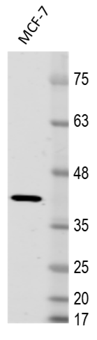
Application: Western BlotSample Tested: MCF-7 human breast cancer cell lineSpecies: HumanVerified Customer | Posted 03/22/2017MCF-7 cell lysate probed with 1:2,500 Mouse anti-Human BMI-1 Antibody. Buffer: 5% BSA in PBS. Secondary Ab: anti-Mouse IgG 1:5,000.

There are no reviews that match your criteria.
Protocols
Find general support by application which include: protocols, troubleshooting, illustrated assays, videos and webinars.
- 7-Amino Actinomycin D (7-AAD) Cell Viability Flow Cytometry Protocol
- Cellular Response to Hypoxia Protocols
- ChIP Protocol Video
- Chromatin Immunoprecipitation (ChIP) Protocol
- Chromatin Immunoprecipitation Protocol
- Extracellular Membrane Flow Cytometry Protocol
- Flow Cytometry Protocol for Cell Surface Markers
- Flow Cytometry Protocol for Staining Membrane Associated Proteins
- Flow Cytometry Staining Protocols
- Flow Cytometry Troubleshooting Guide
- Intracellular Flow Cytometry Protocol Using Alcohol (Methanol)
- Intracellular Flow Cytometry Protocol Using Detergents
- Intracellular Nuclear Staining Flow Cytometry Protocol Using Detergents
- Intracellular Staining Flow Cytometry Protocol Using Alcohol Permeabilization
- Intracellular Staining Flow Cytometry Protocol Using Detergents to Permeabilize Cells
- Propidium Iodide Cell Viability Flow Cytometry Protocol
- Protocol for Liperfluo
- Protocol for the Characterization of Human Th22 Cells
- Protocol for the Characterization of Human Th9 Cells
- Protocol: Annexin V and PI Staining by Flow Cytometry
- Protocol: Annexin V and PI Staining for Apoptosis by Flow Cytometry
- R&D Systems Quality Control Western Blot Protocol
- Troubleshooting Guide: Fluorokine Flow Cytometry Kits
- Troubleshooting Guide: Western Blot Figures
- Western Blot Conditions
- Western Blot Protocol
- Western Blot Protocol for Cell Lysates
- Western Blot Troubleshooting
- Western Blot Troubleshooting Guide
- View all Protocols, Troubleshooting, Illustrated assays and Webinars

