Cellular responses to bone morphogenetic proteins (BMPs) have been shown to be mediated by the formation of hetero-oligomeric complexes of the type I and type II serine/threonine kinase receptors. BMP receptor IB (BMPR-IB), also known as activin receptor-like kinase (ALK)-6, is one of seven known type I serine/threonine kinases that are required for the signal transduction of TGF-beta family cytokines. In contrast to the TGF-beta receptor system in which the type I receptor does not bind TGF-beta in the absence of the type II receptor, type I receptors involved in BMP signaling (including BMPR-IA, BMPR-IB/ALK-6, and ActR-I/ALK-2) can independently bind the various BMP family proteins in the absence of type II receptors. Recombinant soluble BMPR-IB binds BMP-4 with high-affinity in solution and is a potent BMP-4 antagonist in vitro. BMPR-IB is expressed in various tissues during embryogenesis. In adult tissues, BMPR-IB is only found in the brain. The extracellular domain of BMPR-IB shares little amino acid sequence identity with the other mammalian ALK type I receptor kinases, but the cysteine residues are conserved. Human and mouse BMPR-IB are highly conserved and share 98% amino acid sequence identity.
Human BMPR-IB/ALK-6 Antibody (477914)
R&D Systems | Catalog # MAB5051

Key Product Details
Species Reactivity
Validated:
Human
Cited:
Human
Applications
Validated:
Flow Cytometry, Immunocytochemistry, CyTOF-ready
Cited:
Flow Cytometry, Immunocytochemistry
Label
Unconjugated
Antibody Source
Monoclonal Mouse IgG2B Clone # 477914
Loading...
Product Specifications
Immunogen
Mouse myeloma cell line NS0-derived recombinant human BMPR‑IB/ALK‑6
Lys14-Arg126
Accession # O00238
Lys14-Arg126
Accession # O00238
Specificity
Detects human BMPR‑IB/ALK‑6 in direct ELISAs. In direct ELISAs, no cross-reactivity with recombinant mouse BMPR-IB is observed.
Clonality
Monoclonal
Host
Mouse
Isotype
IgG2B
Scientific Data Images for Human BMPR-IB/ALK-6 Antibody (477914)
Detection of BMPR‑IB/ALK‑6 in PC‑3 Human Cell Line by Flow Cytometry.
PC-3 human prostate cancer cell line was stained with Mouse Anti-Human BMPR-IB/ ALK-6 Monoclonal Antibody (Catalog # MAB5051, filled histogram) or isotype control antibody (Catalog # MAB0041, open histogram), followed by Allophycocyanin-conjugated Anti-Mouse IgG F(ab')2Secondary Antibody (Catalog # F0101B).
Detection of BMPR-IB/ALK-6 in Human iPS cells differentiated to Mesoderm by Flow Cytometry.
Human iPS cells differentiated to mesoderm (using Catalog # SC030B) were stained with Mouse Anti-Human BMPR-IB/ALK-6 Monoclonal Antibody (Catalog # MAB5051, filled histogram) or isotype control antibody (Catalog # MAB0041, open histogram) followed by anti-Mouse IgG PE-conjugated secondary antibosy (Catalog # F0101B). View our protocol for Staining Membrane-associated Proteins.
BMPR‑IB/ALK‑6 in PC‑3 Human Cell Line.
BMPR-IB/ALK-6 was detected in immersion fixed PC-3 human prostate cancer cell line using Mouse Anti-Human BMPR-IB/ALK-6 Monoclonal Antibody (Catalog # MAB5051) at 10 µg/mL for 3 hours at room temperature. Cells were stained using the NorthernLights™ 557-conjugated Anti-Mouse IgG Secondary Antibody (red; Catalog # NL007) and counterstained with DAPI (blue). Specific staining was localized to the cytoplasm and cell surface. View our protocol for Fluorescent ICC Staining of Cells on Coverslips.Applications for Human BMPR-IB/ALK-6 Antibody (477914)
Application
Recommended Usage
CyTOF-ready
Ready to be labeled using established conjugation methods. No BSA or other carrier proteins that could interfere with conjugation.
Flow Cytometry
0.25 µg/106 cells
Sample: PC‑3 human prostate cancer cell line and human iPS cells differentiated to mesoderm (using Catalog # SC030B)
Sample: PC‑3 human prostate cancer cell line and human iPS cells differentiated to mesoderm (using Catalog # SC030B)
Immunocytochemistry
8-25 µg/mL
Sample: Immersion fixed PC-3 human prostate cancer cell line
Sample: Immersion fixed PC-3 human prostate cancer cell line
Reviewed Applications
Read 1 review rated 5 using MAB5051 in the following applications:
Flow Cytometry Panel Builder
Bio-Techne Knows Flow Cytometry
Save time and reduce costly mistakes by quickly finding compatible reagents using the Panel Builder Tool.
Advanced Features
- Spectra Viewer - Custom analysis of spectra from multiple fluorochromes
- Spillover Popups - Visualize the spectra of individual fluorochromes
- Antigen Density Selector - Match fluorochrome brightness with antigen density

Formulation, Preparation, and Storage
Purification
Protein A or G purified from hybridoma culture supernatant
Reconstitution
Reconstitute at 0.5 mg/mL in sterile PBS. For liquid material, refer to CoA for concentration.
Loading...
Formulation
Lyophilized from a 0.2 μm filtered solution in PBS with Trehalose. *Small pack size (SP) is supplied either lyophilized or as a 0.2 µm filtered solution in PBS.
Shipping
Lyophilized product is shipped at ambient temperature. Liquid small pack size (-SP) is shipped with polar packs. Upon receipt, store immediately at the temperature recommended below.
Stability & Storage
Use a manual defrost freezer and avoid repeated freeze-thaw cycles.
- 12 months from date of receipt, -20 to -70 °C as supplied.
- 1 month, 2 to 8 °C under sterile conditions after reconstitution.
- 6 months, -20 to -70 °C under sterile conditions after reconstitution.
Calculators
Background: BMPR-IB/ALK-6
References
- Kawabata, M. et al. (1998) Cytokine and Growth Factor Reviews 9:49.
- Ebendal, T. et al. (1998) J. Neuroscience Research 51:139.
Long Name
Bone Morphogenetic Protein Receptor IB
Alternate Names
ALK-6, BMPR1B, BMPRIB, CDw293
Gene Symbol
BMPR1B
UniProt
Additional BMPR-IB/ALK-6 Products
Product Documents for Human BMPR-IB/ALK-6 Antibody (477914)
Certificate of Analysis
To download a Certificate of Analysis, please enter a lot or batch number in the search box below.
Note: Certificate of Analysis not available for kit components.
Product Specific Notices for Human BMPR-IB/ALK-6 Antibody (477914)
For research use only
Related Research Areas
Citations for Human BMPR-IB/ALK-6 Antibody (477914)
 Powered by Bioz
Customer Reviews for Human BMPR-IB/ALK-6 Antibody (477914) (1)
5 out of 5
1 Customer Rating
Have you used Human BMPR-IB/ALK-6 Antibody (477914)?
Submit a review and receive an Amazon gift card!
$25/€18/£15/$25CAN/¥2500 Yen for a review with an image
$10/€7/£6/$10CAN/¥1110 Yen for a review without an image
Submit a review
Customer Images
Showing
1
-
1 of
1 review
Showing All
Filter By:
-
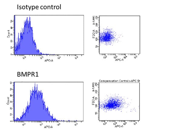
Application: Flow CytometrySample Tested: Cells from neural differentiation of hiPSCsSpecies: HumanVerified Customer | Posted 10/20/2017The cells were stained with 1µl of anti-BMPR1 antibody in 100 µl FACS staining buffer for 30 min at 4C. After washing, the cells were incubated with 1µl of anti-mouse IgG2b-APC antibody in 100 µl FACS staining buffer for 20 min at 4C.

There are no reviews that match your criteria.
Protocols
Find general support by application which include: protocols, troubleshooting, illustrated assays, videos and webinars.
- 7-Amino Actinomycin D (7-AAD) Cell Viability Flow Cytometry Protocol
- Appropriate Fixation of IHC/ICC Samples
- Cellular Response to Hypoxia Protocols
- ClariTSA™ Fluorophore Kits
- Detection & Visualization of Antibody Binding
- Extracellular Membrane Flow Cytometry Protocol
- Flow Cytometry Protocol for Cell Surface Markers
- Flow Cytometry Protocol for Staining Membrane Associated Proteins
- Flow Cytometry Staining Protocols
- Flow Cytometry Troubleshooting Guide
- ICC Cell Smear Protocol for Suspension Cells
- ICC Immunocytochemistry Protocol Videos
- ICC for Adherent Cells
- Immunocytochemistry (ICC) Protocol
- Immunocytochemistry Troubleshooting
- Immunofluorescence of Organoids Embedded in Cultrex Basement Membrane Extract
- Immunohistochemistry (IHC) and Immunocytochemistry (ICC) Protocols
- Intracellular Flow Cytometry Protocol Using Alcohol (Methanol)
- Intracellular Flow Cytometry Protocol Using Detergents
- Intracellular Nuclear Staining Flow Cytometry Protocol Using Detergents
- Intracellular Staining Flow Cytometry Protocol Using Alcohol Permeabilization
- Intracellular Staining Flow Cytometry Protocol Using Detergents to Permeabilize Cells
- Preparing Samples for IHC/ICC Experiments
- Preventing Non-Specific Staining (Non-Specific Binding)
- Primary Antibody Selection & Optimization
- Propidium Iodide Cell Viability Flow Cytometry Protocol
- Protocol for Liperfluo
- Protocol for VisUCyte™ HRP Polymer Detection Reagent
- Protocol for the Characterization of Human Th22 Cells
- Protocol for the Characterization of Human Th9 Cells
- Protocol for the Fluorescent ICC Staining of Cell Smears - Graphic
- Protocol for the Fluorescent ICC Staining of Cultured Cells on Coverslips - Graphic
- Protocol for the Preparation and Fluorescent ICC Staining of Cells on Coverslips
- Protocol for the Preparation and Fluorescent ICC Staining of Non-adherent Cells
- Protocol for the Preparation and Fluorescent ICC Staining of Stem Cells on Coverslips
- Protocol for the Preparation of a Cell Smear for Non-adherent Cell ICC - Graphic
- Protocol: Annexin V and PI Staining by Flow Cytometry
- Protocol: Annexin V and PI Staining for Apoptosis by Flow Cytometry
- TUNEL and Active Caspase-3 Detection by IHC/ICC Protocol
- The Importance of IHC/ICC Controls
- Troubleshooting Guide: Fluorokine Flow Cytometry Kits
- View all Protocols, Troubleshooting, Illustrated assays and Webinars
Loading...