Carbonic Anhydrase (CA) catalyzes the reversible reaction of CO2 + H2O = HCO3- + H+, which is fundamental to many processes such as respiration, renal tubular acidification and bone resorption (1-3). Topics in the CA meeting (6th International Conference on the CAs, June 20-25, 2003, in Slovakia) ranged from use of CAs as markers for tumor and hypoxia in clinic, as nutritional supplement in milk, and as a tool for CO2 removal and mosquito control in industry. CA9, also known as membrane antigen MN and renal cell carcinoma (RCC)-associated antigen G250, is a transmembrane enzyme expressed primarily in carcinoma cells. It is one of the best markers for hypoxia and for RCC (4, 5). Recombinant human CA9 corresponds to the extracellular portion of human CA9.
Human Carbonic Anhydrase IX/CA9 Antibody
R&D Systems | Catalog # AF2188

Key Product Details
Species Reactivity
Validated:
Human
Cited:
Human, Mouse, Xenograft
Applications
Validated:
Immunohistochemistry, Western Blot, Flow Cytometry, Immunocytochemistry, Immunoprecipitation, CyTOF-reported
Cited:
Immunohistochemistry, Immunohistochemistry-Paraffin, Western Blot, Immunocytochemistry, CyTof, IF/ICC, IF/IHC, Lateral Flow Assay
Label
Unconjugated
Antibody Source
Polyclonal Goat IgG
Loading...
Product Specifications
Immunogen
Mouse myeloma cell line NS0-derived recombinant human Carbonic Anhydrase IX
Pro59-Asp414
Accession # Q16790
Pro59-Asp414
Accession # Q16790
Specificity
Detects human Carbonic Anhydrase IX (CA9) in direct ELISAs and Western blots. In direct ELISAs, approximately 10% cross-reactivity with recombinant mouse CA9 is observed and less than 1% cross-reactivity with recombinant human CA1, 3, 4, 8, 10, 12, and 14 is observed.
Clonality
Polyclonal
Host
Goat
Isotype
IgG
Scientific Data Images for Human Carbonic Anhydrase IX/CA9 Antibody
Detection of Human Carbonic Anhydrase IX/CA9 by Western Blot.
Western blot shows lysates of U-87 MG human glioblastoma/ astrocytoma cell line. PVDF membrane was probed with 1 µg/mL of Goat Anti-Human Carbonic Anhydrase IX/CA9 Antigen Affinity-purified Polyclonal Antibody (Catalog # AF2188) followed by HRP-conjugated Anti-Goat IgG Secondary Antibody (HAF109). A specific band was detected for Carbonic Anhydrase IX/CA9 at approximately 58 kDa (as indicated). This experiment was conducted under reducing conditions and using Immunoblot Buffer Group 8.
Carbonic Anhydrase IX/CA9 in A431 Human Cell Line.
Carbonic Anhydrase IX/CA9 was detected in immersion fixed A431 human epithelial carcinoma cell line using Goat Anti-Human Carbonic Anhydrase IX/CA9 Antigen Affinity-purified Polyclonal Antibody (Catalog # AF2188) at 3 µg/mL for 3 hours at room temperature. Cells were stained using the NorthernLights™ 557-conjugated Anti-Goat IgG Secondary Antibody (red; NL001) and counterstained with DAPI (blue). Specific staining was localized to cytoplasm. View our protocol for Fluorescent ICC Staining of Cells on Coverslips.
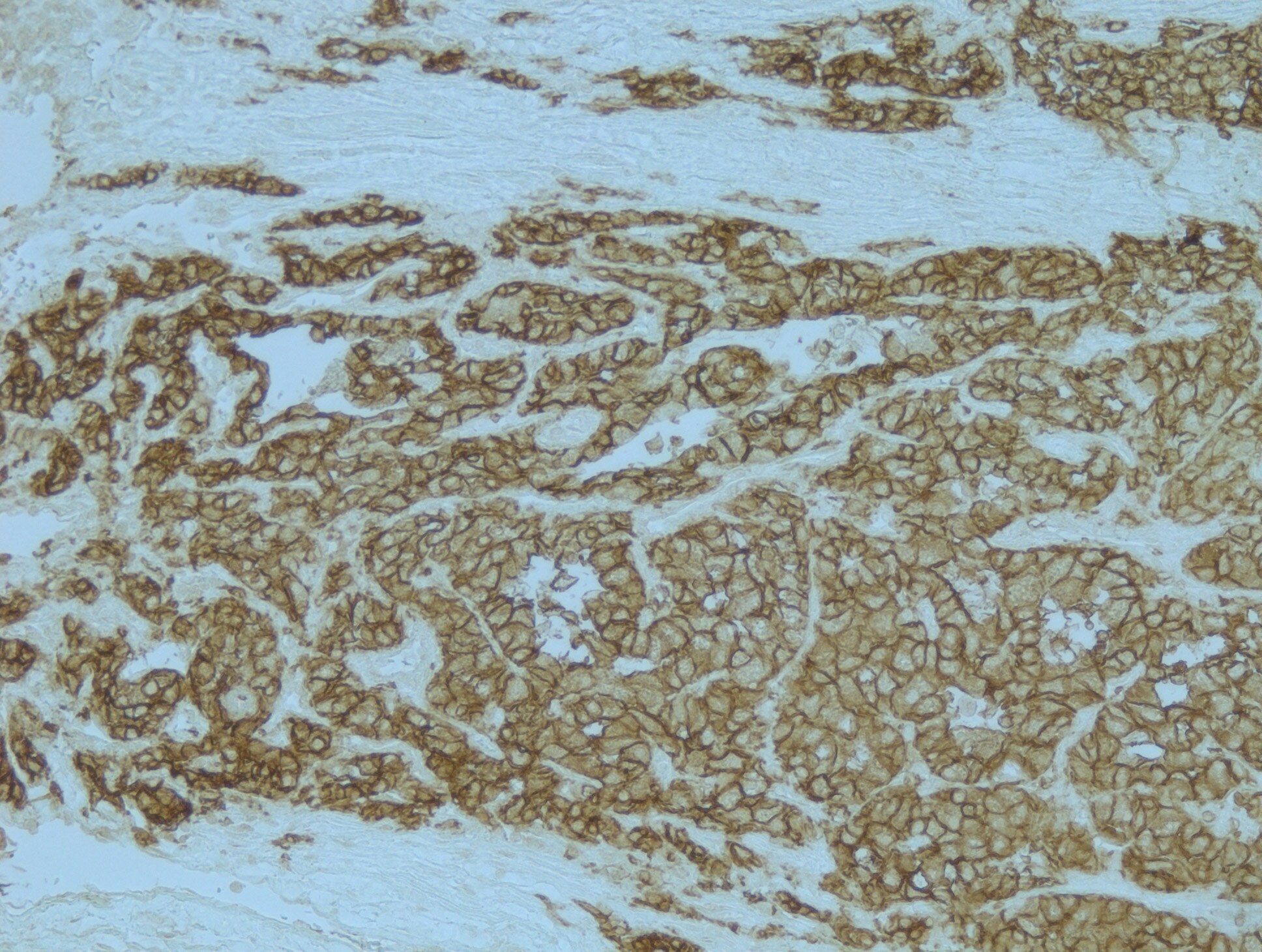
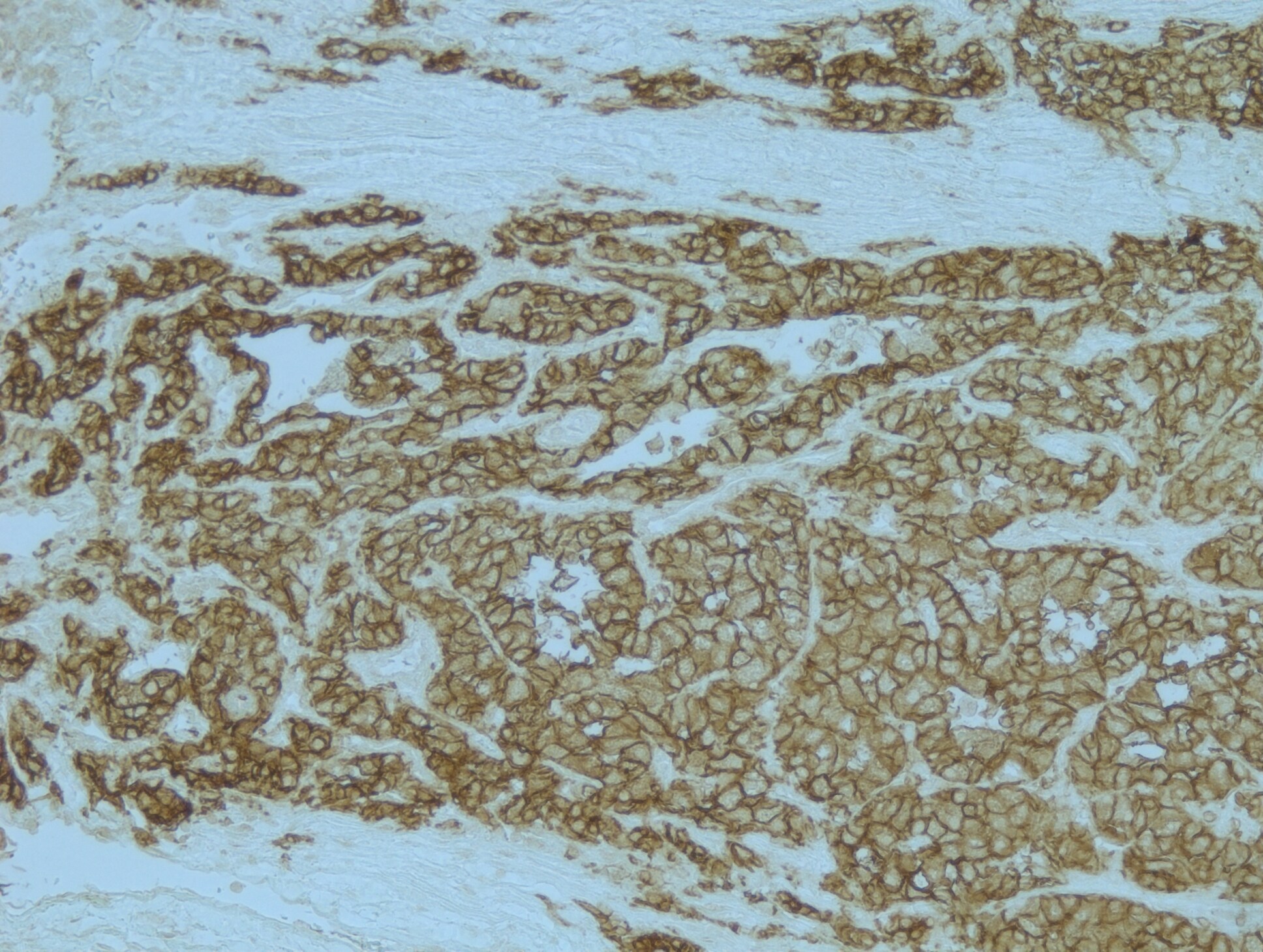
Carbonic Anhydrase IX/CA9 in Human Colon Cancer Tissue.
Carbonic Anhydrase IX/CA9 was detected in immersion fixed paraffin-embedded sections of human colon cancer tissue using Goat Anti-Human Carbonic Anhydrase IX/CA9 Antigen Affinity-purified Polyclonal Antibody (Catalog # AF2188) at 15 µg/mL overnight at 4 °C. Tissue was stained using the Anti-Goat HRP-DAB Cell & Tissue Staining Kit (brown; CTS008) and counterstained with hematoxylin (blue). Specific labeling was localized to the plasma membrane of epithelial cells. View our protocol for Chromogenic IHC Staining of Paraffin-embedded Tissue Sections.
Detection of Carbonic Anhydrase IX/CA9 by Western Blot
Beta-catenin enhances the breast cancer stem cell phenotype in response to hypoxia independently of its nuclear transcriptional activity. A, Western analysis (WB) of beta-catenin, SNAI2 and CA9 protein levels in Ctrl/shBeta MCF7 cells upon Nor/1%pO2 conditions; B, MS forming assay in stable beta-catenin silenced (shBeta) MCF7 cells upon Nor/1%pO2 conditions; C, Cytofluorimetric analysis of CD44high/CD24low stem/progenitor population in ctrl/shBeta MCF7 cells upon Nor/1%pO2 conditions; D, Real-Time PCR analysis of ESR1 mRNA level in Ctrl/shBeta MCF7 cells upon Nor/1%pO2 conditions; E, Immunofluorescence (IF) analysis of Beta-catenin in Nor/1%pO2 MCF7 cells; F, WB analysis of beta-catenin in Nor/1%pO2 MCF7 cells cytoplasmic and nuclear fractions (lamin B and beta-tubulin were used as fractionation controls); G, beta-catenin/TCF transcriptional reporter (TOPFLASH) assay in MCF7 cells and T-MS under Nor/1%pO2. Data are presented as mean +/- s.d.; p values refers to t test. n=3, unless otherwise specified. Image collected and cropped by CiteAb from the following open publication (https://pubmed.ncbi.nlm.nih.gov/24260469), licensed under a CC-BY license. Not internally tested by R&D Systems.
Detection of Carbonic Anhydrase IX/CA9 in U87-MG cells cells by Flow Cytometry.
U87-MG cells were stained with Goat Anti-Human Carbonic Anhydrase IX/CA9 Antigen Affinity-purified Polyclonal Antibody (Catalog # AF2188, filled histogram) or isotype control antibody (Catalog # AB-108-C, open histogram), followed by Phycoerythrin-conjugated Anti-Goat IgG Secondary Antibody (Catalog # F0107). View our protocol for Staining Membrane-associated Proteins.
Detection of Carbonic Anhydrase IX/CA9 by Western Blot
In normoxic basal-like breast cancer cells, cytoplasmic beta-catenin promotes stem cell features in vitro and tumor growth in vivo via constitutive stabilization of CA9 and SNAI2 mRNAs.A, Beta-catenin IF analysis in luminal MCF7 cells and in basal-like MDA-MB-468 and MDA-MB-231 cells; B, Cytofluorimetric analysis of the CD44high/CD24low population in ctrl/shBeta MDA-MB-468 and MDA-MB-231 cells; C, Ctrl/shBeta MDA-MB-468 10-weeks mammary fat pad xenograft assay (n=5, each group); representative pictures of xenograft tissues hematoxylin-eosin staining are include; D, WB analysis of CA9, SNAI2 and beta-catenin protein levels in ctrl/shBeta normoxic MDA-MB-468 and MDA-MB-231 cells; E, CA9 and SNAI2 mRNA stability assay following inhibition of Polymerase 2 transcriptional activity by actinomycin D (100ng/ml) in Ctrl/shBeta MDA-MB-468 and MDA-MB-231 cells; F, Four weeks growth curve of ctrl/shSNAI2 MDA-MB-231 subcutaneous xenograft assay (n=6, each group); G, Hematoxylin-eosin, ESR1 and CDH1 immunohistochemical stainings in xenograft tissue sections. Data are presented as mean +/- s.d.; p values refers to t test. n=3, unless otherwise specified. Image collected and cropped by CiteAb from the following open publication (https://pubmed.ncbi.nlm.nih.gov/24260469), licensed under a CC-BY license. Not internally tested by R&D Systems.
Detection of Carbonic Anhydrase IX/CA9 by Immunohistochemistry
Sunitinib induces hypoxia and Carbonic Anhydrase IX (CAIX) expression in primary Triple Negative Breast Cancer (TNBC) tumors. (a) Images of MDA-MB-231 LM2-4Luc+ primary tumor tissue sections harvested at increasing tumor volumes from animals administered either vehicle or 60 mg/kg sunitinib and immunohistochemically stained for the indicated markers. Scale bars: upper panels, 1 mm; lower panels, 200 μm. (b to e) Image-based quantification of (b) CD31+ blood vessels, (c) CAIX expression, (d) pimonidazole and (e) proliferation. Data show the mean ± standard error of the mean (SEM). n = 3 animals/group with 10 images/animal. * p < 0.05, *** p < 0.001. Image collected and cropped by CiteAb from the following open publication (https://pubmed.ncbi.nlm.nih.gov/31319613), licensed under a CC-BY license. Not internally tested by R&D Systems.
Human Carbonic Anhydrase IX / CA9 ELISA Standard Curve
Recombinant Human Carbonic Anhydrase IX/CA9 (Catalog # 2188-CA) was serially diluted and captured by Mouse Anti-Human Carbonic Anhydrase IX/CA9 Monoclonal Antibody (Catalog # MAB2188) coated on a Clear Polystyrene Microplate (Catalog # DY990). Goat Anti-Human Carbonic Anhydrase IX/CA9 Antigen Affinity-purified Polyclonal Antibody (Catalog # AF2188) was biotinylated and incubated with the protein captured on the plate. Detection of the standard curve was achieved by incubating Streptavidin-HRP (Catalog # DY998)Applications for Human Carbonic Anhydrase IX/CA9 Antibody
Application
Recommended Usage
CyTOF-reported
Chevrier, S. et al. (2018) Cell Systems. 6: 612. Ready to be labeled using established conjugation methods. No BSA or other carrier proteins that could interfere with conjugation.
Flow Cytometry
0.25 µg/106 cells
Sample: U87-MG cells
Sample: U87-MG cells
Immunocytochemistry
3-15 µg/mL
Sample: Immersion fixed A431 human epithelial carcinoma cell line
Sample: Immersion fixed A431 human epithelial carcinoma cell line
Immunohistochemistry
5-15 µg/mL
Sample: Immersion fixed paraffin-embedded sections of human colon cancer tissue
Sample: Immersion fixed paraffin-embedded sections of human colon cancer tissue
Immunoprecipitation
25 µg/mL
Sample: Conditioned cell culture medium spiked with Recombinant Human Carbonic Anhydrase IX (Catalog # 2188-CA), see our available Western blot detection antibodies
Sample: Conditioned cell culture medium spiked with Recombinant Human Carbonic Anhydrase IX (Catalog # 2188-CA), see our available Western blot detection antibodies
Western Blot
1 µg/mL
Sample: U‑87 MG human glioblastoma/astrocytoma cell line
Sample: U‑87 MG human glioblastoma/astrocytoma cell line
Reviewed Applications
Read 2 reviews rated 5 using AF2188 in the following applications:
Flow Cytometry Panel Builder
Bio-Techne Knows Flow Cytometry
Save time and reduce costly mistakes by quickly finding compatible reagents using the Panel Builder Tool.
Advanced Features
- Spectra Viewer - Custom analysis of spectra from multiple fluorochromes
- Spillover Popups - Visualize the spectra of individual fluorochromes
- Antigen Density Selector - Match fluorochrome brightness with antigen density

Formulation, Preparation, and Storage
Purification
Antigen Affinity-purified
Reconstitution
Reconstitute at 0.2 mg/mL in sterile PBS. For liquid material, refer to CoA for concentration.
Loading...
Formulation
Lyophilized from a 0.2 μm filtered solution in PBS with Trehalose. *Small pack size (SP) is supplied either lyophilized or as a 0.2 µm filtered solution in PBS.
Shipping
Lyophilized product is shipped at ambient temperature. Liquid small pack size (-SP) is shipped with polar packs. Upon receipt, store immediately at the temperature recommended below.
Stability & Storage
Use a manual defrost freezer and avoid repeated freeze-thaw cycles.
- 12 months from date of receipt, -20 to -70 °C as supplied.
- 1 month, 2 to 8 °C under sterile conditions after reconstitution.
- 6 months, -20 to -70 °C under sterile conditions after reconstitution.
Calculators
Background: Carbonic Anhydrase IX/CA9
References
- Pastorek, J. et al. (1994) Oncogene 9: 2877.
- Opavsky, R. et al. (1996) Genomics 33: 480.
- Hewett-Emmett, D. and R.E. Tashian (1996) Mol. Phylogenet. Evol. 5:50.
- Kaluzova, M. et al. (2004) Mol. Cell Biol. 24:5757.
- Mukouyama, H. et al. (2004) Clin. Cancer Res. 10:1421.
Alternate Names
CA9, G250, MN, RCC
Gene Symbol
CA9
UniProt
Additional Carbonic Anhydrase IX/CA9 Products
Product Documents for Human Carbonic Anhydrase IX/CA9 Antibody
Certificate of Analysis
To download a Certificate of Analysis, please enter a lot or batch number in the search box below.
Note: Certificate of Analysis not available for kit components.
Product Specific Notices for Human Carbonic Anhydrase IX/CA9 Antibody
For research use only
Related Research Areas
Citations for Human Carbonic Anhydrase IX/CA9 Antibody
 Powered by Bioz
Customer Reviews for Human Carbonic Anhydrase IX/CA9 Antibody (2)
5 out of 5
2 Customer Ratings
Have you used Human Carbonic Anhydrase IX/CA9 Antibody?
Submit a review and receive an Amazon gift card!
$25/€18/£15/$25CAN/¥2500 Yen for a review with an image
$10/€7/£6/$10CAN/¥1110 Yen for a review without an image
Submit a review
Customer Images
Showing
1
-
2 of
2 reviews
Showing All
Filter By:
-
Application: ImmunohistochemistrySample Tested: Renal cancer tissueSpecies: HumanVerified Customer | Posted 05/21/2018Immunohistochemistry was performed on FFPE tissue section. Heat induced antigen retrieval was performed by heating section in citrate buffer (pH 6) at 95C for 20 minutes.

-
Application: Immunohistochemistry-ParaffinSample Tested: Renal Cell Carcinoma tissuesSpecies: HumanVerified Customer | Posted 05/18/2018Heat mediated antigen retrieval performed in citrate buffer (pH 6) for 20 minutes.
There are no reviews that match your criteria.
Protocols
Find general support by application which include: protocols, troubleshooting, illustrated assays, videos and webinars.
- 7-Amino Actinomycin D (7-AAD) Cell Viability Flow Cytometry Protocol
- Antigen Retrieval Protocol (PIER)
- Antigen Retrieval for Frozen Sections Protocol
- Appropriate Fixation of IHC/ICC Samples
- Cellular Response to Hypoxia Protocols
- Chromogenic IHC Staining of Formalin-Fixed Paraffin-Embedded (FFPE) Tissue Protocol
- Chromogenic Immunohistochemistry Staining of Frozen Tissue
- ClariTSA™ Fluorophore Kits
- Detection & Visualization of Antibody Binding
- Extracellular Membrane Flow Cytometry Protocol
- Flow Cytometry Protocol for Cell Surface Markers
- Flow Cytometry Protocol for Staining Membrane Associated Proteins
- Flow Cytometry Staining Protocols
- Flow Cytometry Troubleshooting Guide
- Fluorescent IHC Staining of Frozen Tissue Protocol
- Graphic Protocol for Heat-induced Epitope Retrieval
- Graphic Protocol for the Preparation and Fluorescent IHC Staining of Frozen Tissue Sections
- Graphic Protocol for the Preparation and Fluorescent IHC Staining of Paraffin-embedded Tissue Sections
- Graphic Protocol for the Preparation of Gelatin-coated Slides for Histological Tissue Sections
- ICC Cell Smear Protocol for Suspension Cells
- ICC Immunocytochemistry Protocol Videos
- ICC for Adherent Cells
- IHC Sample Preparation (Frozen sections vs Paraffin)
- Immunocytochemistry (ICC) Protocol
- Immunocytochemistry Troubleshooting
- Immunofluorescence of Organoids Embedded in Cultrex Basement Membrane Extract
- Immunofluorescent IHC Staining of Formalin-Fixed Paraffin-Embedded (FFPE) Tissue Protocol
- Immunohistochemistry (IHC) and Immunocytochemistry (ICC) Protocols
- Immunohistochemistry Frozen Troubleshooting
- Immunohistochemistry Paraffin Troubleshooting
- Immunoprecipitation Protocol
- Intracellular Flow Cytometry Protocol Using Alcohol (Methanol)
- Intracellular Flow Cytometry Protocol Using Detergents
- Intracellular Nuclear Staining Flow Cytometry Protocol Using Detergents
- Intracellular Staining Flow Cytometry Protocol Using Alcohol Permeabilization
- Intracellular Staining Flow Cytometry Protocol Using Detergents to Permeabilize Cells
- Preparing Samples for IHC/ICC Experiments
- Preventing Non-Specific Staining (Non-Specific Binding)
- Primary Antibody Selection & Optimization
- Propidium Iodide Cell Viability Flow Cytometry Protocol
- Protocol for Heat-Induced Epitope Retrieval (HIER)
- Protocol for Liperfluo
- Protocol for Making a 4% Formaldehyde Solution in PBS
- Protocol for VisUCyte™ HRP Polymer Detection Reagent
- Protocol for the Characterization of Human Th22 Cells
- Protocol for the Characterization of Human Th9 Cells
- Protocol for the Fluorescent ICC Staining of Cell Smears - Graphic
- Protocol for the Fluorescent ICC Staining of Cultured Cells on Coverslips - Graphic
- Protocol for the Preparation & Fixation of Cells on Coverslips
- Protocol for the Preparation and Chromogenic IHC Staining of Frozen Tissue Sections
- Protocol for the Preparation and Chromogenic IHC Staining of Frozen Tissue Sections - Graphic
- Protocol for the Preparation and Chromogenic IHC Staining of Paraffin-embedded Tissue Sections
- Protocol for the Preparation and Chromogenic IHC Staining of Paraffin-embedded Tissue Sections - Graphic
- Protocol for the Preparation and Fluorescent ICC Staining of Cells on Coverslips
- Protocol for the Preparation and Fluorescent ICC Staining of Non-adherent Cells
- Protocol for the Preparation and Fluorescent ICC Staining of Stem Cells on Coverslips
- Protocol for the Preparation and Fluorescent IHC Staining of Frozen Tissue Sections
- Protocol for the Preparation and Fluorescent IHC Staining of Paraffin-embedded Tissue Sections
- Protocol for the Preparation of Gelatin-coated Slides for Histological Tissue Sections
- Protocol for the Preparation of a Cell Smear for Non-adherent Cell ICC - Graphic
- Protocol: Annexin V and PI Staining by Flow Cytometry
- Protocol: Annexin V and PI Staining for Apoptosis by Flow Cytometry
- R&D Systems Quality Control Western Blot Protocol
- TUNEL and Active Caspase-3 Detection by IHC/ICC Protocol
- The Importance of IHC/ICC Controls
- Troubleshooting Guide: Fluorokine Flow Cytometry Kits
- Troubleshooting Guide: Immunohistochemistry
- Troubleshooting Guide: Western Blot Figures
- Western Blot Conditions
- Western Blot Protocol
- Western Blot Protocol for Cell Lysates
- Western Blot Troubleshooting
- Western Blot Troubleshooting Guide
- View all Protocols, Troubleshooting, Illustrated assays and Webinars
Loading...