Caspase-1, also known as IL-1 beta -converting enzyme (ICE), is an aspartic protease that plays a key role in the inflammatory response and apoptosis. Caspase-1 precursor (about 50kDa) can be cleaved and the active enzyme consists of a complex of two 20 kDa (aa 120-297) and two 10 kDa (aa 317-404) subunits which associate following cleavage of inactive precursors. Caspase-1 is required for proteolytic cleavage of the IL-1 beta precursor to form the active proinflammatory cytokine. Alternate splicing generates several additional Caspase-1 isoforms with deletions in the propeptide regions or also in the mature subunits. Within the large subunit, human Caspase 1 shares 61% aa sequence identity with mouse and rat Caspase-1.
Human Caspase-1 Antibody (661228)
R&D Systems | Catalog # MAB6215

Key Product Details
Species Reactivity
Validated:
Human
Cited:
Human, Mouse, Rat, Primate - Cercopithecus aethiops (African Green Monkey), Transgenic Mouse
Applications
Validated:
Western Blot, Immunocytochemistry
Cited:
Immunohistochemistry-Paraffin, Western Blot, Immunoprecipitation
Label
Unconjugated
Antibody Source
Monoclonal Mouse IgG2A Clone # 661228
Loading...
Product Specifications
Immunogen
E.coli-derived recombinant human Caspase-1
Asn120-Asp297
Accession # P29466
Asn120-Asp297
Accession # P29466
Specificity
Detects human Caspase-1 in direct ELISAs and Western blots.
Clonality
Monoclonal
Host
Mouse
Isotype
IgG2A
Scientific Data Images for Human Caspase-1 Antibody (661228)
Detection of Human, Mouse, and Rat Caspase‑1 by Western Blot.
Western blot shows lysates of A431 human epithelial carcinoma cell line, NIH-3T3 mouse embryonic fibroblast cell line, and Rat-2 rat embryonic fibroblast cell line. PVDF membrane was probed with 0.1 µg/mL of Mouse Anti-Human Caspase-1 Monoclonal Antibody (Catalog # MAB6215) followed by HRP-conjugated Anti-Mouse IgG Secondary Antibody (Catalog # HAF007). A specific band was detected for Caspase-1 at approximately 45 kDa (as indicated). This experiment was conducted under reducing conditions and using Immunoblot Buffer Group 2.
Caspase‑1 in THP‑1 Human Cell Line.
Caspase-1 was detected in immersion fixed THP-1 human acute monocytic leukemia cell line using Mouse Anti-Human Caspase-1 Monoclonal Antibody (Catalog # MAB6215) at 15 µg/mL for 3 hours at room temperature. Cells were stained using the Northern-Lights™ 557-conjugated Anti-Mouse IgG Secondary Antibody (yellow; Catalog # NL007) and counterstained with DAPI (blue). Specific staining was localized to cytoplasm. View our protocol for Fluorescent ICC Staining of Non-adherent Cells.Applications for Human Caspase-1 Antibody (661228)
Application
Recommended Usage
Immunocytochemistry
8-25 µg/mL
Sample: Immersion fixed THP‑1 human acute monocytic leukemia cell line
Sample: Immersion fixed THP‑1 human acute monocytic leukemia cell line
Western Blot
0.1 µg/mL
Sample: A431 human epithelial carcinoma cell line, NIH‑3T3 mouse embryonic fibroblast cell line, and Rat‑2 rat embryonic fibroblast cell line
Sample: A431 human epithelial carcinoma cell line, NIH‑3T3 mouse embryonic fibroblast cell line, and Rat‑2 rat embryonic fibroblast cell line
Reviewed Applications
Read 1 review rated 5 using MAB6215 in the following applications:
Formulation, Preparation, and Storage
Purification
Protein A or G purified from hybridoma culture supernatant
Reconstitution
Sterile PBS to a final concentration of 0.5 mg/mL. For liquid material, refer to CoA for concentration.
Loading...
Formulation
Lyophilized from a 0.2 μm filtered solution in PBS with Trehalose. *Small pack size (SP) is supplied either lyophilized or as a 0.2 µm filtered solution in PBS.
Shipping
Lyophilized product is shipped at ambient temperature. Liquid small pack size (-SP) is shipped with polar packs. Upon receipt, store immediately at the temperature recommended below.
Stability & Storage
Use a manual defrost freezer and avoid repeated freeze-thaw cycles.
- 12 months from date of receipt, -20 to -70 °C as supplied.
- 1 month, 2 to 8 °C under sterile conditions after reconstitution.
- 6 months, -20 to -70 °C under sterile conditions after reconstitution.
Calculators
Background: Caspase-1
Additional Caspase-1 Products
Product Documents for Human Caspase-1 Antibody (661228)
Certificate of Analysis
To download a Certificate of Analysis, please enter a lot or batch number in the search box below.
Note: Certificate of Analysis not available for kit components.
Product Specific Notices for Human Caspase-1 Antibody (661228)
For research use only
Related Research Areas
Citations for Human Caspase-1 Antibody (661228)
 Powered by Bioz
Customer Reviews for Human Caspase-1 Antibody (661228) (1)
5 out of 5
1 Customer Rating
Have you used Human Caspase-1 Antibody (661228)?
Submit a review and receive an Amazon gift card!
$25/€18/£15/$25CAN/¥2500 Yen for a review with an image
$10/€7/£6/$10CAN/¥1110 Yen for a review without an image
Submit a review
Customer Images
Showing
1
-
1 of
1 review
Showing All
Filter By:
-
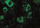
Application: Immunocytochemistry/ImmunofluorescenceSample Tested: Spinal motor neuronsSpecies: HumanVerified Customer | Posted 10/06/2021

There are no reviews that match your criteria.
Protocols
Find general support by application which include: protocols, troubleshooting, illustrated assays, videos and webinars.
- Appropriate Fixation of IHC/ICC Samples
- Cellular Response to Hypoxia Protocols
- ClariTSA™ Fluorophore Kits
- Detection & Visualization of Antibody Binding
- ICC Cell Smear Protocol for Suspension Cells
- ICC Immunocytochemistry Protocol Videos
- ICC for Adherent Cells
- Immunocytochemistry (ICC) Protocol
- Immunocytochemistry Troubleshooting
- Immunofluorescence of Organoids Embedded in Cultrex Basement Membrane Extract
- Immunohistochemistry (IHC) and Immunocytochemistry (ICC) Protocols
- Preparing Samples for IHC/ICC Experiments
- Preventing Non-Specific Staining (Non-Specific Binding)
- Primary Antibody Selection & Optimization
- Protocol for VisUCyte™ HRP Polymer Detection Reagent
- Protocol for the Fluorescent ICC Staining of Cell Smears - Graphic
- Protocol for the Fluorescent ICC Staining of Cultured Cells on Coverslips - Graphic
- Protocol for the Preparation and Fluorescent ICC Staining of Cells on Coverslips
- Protocol for the Preparation and Fluorescent ICC Staining of Non-adherent Cells
- Protocol for the Preparation and Fluorescent ICC Staining of Stem Cells on Coverslips
- Protocol for the Preparation of a Cell Smear for Non-adherent Cell ICC - Graphic
- R&D Systems Quality Control Western Blot Protocol
- TUNEL and Active Caspase-3 Detection by IHC/ICC Protocol
- The Importance of IHC/ICC Controls
- Troubleshooting Guide: Western Blot Figures
- Western Blot Conditions
- Western Blot Protocol
- Western Blot Protocol for Cell Lysates
- Western Blot Troubleshooting
- Western Blot Troubleshooting Guide
- View all Protocols, Troubleshooting, Illustrated assays and Webinars
Loading...
Associated Pathways

