Caspase-14 belongs to the evolutionarily conserved caspase family of cysteinyl aspartate-specific proteinases that frequently play a central role in apoptosis. Caspases exist as inactive proenzymes that undergo proteolytic processing at conserved aspartic residues to produce large and small subunits that dimerize to form an active enzyme. Caspase-14 is processed to form p19 and p10 subunits, but unlike many caspases, this activation is believed to be non-apoptotic. Expressed within hair follicles and sebaceous glands in the epidermis, Caspase-14 processing is implicated in terminal keratinocyte differentiation and cornification. Caspase-14 expression may also protect against psoriasis and epidermal UVB photodamage. Full-length human Caspase-14 is 242 amino acids (aa) in length, and shares 71% and 72% aa sequence identity with mouse and rat Caspase-14, respectively.
Human Caspase-14 Antibody (868715)
R&D Systems | Catalog # MAB8215

Key Product Details
Species Reactivity
Human
Applications
Immunohistochemistry, Western Blot, Simple Western
Label
Unconjugated
Antibody Source
Monoclonal Mouse IgG2B Clone # 868715
Loading...
Product Specifications
Immunogen
E. coli-derived recombinant human Caspase-14
Ser2-Gln242
Accession # P31944
Ser2-Gln242
Accession # P31944
Specificity
Detects human Caspase-14 in direct ELISA and Western Blot.
Clonality
Monoclonal
Host
Mouse
Isotype
IgG2B
Scientific Data Images for Human Caspase-14 Antibody (868715)
Detection of Human Caspase-14 by Western Blot.
Western blot shows lysates of human skin tissue. PVDF membrane was probed with 0.2 µg/mL of Mouse Anti-Human Caspase-14 Monoclonal Antibody (Catalog # MAB8215) followed by HRP-conjugated Anti-Mouse IgG Secondary Antibody (Catalog # HAF018). Specific bands were detected for full length Caspase-14 at approximately 28-30 kDa and the p10 subunit at approximately 10 kDa (as indicated). This experiment was conducted under reducing conditions and using Immunoblot Buffer Group 1.
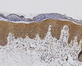
Caspase-14 in Human Epidermis.
Caspase-14 was detected in immersion fixed paraffin-embedded sections of human epidermis using Mouse Anti-Human Caspase-14 Monoclonal Antibody (Catalog # MAB8215) at 15 µg/mL overnight at 4 °C. Tissue was stained using the Anti-Mouse HRP-DAB Cell & Tissue Staining Kit (brown; Catalog # CTS002) and counterstained with hematoxylin (blue). Specific staining was localized to the nuclei of suprabasal keratinocytes. View our protocol for Chromogenic IHC Staining of Paraffin-embedded Tissue Sections.
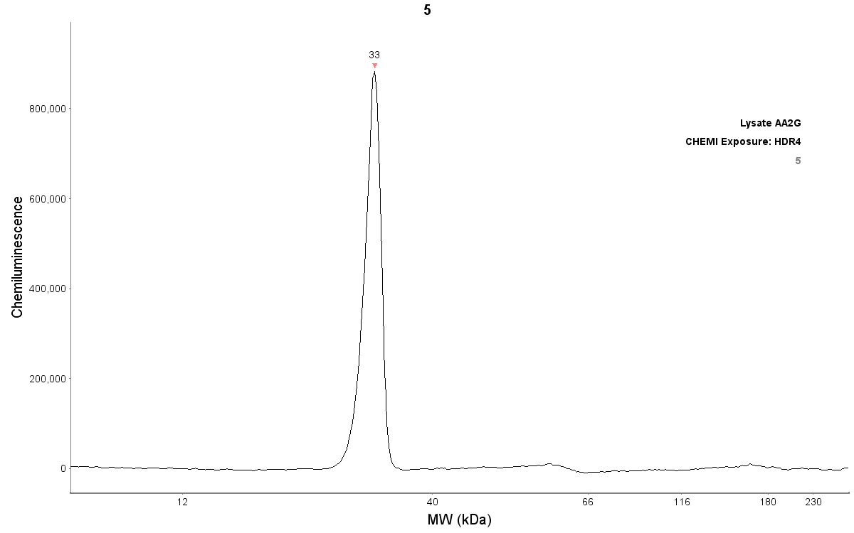
Detection of Human Caspase-14 by Simple WesternTM.
Simple Western lane view shows lysates of human skin tissue, loaded at 0.5 mg/mL. Specific bands were detected for full length Caspase-14 at approximately 36 kDa and the p10 subunit at approximately 12 kDa (as indicated) using 2 µg/mL of Mouse Anti-Human Caspase-14 Monoclonal Antibody (Catalog # MAB8215). This experiment was conducted under reducing conditions and using the 12-230 kDa separation system.Applications for Human Caspase-14 Antibody (868715)
Application
Recommended Usage
Immunohistochemistry
8-25 µg/mL
Sample: Immersion fixed paraffin-embedded sections of human epidermis
Sample: Immersion fixed paraffin-embedded sections of human epidermis
Simple Western
2 µg/mL
Sample: Human skin tissue
Sample: Human skin tissue
Western Blot
0.2 µg/mL
Sample: Human skin tissue
Sample: Human skin tissue
Reviewed Applications
Read 2 reviews rated 5 using MAB8215 in the following applications:
Formulation, Preparation, and Storage
Purification
Protein A or G purified from hybridoma culture supernatant
Reconstitution
Reconstitute at 0.5 mg/mL in sterile PBS. For liquid material, refer to CoA for concentration.
Loading...
Formulation
Lyophilized from a 0.2 μm filtered solution in PBS with Trehalose. *Small pack size (SP) is supplied either lyophilized or as a 0.2 µm filtered solution in PBS.
Shipping
Lyophilized product is shipped at ambient temperature. Liquid small pack size (-SP) is shipped with polar packs. Upon receipt, store immediately at the temperature recommended below.
Stability & Storage
Use a manual defrost freezer and avoid repeated freeze-thaw cycles.
- 12 months from date of receipt, -20 to -70 °C as supplied.
- 1 month, 2 to 8 °C under sterile conditions after reconstitution.
- 6 months, -20 to -70 °C under sterile conditions after reconstitution.
Calculators
Background: Caspase-14
Alternate Names
CASP14, Caspase14
Gene Symbol
CASP14
UniProt
Additional Caspase-14 Products
Product Documents for Human Caspase-14 Antibody (868715)
Certificate of Analysis
To download a Certificate of Analysis, please enter a lot or batch number in the search box below.
Note: Certificate of Analysis not available for kit components.
Product Specific Notices for Human Caspase-14 Antibody (868715)
For research use only
Related Research Areas
Citations for Human Caspase-14 Antibody (868715)
 Powered by Bioz
Customer Reviews for Human Caspase-14 Antibody (868715) (2)
5 out of 5
2 Customer Ratings
Have you used Human Caspase-14 Antibody (868715)?
Submit a review and receive an Amazon gift card!
$25/€18/£15/$25CAN/¥2500 Yen for a review with an image
$10/€7/£6/$10CAN/¥1110 Yen for a review without an image
Submit a review
Customer Images
Showing
1
-
2 of
2 reviews
Showing All
Filter By:
-
Application: ImmunohistochemistrySample Tested: EpidermisSpecies: HumanVerified Customer | Posted 11/11/2022

-
Application: Simple WesternSample Tested: Skin tissueSpecies: HumanVerified Customer | Posted 06/17/2019Detection of caspase-14 by Simple Western (JESS) in reconstructed human epidermis lysate (about 200 µg/mL protein concentration). Antibody dilution: 1/50
There are no reviews that match your criteria.
Protocols
Find general support by application which include: protocols, troubleshooting, illustrated assays, videos and webinars.
- Antigen Retrieval Protocol (PIER)
- Antigen Retrieval for Frozen Sections Protocol
- Appropriate Fixation of IHC/ICC Samples
- Cellular Response to Hypoxia Protocols
- Chromogenic IHC Staining of Formalin-Fixed Paraffin-Embedded (FFPE) Tissue Protocol
- Chromogenic Immunohistochemistry Staining of Frozen Tissue
- ClariTSA™ Fluorophore Kits
- Detection & Visualization of Antibody Binding
- Fluorescent IHC Staining of Frozen Tissue Protocol
- Graphic Protocol for Heat-induced Epitope Retrieval
- Graphic Protocol for the Preparation and Fluorescent IHC Staining of Frozen Tissue Sections
- Graphic Protocol for the Preparation and Fluorescent IHC Staining of Paraffin-embedded Tissue Sections
- Graphic Protocol for the Preparation of Gelatin-coated Slides for Histological Tissue Sections
- IHC Sample Preparation (Frozen sections vs Paraffin)
- Immunofluorescent IHC Staining of Formalin-Fixed Paraffin-Embedded (FFPE) Tissue Protocol
- Immunohistochemistry (IHC) and Immunocytochemistry (ICC) Protocols
- Immunohistochemistry Frozen Troubleshooting
- Immunohistochemistry Paraffin Troubleshooting
- Preparing Samples for IHC/ICC Experiments
- Preventing Non-Specific Staining (Non-Specific Binding)
- Primary Antibody Selection & Optimization
- Protocol for Heat-Induced Epitope Retrieval (HIER)
- Protocol for Making a 4% Formaldehyde Solution in PBS
- Protocol for VisUCyte™ HRP Polymer Detection Reagent
- Protocol for the Preparation & Fixation of Cells on Coverslips
- Protocol for the Preparation and Chromogenic IHC Staining of Frozen Tissue Sections
- Protocol for the Preparation and Chromogenic IHC Staining of Frozen Tissue Sections - Graphic
- Protocol for the Preparation and Chromogenic IHC Staining of Paraffin-embedded Tissue Sections
- Protocol for the Preparation and Chromogenic IHC Staining of Paraffin-embedded Tissue Sections - Graphic
- Protocol for the Preparation and Fluorescent IHC Staining of Frozen Tissue Sections
- Protocol for the Preparation and Fluorescent IHC Staining of Paraffin-embedded Tissue Sections
- Protocol for the Preparation of Gelatin-coated Slides for Histological Tissue Sections
- R&D Systems Quality Control Western Blot Protocol
- TUNEL and Active Caspase-3 Detection by IHC/ICC Protocol
- The Importance of IHC/ICC Controls
- Troubleshooting Guide: Immunohistochemistry
- Troubleshooting Guide: Western Blot Figures
- Western Blot Conditions
- Western Blot Protocol
- Western Blot Protocol for Cell Lysates
- Western Blot Troubleshooting
- Western Blot Troubleshooting Guide
- View all Protocols, Troubleshooting, Illustrated assays and Webinars
Loading...