
Key Product Details
Species Reactivity
Applications
Label
Antibody Source
Product Specifications
Immunogen
Thr36-Asn233
Accession # Q14002
Specificity
Clonality
Host
Isotype
Scientific Data Images for Human CEACAM-7 Antibody (962703)
Detection of CEACAM‑7 in HEK293 Human Cell Line Transfected with Human CEACAM-7 and eGFP by Flow Cytometry.
HEK293 human embryonic kidney cell line transfected with human CEACAM-7 and eGFP was stained with either (A) Mouse Anti-Human CEACAM-7 Monoclonal Antibody (Catalog # MAB44781) or (B) Mouse IgG1 Isotype Control (Catalog # MAB002) followed by APC-conjugated Anti-Mouse IgG Secondary Antibody(Catalog # F0101B). View our protocol for Staining Membrane-associated Proteins.
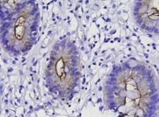
CEACAM‑7 in Human Colon Cancer Tissue.
CEACAM-7 was detected in immersion fixed paraffin-embedded sections of human colon cancer tissue using Mouse Anti-Human CEACAM-7 Monoclonal Antibody (Catalog # MAB44781) at 5 µg/mL for 1 hour at room temperature followed by incubation with the Anti-Mouse IgG VisUCyte™ HRP Polymer Antibody (Catalog # VC001). Tissue was stained using DAB (brown) and counterstained with hematoxylin (blue). Specific staining was localized to cytoplasm. View our protocol for IHC Staining with VisUCyte HRP Polymer Detection Reagents.
Human CEACAM‑7 ELISA Standard Curve.
Recombinant Human CEACAM-7 protein was serially diluted 2-fold and captured by Mouse Anti-Human CEACAM-7 Monoclonal Antibody (Catalog # MAB44782) coated on a Clear Polystyrene Microplate (Catalog # DY990). Mouse Anti-Human CEACAM-7 Monoclonal Antibody (Catalog # MAB44781) was biotinylated and incubated with the protein captured on the plate. Detection of the standard curve was achieved by incubating Streptavidin-HRP (Catalog # DY998) followed by Substrate Solution (Catalog # DY999) and stopping the enzymatic reaction with Stop Solution (Catalog # DY994).Applications for Human CEACAM-7 Antibody (962703)
CyTOF-ready
ELISA
This antibody functions as an ELISA detection antibody when paired with Mouse Anti-Human CEACAM‑7 Monoclonal Antibody (Catalog # MAB44782).
This product is intended for assay development on various assay platforms requiring antibody pairs. We recommend the Human CEACAM-7 DuoSet ELISA Kit (Catalog # DY4478-05) for convenient development of a sandwich ELISA.
Flow Cytometry
Sample: HEK293 Human Cell Line Transfected with Human CEACAM-7 and eGFP
Immunohistochemistry
Sample: Immersion fixed paraffin-embedded sections of human colon cancer tissue
Reviewed Applications
Read 1 review rated 5 using MAB44781 in the following applications:
Flow Cytometry Panel Builder
Bio-Techne Knows Flow Cytometry
Save time and reduce costly mistakes by quickly finding compatible reagents using the Panel Builder Tool.
Advanced Features
- Spectra Viewer - Custom analysis of spectra from multiple fluorochromes
- Spillover Popups - Visualize the spectra of individual fluorochromes
- Antigen Density Selector - Match fluorochrome brightness with antigen density

Formulation, Preparation, and Storage
Purification
Reconstitution
Reconstitute at 0.5 mg/mL in sterile PBS. For liquid material, refer to CoA for concentration.
Formulation
Shipping
Stability & Storage
- 12 months from date of receipt, -20 to -70 °C as supplied.
- 1 month, 2 to 8 °C under sterile conditions after reconstitution.
- 6 months, -20 to -70 °C under sterile conditions after reconstitution.
Calculators
Background: CEACAM-7
References
- Tchoupa, A.K. et al. (2014) Cell Commun. Signal. 12:27.
- Thompson, J. et al. (1994) J. Biol. Chem. 269:32924.
- Thompson, J. et al. (1997) Cancer Res. 57:1776.
- Scholzel, S. et al. (2000) Am. J. Pathol. 156:595.
- Nollau, P. et al. (1997) Cancer Res. 57:2354.
- Nollau, P. et al. (1997) Am. J. Pathol. 151:521.
- Zhou, J. et al. (2011) World J. Surgical Oncol. 9:172.
- Markel, G. et al. (2004) J. Immunol. 173:3732.
- Oikawa, S. et al. (1992) Biochem. Biophys. Res. Commun. 186:881.
Long Name
Alternate Names
Entrez Gene IDs
Gene Symbol
UniProt
Additional CEACAM-7 Products
Product Documents for Human CEACAM-7 Antibody (962703)
Certificate of Analysis
To download a Certificate of Analysis, please enter a lot or batch number in the search box below.
Note: Certificate of Analysis not available for kit components.
Product Specific Notices for Human CEACAM-7 Antibody (962703)
For research use only
Related Research Areas
Customer Reviews for Human CEACAM-7 Antibody (962703) (1)
Have you used Human CEACAM-7 Antibody (962703)?
Submit a review and receive an Amazon gift card!
$25/€18/£15/$25CAN/¥2500 Yen for a review with an image
$10/€7/£6/$10CAN/¥1110 Yen for a review without an image
Submit a review
Customer Images
-
Application: ImmunohistochemistrySample Tested: Gastric carcinomaSpecies: HumanVerified Customer | Posted 11/30/2021

There are no reviews that match your criteria.
Protocols
Find general support by application which include: protocols, troubleshooting, illustrated assays, videos and webinars.
- 7-Amino Actinomycin D (7-AAD) Cell Viability Flow Cytometry Protocol
- Antigen Retrieval Protocol (PIER)
- Antigen Retrieval for Frozen Sections Protocol
- Appropriate Fixation of IHC/ICC Samples
- Cellular Response to Hypoxia Protocols
- Chromogenic IHC Staining of Formalin-Fixed Paraffin-Embedded (FFPE) Tissue Protocol
- Chromogenic Immunohistochemistry Staining of Frozen Tissue
- ClariTSA™ Fluorophore Kits
- Detection & Visualization of Antibody Binding
- ELISA Sample Preparation & Collection Guide
- ELISA Troubleshooting Guide
- Extracellular Membrane Flow Cytometry Protocol
- Flow Cytometry Protocol for Cell Surface Markers
- Flow Cytometry Protocol for Staining Membrane Associated Proteins
- Flow Cytometry Staining Protocols
- Flow Cytometry Troubleshooting Guide
- Fluorescent IHC Staining of Frozen Tissue Protocol
- Graphic Protocol for Heat-induced Epitope Retrieval
- Graphic Protocol for the Preparation and Fluorescent IHC Staining of Frozen Tissue Sections
- Graphic Protocol for the Preparation and Fluorescent IHC Staining of Paraffin-embedded Tissue Sections
- Graphic Protocol for the Preparation of Gelatin-coated Slides for Histological Tissue Sections
- How to Run an R&D Systems DuoSet ELISA
- How to Run an R&D Systems Quantikine ELISA
- How to Run an R&D Systems Quantikine™ QuicKit™ ELISA
- IHC Sample Preparation (Frozen sections vs Paraffin)
- Immunofluorescent IHC Staining of Formalin-Fixed Paraffin-Embedded (FFPE) Tissue Protocol
- Immunohistochemistry (IHC) and Immunocytochemistry (ICC) Protocols
- Immunohistochemistry Frozen Troubleshooting
- Immunohistochemistry Paraffin Troubleshooting
- Intracellular Flow Cytometry Protocol Using Alcohol (Methanol)
- Intracellular Flow Cytometry Protocol Using Detergents
- Intracellular Nuclear Staining Flow Cytometry Protocol Using Detergents
- Intracellular Staining Flow Cytometry Protocol Using Alcohol Permeabilization
- Intracellular Staining Flow Cytometry Protocol Using Detergents to Permeabilize Cells
- Preparing Samples for IHC/ICC Experiments
- Preventing Non-Specific Staining (Non-Specific Binding)
- Primary Antibody Selection & Optimization
- Propidium Iodide Cell Viability Flow Cytometry Protocol
- Protocol for Heat-Induced Epitope Retrieval (HIER)
- Protocol for Liperfluo
- Protocol for Making a 4% Formaldehyde Solution in PBS
- Protocol for VisUCyte™ HRP Polymer Detection Reagent
- Protocol for the Characterization of Human Th22 Cells
- Protocol for the Characterization of Human Th9 Cells
- Protocol for the Preparation & Fixation of Cells on Coverslips
- Protocol for the Preparation and Chromogenic IHC Staining of Frozen Tissue Sections
- Protocol for the Preparation and Chromogenic IHC Staining of Frozen Tissue Sections - Graphic
- Protocol for the Preparation and Chromogenic IHC Staining of Paraffin-embedded Tissue Sections
- Protocol for the Preparation and Chromogenic IHC Staining of Paraffin-embedded Tissue Sections - Graphic
- Protocol for the Preparation and Fluorescent IHC Staining of Frozen Tissue Sections
- Protocol for the Preparation and Fluorescent IHC Staining of Paraffin-embedded Tissue Sections
- Protocol for the Preparation of Gelatin-coated Slides for Histological Tissue Sections
- Protocol: Annexin V and PI Staining by Flow Cytometry
- Protocol: Annexin V and PI Staining for Apoptosis by Flow Cytometry
- Quantikine HS ELISA Kit Assay Principle, Alkaline Phosphatase
- Quantikine HS ELISA Kit Principle, Streptavidin-HRP Polymer
- Sandwich ELISA (Colorimetric) – Biotin/Streptavidin Detection Protocol
- Sandwich ELISA (Colorimetric) – Direct Detection Protocol
- TUNEL and Active Caspase-3 Detection by IHC/ICC Protocol
- The Importance of IHC/ICC Controls
- Troubleshooting Guide: ELISA
- Troubleshooting Guide: Fluorokine Flow Cytometry Kits
- Troubleshooting Guide: Immunohistochemistry
- View all Protocols, Troubleshooting, Illustrated assays and Webinars