Human Chorionic Gonadotropin, alpha chain (CGA) is a 92 aa glycopeptide that functions as the shared alpha subunit of the heterodimeric peptide hormones choriogonadotropin, leutinizing hormone, thryoid stimulating hormone, and follicle stimulating hormone. CGA circulates as a free molecule and in noncovalent complexes with the unique beta subunits of those hormones. CGA is secreted by the pituitary and placenta. Mature human CGA shares 69%‑73% aa sequence identity with canine, equine, feline, mouse, porcine, and rat CGA.
Human Chorionic Gonadotropin alpha Chain (HCG alpha) Antibody (381012)
R&D Systems | Catalog # MAB4169
 in Human Placenta.")
Key Product Details
Species Reactivity
Human
Applications
Immunohistochemistry, Western Blot
Label
Unconjugated
Antibody Source
Monoclonal Mouse IgG2A Clone # 381012
Loading...
Product Specifications
Immunogen
Mouse myeloma cell line NS0-derived recombinant human Chorionic Gonadotropin, alpha Chain ( alpha HCG)
Ala25-Ser116
Accession # P01215
Ala25-Ser116
Accession # P01215
Specificity
Detects human Chorionic Gonadotropin, alpha Chain ( alpha HCG) in direct ELISAs and Western blots. In direct ELISAs and Western blots, no cross‑reactivity with recombinant human FSH beta or recombinant rat FSH beta is observed.
Clonality
Monoclonal
Host
Mouse
Isotype
IgG2A
Scientific Data Images
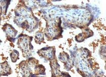
Chorionic Gonadotropin, alpha Chain ( alpha HCG) in Human Placenta.
Chorionic Gonadotropin, a Chain (a HCG) was detected in immersion fixed paraffin-embedded sections of human placenta using Mouse Anti-Human Chorionic Gonadotropin, a Chain (a HCG) Monoclonal Antibody (Catalog # MAB4169) at 15 µg/mL overnight at 4 °C. Tissue was stained using the Anti-Mouse HRP-DAB Cell & Tissue Staining Kit (brown; Catalog # CTS002) and counterstained with hematoxylin (blue). Lower panel shows a lack of labeling if primary antibodies are omitted and tissue is stained only with secondary antibody followed by incubation with detection reagents. View our protocol for Chromogenic IHC Staining of Paraffin-embedded Tissue Sections.Applications
Application
Recommended Usage
Immunohistochemistry
8-25 µg/mL
Sample: Immersion fixed paraffin-embedded sections of human placenta subjected to Antigen Retrieval Reagent-Basic (Catalog # CTS013)
Sample: Immersion fixed paraffin-embedded sections of human placenta subjected to Antigen Retrieval Reagent-Basic (Catalog # CTS013)
Western Blot
1 µg/mL
Sample: Recombinant Human Chorionic Gonadotropin, alpha Chain ( alpha HCG)
Sample: Recombinant Human Chorionic Gonadotropin, alpha Chain ( alpha HCG)
Reviewed Applications
Read 2 reviews rated 4.5 using MAB4169 in the following applications:
Formulation, Preparation, and Storage
Purification
Protein A or G purified from hybridoma culture supernatant
Reconstitution
Reconstitute at 0.5 mg/mL in sterile PBS. For liquid material, refer to CoA for concentration.
Loading...
Formulation
Lyophilized from a 0.2 μm filtered solution in PBS with Trehalose. *Small pack size (SP) is supplied either lyophilized or as a 0.2 µm filtered solution in PBS.
Shipping
Lyophilized product is shipped at ambient temperature. Liquid small pack size (-SP) is shipped with polar packs. Upon receipt, store immediately at the temperature recommended below.
Stability & Storage
Use a manual defrost freezer and avoid repeated freeze-thaw cycles.
- 12 months from date of receipt, -20 to -70 °C as supplied.
- 1 month, 2 to 8 °C under sterile conditions after reconstitution.
- 6 months, -20 to -70 °C under sterile conditions after reconstitution.
Calculators
Background: Chorionic Gonadotropin alpha Chain (HCG alpha)
Alternate Names
alpha HCG, CGA, FSHA, GPHa1, LHA, TSHA
Entrez Gene IDs
1081 (Human)
Gene Symbol
CGA
UniProt
Additional Chorionic Gonadotropin alpha Chain (HCG alpha) Products
- All Products for Chorionic Gonadotropin alpha Chain (HCG alpha)
- Chorionic Gonadotropin alpha Chain (HCG alpha) ELISA Kits
- Chorionic Gonadotropin alpha Chain (HCG alpha) Lysates
- Chorionic Gonadotropin alpha Chain (HCG alpha) Primary Antibodies
- Chorionic Gonadotropin alpha Chain (HCG alpha) Proteins and Enzymes
Product Documents
Certificate of Analysis
To download a Certificate of Analysis, please enter a lot or batch number in the search box below.
Note: Certificate of Analysis not available for kit components.
Product Specific Notices
For research use only
Related Research Areas
Customer Reviews (2)
4.5 out of 5
2 Customer Ratings
Have you used Human Chorionic Gonadotropin alpha Chain (HCG alpha) Antibody (381012)?
Submit a review and receive an Amazon gift card!
$25/€18/£15/$25CAN/¥2500 Yen for a review with an image
$10/€7/£6/$10CAN/¥1110 Yen for a review without an image
Submit a review
Customer Images
Showing
1
-
2 of
2 reviews
Showing All
Filter By:
-
Application: ImmunohistochemistrySample Tested: Placental tissueSpecies: HumanVerified Customer | Posted 05/03/2022incubation 15 µg/mL overnight at 4 °C

-
Application: Immunocytochemistry/ImmunofluorescenceSample Tested: embryonic stem cells and induced pluripotent stem cellsSpecies: HumanVerified Customer | Posted 12/15/2016We use the monoclonal antibody to detect human Chorionic Gonadotropin, alpha (CGA) Chain expressed in human embryonic stem cells and induced pluripotent stem cells (PSC) derived model for studying human placental trophoblasts in vitro. This antibody has been used with immunofluorescent methods to identify precursors of syncytiotrophoblasts emerging from PSC differentiation. The trophoblasts generated from human PSC most likely represents the primitive placenta encountered in early pregnancy soon after the human trophoblasts invades into the uterine wall.
There are no reviews that match your criteria.
Protocols
Find general support by application which include: protocols, troubleshooting, illustrated assays, videos and webinars.
- Antigen Retrieval Protocol (PIER)
- Antigen Retrieval for Frozen Sections Protocol
- Appropriate Fixation of IHC/ICC Samples
- Cellular Response to Hypoxia Protocols
- Chromogenic IHC Staining of Formalin-Fixed Paraffin-Embedded (FFPE) Tissue Protocol
- Chromogenic Immunohistochemistry Staining of Frozen Tissue
- ClariTSA™ Fluorophore Kits
- Detection & Visualization of Antibody Binding
- Fluorescent IHC Staining of Frozen Tissue Protocol
- Graphic Protocol for Heat-induced Epitope Retrieval
- Graphic Protocol for the Preparation and Fluorescent IHC Staining of Frozen Tissue Sections
- Graphic Protocol for the Preparation and Fluorescent IHC Staining of Paraffin-embedded Tissue Sections
- Graphic Protocol for the Preparation of Gelatin-coated Slides for Histological Tissue Sections
- IHC Sample Preparation (Frozen sections vs Paraffin)
- Immunofluorescent IHC Staining of Formalin-Fixed Paraffin-Embedded (FFPE) Tissue Protocol
- Immunohistochemistry (IHC) and Immunocytochemistry (ICC) Protocols
- Immunohistochemistry Frozen Troubleshooting
- Immunohistochemistry Paraffin Troubleshooting
- Preparing Samples for IHC/ICC Experiments
- Preventing Non-Specific Staining (Non-Specific Binding)
- Primary Antibody Selection & Optimization
- Protocol for Heat-Induced Epitope Retrieval (HIER)
- Protocol for Making a 4% Formaldehyde Solution in PBS
- Protocol for VisUCyte™ HRP Polymer Detection Reagent
- Protocol for the Preparation & Fixation of Cells on Coverslips
- Protocol for the Preparation and Chromogenic IHC Staining of Frozen Tissue Sections
- Protocol for the Preparation and Chromogenic IHC Staining of Frozen Tissue Sections - Graphic
- Protocol for the Preparation and Chromogenic IHC Staining of Paraffin-embedded Tissue Sections
- Protocol for the Preparation and Chromogenic IHC Staining of Paraffin-embedded Tissue Sections - Graphic
- Protocol for the Preparation and Fluorescent IHC Staining of Frozen Tissue Sections
- Protocol for the Preparation and Fluorescent IHC Staining of Paraffin-embedded Tissue Sections
- Protocol for the Preparation of Gelatin-coated Slides for Histological Tissue Sections
- R&D Systems Quality Control Western Blot Protocol
- TUNEL and Active Caspase-3 Detection by IHC/ICC Protocol
- The Importance of IHC/ICC Controls
- Troubleshooting Guide: Immunohistochemistry
- Troubleshooting Guide: Western Blot Figures
- Western Blot Conditions
- Western Blot Protocol
- Western Blot Protocol for Cell Lysates
- Western Blot Troubleshooting
- Western Blot Troubleshooting Guide
- View all Protocols, Troubleshooting, Illustrated assays and Webinars
Loading...