CTLA-4 and CD28, together with their ligands B7-1 and B7-2, constitute one of the dominant costimulatory pathways that regulate T- and B-cell responses. CTLA-4 and CD28 are structurally homologous molecules that are members of the immunoglobulin (Ig) gene superfamily. Both CTLA-4 and CD28 are composed of a single
Ig V‑like extracellular domain, a transmembrane domain and an intracellular domain. CTLA-4 and CD28 are both expressed on the cell surface as disulfide-linked homodimers or as monomers. The genes encoding these two molecules are closely linked on human chromosome 2. CTLA-4 was originally identified as a gene that was specifically expressed by cytotoxic T lymphocytes. However, CTLA-4 transcripts have since been found in both Th1 and Th2, and CD4+ and CD8+ T cell clones. Whereas CD28 expression is constitutive on the surfaces of 95% of CD4+ T cells and 50% of CD8+ T cells and is down regulated upon T cell activation, CTLA-4 expression is upregulated rapidly following T cell activation and peaks approximately 24 hours following activation. Although both CTLA-4 and CD28 can bind to the same ligands, CTLA-4 binds to B7-1 and B7-2 with 20‑100‑fold higher affinity than CD28. The physiological role of CTLA-4 in T cell costimulation is currently being studied.

Key Product Details
Validated by
Biological Validation
Species Reactivity
Validated:
Human
Cited:
Human, Rat, Bovine, Xenograft
Applications
Validated:
Western Blot, Flow Cytometry, Immunocytochemistry, CyTOF-ready
Cited:
Immunohistochemistry, Immunohistochemistry-Paraffin, Immunohistochemistry-Frozen, Flow Cytometry, In vivo assay
Label
Unconjugated
Antibody Source
Polyclonal Goat IgG
Loading...
Product Specifications
Immunogen
S. frugiperda insect ovarian cell line Sf 21-derived recombinant human CTLA-4
Ala37-Phe162
Accession # Q6GR94
Ala37-Phe162
Accession # Q6GR94
Specificity
Detects human CTLA-4 in direct ELISAs and Western blots. In Western blots, approximately 25% cross-reactivity with recombinant mouse CTLA-4 is observed.
Clonality
Polyclonal
Host
Goat
Isotype
IgG
Scientific Data Images for Human CTLA-4 Antibody
Detection of Human CTLA‑4 by Western Blot.
Western blot shows lysates of NS0 mouse myeloma cell line either mock transfected or transfected with human CTLA-4. PVDF membrane was probed with 0.5 µg/mL of Goat Anti-Human CTLA-4 Antigen Affinity-purified Polyclonal Antibody (Catalog # AF-386-PB) followed by HRP-conjugated Anti-Goat IgG Secondary Antibody (Catalog # HAF017). A specific band was detected for CTLA-4 at approximately 50 kDa (as indicated). This experiment was conducted under reducing conditions and using Immunoblot Buffer Group 1.
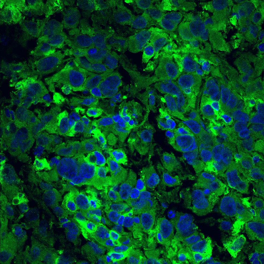
CTLA‑4 in Human Peripheral Blood Mononuclear Cells.
CTLA-4 was detected in immersion fixed human peripheral blood mononuclear cells treated with treated with PMA and calcium ionomycin using Goat Anti-Human CTLA-4 Antigen Affinity-purified Polyclonal Antibody (Catalog # AF-386-PB) at 15 µg/mL for 3 hours at room temperature. Cells were stained using the NorthernLights™ 557-conjugated Anti-Goat IgG Secondary Antibody (red; Catalog # NL001) and counterstained with DAPI (blue). Specific staining was localized to cell surfaces. View our protocol for Fluorescent ICC Staining of Non-adherent Cells.
Detection of CTLA‑4 in NS0 Mouse Cell Line Co-transfected with CTLA-4 and eGFP by Flow Cytometry.
NS0 mouse myeloma cell line co-transfected with human CTLA-4 and eGFP was stained with either (A) Goat Anti-Human CTLA-4 Antigen Affinity-purified Polyclonal Antibody (Catalog # AF-386-PB) or (B) Normal Goat IgG Control (Catalog # AB-108-C) followed by Allophycocyanin-conjugated Anti-Goat IgG Secondary Antibody (Catalog # F0108).Applications for Human CTLA-4 Antibody
Application
Recommended Usage
CyTOF-ready
Ready to be labeled using established conjugation methods. No BSA or other carrier proteins that could interfere with conjugation.
Flow Cytometry
0.25 µg/106 cells
Sample: NS0 mouse myeloma cell line co-transfected with human CTLA-4 and eGFP
Sample: NS0 mouse myeloma cell line co-transfected with human CTLA-4 and eGFP
Immunocytochemistry
5-15 µg/mL
Sample: Immersion fixed human peripheral blood mononuclear cells treated with PMA and calcium ionomycin
Sample: Immersion fixed human peripheral blood mononuclear cells treated with PMA and calcium ionomycin
Western Blot
0.5 µg/mL
Sample: NS0 mouse myeloma cell line transfected with human CTLA-4
Sample: NS0 mouse myeloma cell line transfected with human CTLA-4
Reviewed Applications
Read 2 reviews rated 5 using AF-386-PB in the following applications:
Flow Cytometry Panel Builder
Bio-Techne Knows Flow Cytometry
Save time and reduce costly mistakes by quickly finding compatible reagents using the Panel Builder Tool.
Advanced Features
- Spectra Viewer - Custom analysis of spectra from multiple fluorochromes
- Spillover Popups - Visualize the spectra of individual fluorochromes
- Antigen Density Selector - Match fluorochrome brightness with antigen density

Formulation, Preparation, and Storage
Purification
Antigen Affinity-purified
Reconstitution
Reconstitute at 0.2 mg/mL in sterile PBS. For liquid material, refer to CoA for concentration.
Loading...
Formulation
Lyophilized from a 0.2 μm filtered solution in PBS with Trehalose. *Small pack size (SP) is supplied either lyophilized or as a 0.2 µm filtered solution in PBS.
Shipping
Lyophilized product is shipped at ambient temperature. Liquid small pack size (-SP) is shipped with polar packs. Upon receipt, store immediately at the temperature recommended below.
Stability & Storage
Use a manual defrost freezer and avoid repeated freeze-thaw cycles.
- 12 months from date of receipt, -20 to -70 °C as supplied.
- 1 month, 2 to 8 °C under sterile conditions after reconstitution.
- 6 months, -20 to -70 °C under sterile conditions after reconstitution.
Calculators
Background: CTLA-4
References
- Lenschow, D.J. et al. (1996) Annu. Rev. Immunol. 14:233.
- Hathcock, K.S. and R.J. Hodes (1996) Advances in Immunol. 62:131.
- Ward, S.G. (1996) Biochem. J. 318:361.
Long Name
Cytotoxic T-lymphocyte-associated Molecule 4
Alternate Names
CD152, CTLA4
Gene Symbol
CTLA4
UniProt
Additional CTLA-4 Products
Product Documents for Human CTLA-4 Antibody
Certificate of Analysis
To download a Certificate of Analysis, please enter a lot or batch number in the search box below.
Note: Certificate of Analysis not available for kit components.
Product Specific Notices for Human CTLA-4 Antibody
For research use only
Citations for Human CTLA-4 Antibody
 Powered by Bioz
Customer Reviews for Human CTLA-4 Antibody (2)
5 out of 5
2 Customer Ratings
Have you used Human CTLA-4 Antibody?
Submit a review and receive an Amazon gift card!
$25/€18/£15/$25CAN/¥2500 Yen for a review with an image
$10/€7/£6/$10CAN/¥1110 Yen for a review without an image
Submit a review
Customer Images
Showing
1
-
2 of
2 reviews
Showing All
Filter By:
-
Application: ImmunocytochemistrySample Tested: Breast cancer tissue and Adult lungSpecies: HumanVerified Customer | Posted 06/21/2017
-
Application: ImmunofluorescenceSample Tested: Human lung cancer cell line MiaPaCa-2Species: HumanVerified Customer | Posted 08/05/2016CTLA-4 Expression in Pancreatic Cancer Cell Line (MiaPaCa-2)

There are no reviews that match your criteria.
Protocols
Find general support by application which include: protocols, troubleshooting, illustrated assays, videos and webinars.
- 7-Amino Actinomycin D (7-AAD) Cell Viability Flow Cytometry Protocol
- Appropriate Fixation of IHC/ICC Samples
- Cellular Response to Hypoxia Protocols
- ClariTSA™ Fluorophore Kits
- Detection & Visualization of Antibody Binding
- Extracellular Membrane Flow Cytometry Protocol
- Flow Cytometry Protocol for Cell Surface Markers
- Flow Cytometry Protocol for Staining Membrane Associated Proteins
- Flow Cytometry Staining Protocols
- Flow Cytometry Troubleshooting Guide
- ICC Cell Smear Protocol for Suspension Cells
- ICC Immunocytochemistry Protocol Videos
- ICC for Adherent Cells
- Immunocytochemistry (ICC) Protocol
- Immunocytochemistry Troubleshooting
- Immunofluorescence of Organoids Embedded in Cultrex Basement Membrane Extract
- Immunohistochemistry (IHC) and Immunocytochemistry (ICC) Protocols
- Intracellular Flow Cytometry Protocol Using Alcohol (Methanol)
- Intracellular Flow Cytometry Protocol Using Detergents
- Intracellular Nuclear Staining Flow Cytometry Protocol Using Detergents
- Intracellular Staining Flow Cytometry Protocol Using Alcohol Permeabilization
- Intracellular Staining Flow Cytometry Protocol Using Detergents to Permeabilize Cells
- Preparing Samples for IHC/ICC Experiments
- Preventing Non-Specific Staining (Non-Specific Binding)
- Primary Antibody Selection & Optimization
- Propidium Iodide Cell Viability Flow Cytometry Protocol
- Protocol for Liperfluo
- Protocol for VisUCyte™ HRP Polymer Detection Reagent
- Protocol for the Characterization of Human Th22 Cells
- Protocol for the Characterization of Human Th9 Cells
- Protocol for the Fluorescent ICC Staining of Cell Smears - Graphic
- Protocol for the Fluorescent ICC Staining of Cultured Cells on Coverslips - Graphic
- Protocol for the Preparation and Fluorescent ICC Staining of Cells on Coverslips
- Protocol for the Preparation and Fluorescent ICC Staining of Non-adherent Cells
- Protocol for the Preparation and Fluorescent ICC Staining of Stem Cells on Coverslips
- Protocol for the Preparation of a Cell Smear for Non-adherent Cell ICC - Graphic
- Protocol: Annexin V and PI Staining by Flow Cytometry
- Protocol: Annexin V and PI Staining for Apoptosis by Flow Cytometry
- R&D Systems Quality Control Western Blot Protocol
- TUNEL and Active Caspase-3 Detection by IHC/ICC Protocol
- The Importance of IHC/ICC Controls
- Troubleshooting Guide: Fluorokine Flow Cytometry Kits
- Troubleshooting Guide: Western Blot Figures
- Western Blot Conditions
- Western Blot Protocol
- Western Blot Protocol for Cell Lysates
- Western Blot Troubleshooting
- Western Blot Troubleshooting Guide
- View all Protocols, Troubleshooting, Illustrated assays and Webinars
Loading...