Desmoglein-1 is one of three members of the desmoglein subfamily of calcium-dependent cadherin cell adhesion molecules. Together with desmocollins, another subfamily within the cadherin superfamily, the desmoglein isoforms form the adhesive components of desmosomes, the cell-cell adhesive structures that are found in epithelial cells. Human Desmoglein-1 is a type I transmembrane glycoprotein of 1049 amino acid (aa) residues with a 23 aa signal peptide and a 26 aa propeptide. It differs from other classic cadherins by having four instead of five cadherin repeat domains in its extracellular region, and a much larger cytoplasmic region containing five desmoglein repeat domains which share homology with the cadherin repeats. Instead of having the HAV adhesion motif found in type I cadherins, Desmoglein-1 has R/YAL as the adhesion motif on its amino-terminal cadherin repeat. The cytoplasmic tail of Desmoglein-1 interacts with desmoplakins, plakoglobin and plakophilins. In turn, these proteins link the Desmoglein-1 with the intermediate filaments. Desmoglein-1 has been shown to be important in establishing cell-cell adhesion and function in the epidermis. In the autoimmune skin disease pemphigus foliaceus, autoantibodies to Desmoglein-1 can cause the loss of keratinocyte adhesion resulting in blisters.
Human Desmoglein-1 Antibody (129204)
R&D Systems | Catalog # MAB944

Key Product Details
Species Reactivity
Validated:
Human
Cited:
Human
Applications
Validated:
Immunohistochemistry, Western Blot
Cited:
Western Blot, Flow Cytometry
Label
Unconjugated
Antibody Source
Monoclonal Mouse IgG2A Clone # 129204
Loading...
Product Specifications
Immunogen
Mouse myeloma cell line NS0-derived recombinant human Desmoglein-1
Glu50-His545
Accession # Q02413
Glu50-His545
Accession # Q02413
Specificity
Detects human Desmoglein-1 in direct ELISAs and Western blots. Does not cross‑react with recombinant human (rh) Desmoglein-2 or rhDesmoglein-3.
Clonality
Monoclonal
Host
Mouse
Isotype
IgG2A
Scientific Data Images for Human Desmoglein-1 Antibody (129204)
Desmoglein‑1 in Human Skin Hyperplasia.
Desmoglein-1 was detected in immersion fixed paraffin-embedded sections of human skin hyperplasia using Mouse Anti-Human Desmoglein-1 Monoclonal Antibody (Catalog # MAB944) at 5 µg/mL for 1 hour at room temperature followed by incubation with the Anti-Mouse IgG VisUCyte™ HRP Polymer Antibody (Catalog # VC001). Tissue was stained using DAB (brown) and counterstained with hematoxylin (blue). Specific staining was localized to keratinocyte membranes. View our protocol for IHC Staining with VisUCyte HRP Polymer Detection Reagents.Applications for Human Desmoglein-1 Antibody (129204)
Application
Recommended Usage
Immunohistochemistry
5-25 µg/mL
Sample: Immersion fixed paraffin-embedded sections of human skin hyperplasia
Sample: Immersion fixed paraffin-embedded sections of human skin hyperplasia
Western Blot
1 µg/mL
Sample: Recombinant Human Desmoglein‑1 Fc Chimera (Catalog # 944-DM)
Sample: Recombinant Human Desmoglein‑1 Fc Chimera (Catalog # 944-DM)
Reviewed Applications
Read 2 reviews rated 4.5 using MAB944 in the following applications:
Formulation, Preparation, and Storage
Purification
Protein A or G purified from hybridoma culture supernatant
Reconstitution
Reconstitute at 0.5 mg/mL in sterile PBS. For liquid material, refer to CoA for concentration.
Loading...
Formulation
Lyophilized from a 0.2 μm filtered solution in PBS with Trehalose. *Small pack size (SP) is supplied either lyophilized or as a 0.2 µm filtered solution in PBS.
Shipping
Lyophilized product is shipped at ambient temperature. Liquid small pack size (-SP) is shipped with polar packs. Upon receipt, store immediately at the temperature recommended below.
Stability & Storage
Use a manual defrost freezer and avoid repeated freeze-thaw cycles.
- 12 months from date of receipt, -20 to -70 °C as supplied.
- 1 month, 2 to 8 °C under sterile conditions after reconstitution.
- 6 months, -20 to -70 °C under sterile conditions after reconstitution.
Calculators
Background: Desmoglein-1
References
- Nollet, R. et al. (2000) J. Mol. Biol. 299:551.
- Elias, P. et al. (2001) J. Cell Biol. 153:243.
Alternate Names
CDHF4, Desmoglein1, DSG1
Entrez Gene IDs
1828 (Human)
Gene Symbol
DSG1
UniProt
Additional Desmoglein-1 Products
Product Documents for Human Desmoglein-1 Antibody (129204)
Certificate of Analysis
To download a Certificate of Analysis, please enter a lot or batch number in the search box below.
Note: Certificate of Analysis not available for kit components.
Product Specific Notices for Human Desmoglein-1 Antibody (129204)
For research use only
Related Research Areas
Citations for Human Desmoglein-1 Antibody (129204)
 Powered by Bioz
Customer Reviews for Human Desmoglein-1 Antibody (129204) (2)
4.5 out of 5
2 Customer Ratings
Have you used Human Desmoglein-1 Antibody (129204)?
Submit a review and receive an Amazon gift card!
$25/€18/£15/$25CAN/¥2500 Yen for a review with an image
$10/€7/£6/$10CAN/¥1110 Yen for a review without an image
Submit a review
Customer Images
Showing
1
-
2 of
2 reviews
Showing All
Filter By:
-
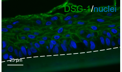
Application: Immunocytochemistry/ImmunofluorescenceSample Tested: Skin tissueSpecies: HumanVerified Customer | Posted 02/27/2020Reconstructed human epidermis tissues were embedded in paraffin. Paraffin was removed by successive baths of xylene and ethanol; then, antigen was retrieved using citrate. A 1:100 dilution of the antibody was used (overnight incubation at 4°C).

-
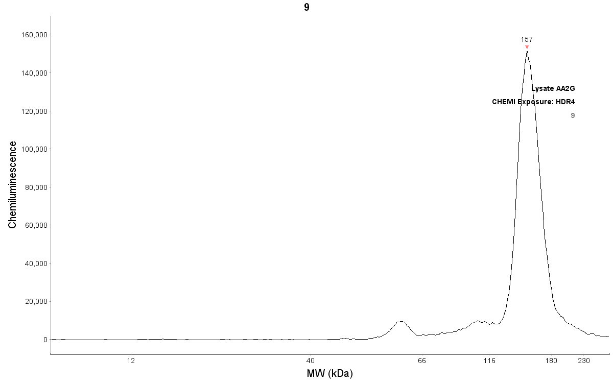
Application: Simple WesternSample Tested: Skin tissueSpecies: HumanVerified Customer | Posted 06/17/2019Demoglein expression in reconstructed human epidermis lysates (about 200 µg/mL protein) Antibody dilution 1:50, works also at 1:100 dilution
There are no reviews that match your criteria.
Protocols
Find general support by application which include: protocols, troubleshooting, illustrated assays, videos and webinars.
- Antigen Retrieval Protocol (PIER)
- Antigen Retrieval for Frozen Sections Protocol
- Appropriate Fixation of IHC/ICC Samples
- Cellular Response to Hypoxia Protocols
- Chromogenic IHC Staining of Formalin-Fixed Paraffin-Embedded (FFPE) Tissue Protocol
- Chromogenic Immunohistochemistry Staining of Frozen Tissue
- ClariTSA™ Fluorophore Kits
- Detection & Visualization of Antibody Binding
- Fluorescent IHC Staining of Frozen Tissue Protocol
- Graphic Protocol for Heat-induced Epitope Retrieval
- Graphic Protocol for the Preparation and Fluorescent IHC Staining of Frozen Tissue Sections
- Graphic Protocol for the Preparation and Fluorescent IHC Staining of Paraffin-embedded Tissue Sections
- Graphic Protocol for the Preparation of Gelatin-coated Slides for Histological Tissue Sections
- IHC Sample Preparation (Frozen sections vs Paraffin)
- Immunofluorescent IHC Staining of Formalin-Fixed Paraffin-Embedded (FFPE) Tissue Protocol
- Immunohistochemistry (IHC) and Immunocytochemistry (ICC) Protocols
- Immunohistochemistry Frozen Troubleshooting
- Immunohistochemistry Paraffin Troubleshooting
- Preparing Samples for IHC/ICC Experiments
- Preventing Non-Specific Staining (Non-Specific Binding)
- Primary Antibody Selection & Optimization
- Protocol for Heat-Induced Epitope Retrieval (HIER)
- Protocol for Making a 4% Formaldehyde Solution in PBS
- Protocol for VisUCyte™ HRP Polymer Detection Reagent
- Protocol for the Preparation & Fixation of Cells on Coverslips
- Protocol for the Preparation and Chromogenic IHC Staining of Frozen Tissue Sections
- Protocol for the Preparation and Chromogenic IHC Staining of Frozen Tissue Sections - Graphic
- Protocol for the Preparation and Chromogenic IHC Staining of Paraffin-embedded Tissue Sections
- Protocol for the Preparation and Chromogenic IHC Staining of Paraffin-embedded Tissue Sections - Graphic
- Protocol for the Preparation and Fluorescent IHC Staining of Frozen Tissue Sections
- Protocol for the Preparation and Fluorescent IHC Staining of Paraffin-embedded Tissue Sections
- Protocol for the Preparation of Gelatin-coated Slides for Histological Tissue Sections
- R&D Systems Quality Control Western Blot Protocol
- TUNEL and Active Caspase-3 Detection by IHC/ICC Protocol
- The Importance of IHC/ICC Controls
- Troubleshooting Guide: Immunohistochemistry
- Troubleshooting Guide: Western Blot Figures
- Western Blot Conditions
- Western Blot Protocol
- Western Blot Protocol for Cell Lysates
- Western Blot Troubleshooting
- Western Blot Troubleshooting Guide
- View all Protocols, Troubleshooting, Illustrated assays and Webinars
Loading...