
Key Product Details
Species Reactivity
Validated:
Human
Cited:
Human, Porcine
Applications
Validated:
Immunohistochemistry, Western Blot, Neutralization
Cited:
Immunohistochemistry, Immunohistochemistry-Paraffin, Western Blot, Neutralization, Control, Functional Assay
Label
Unconjugated
Antibody Source
Monoclonal Mouse IgG1 Clone # 10825
Loading...
Product Specifications
Immunogen
E. coli-derived recombinant human EGF
Specificity
Detects human EGF in direct ELISAs. Detects human EGF and rat EGF in Western blots. In direct ELISAs and Western blots, no cross‑reactivity with recombinant human (rh) HB‑EGF or rhTGF‑ alpha is observed. In Western blots, no cross-reactivity with recombinant mouse EGF is observed.
Clonality
Monoclonal
Host
Mouse
Isotype
IgG1
Endotoxin Level
<0.10 EU per 1 μg of the antibody by the LAL method.
Scientific Data Images for Human EGF Antibody (10825)
Detection of Recombinant Human and Rat EGF by Western Blot.
Western blot shows 100 ng of Recombinant Human EGF (Catalog # 236-EG), Recombinant Mouse EGF (Catalog # 2028-EG) and Recombinant Rat EGF (Catalog # 3214-EG). PVDF Membrane was probed with 1 µg/mL of Mouse Anti-Human EGF Monoclonal Antibody (Catalog # MAB236) followed by HRP-conjugated Anti-Mouse IgG Secondary Antibody (Catalog # HAF007). A specific band was detected for EGF at approximately 10 kDa (as indicated). This experiment was conducted under reducing conditions and using Immunoblot Buffer Group 3.
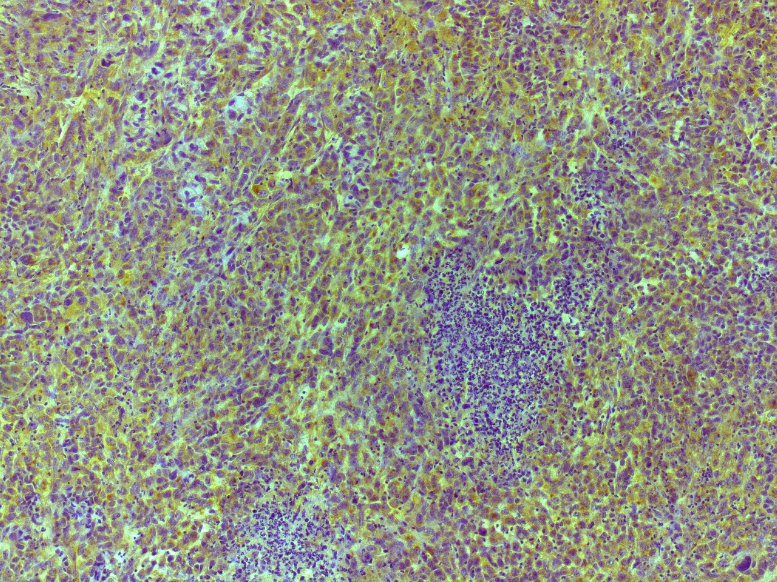
EGF in Human Skin.
EGF was detected in immersion fixed paraffin-embedded sections of human skin using 25 µg/mL Mouse Anti-Human EGF Monoclonal Antibody (Catalog # MAB236) overnight at 4 °C. Tissue was stained with the Anti-Mouse HRP-DAB Cell & Tissue Staining Kit (brown; Catalog # CTS002) and counterstained with hematoxylin (blue). Specific labeling was localized to the cytoplasm of keratinocytes in the stratum granulosum in epidermis. View our protocol for Chromogenic IHC Staining of Paraffin-embedded Tissue Sections.
Cell Proliferation Induced by EGF and Neutralization by Human EGF Antibody.
Recombinant Human EGF (Catalog # 236-EG) stimulates proliferation in the Balb/3T3 mouse embryonic fibroblast cell line in a dose-dependent manner (orange line). Proliferation elicited by Recombinant Human EGF (2 ng/mL) is neutralized (green line) by increasing concentrations of Mouse Anti-Human EGF Monoclonal Antibody (Catalog # MAB236). The ND50 is typically 0.05-0.1 µg/mL.Applications for Human EGF Antibody (10825)
Application
Recommended Usage
Immunohistochemistry
8-25 µg/mL
Sample: Immersion fixed paraffin-embedded sections of human skin
Sample: Immersion fixed paraffin-embedded sections of human skin
Western Blot
1 µg/mL
Sample: Recombinant Human EGF (Catalog # 236-EG)
Sample: Recombinant Human EGF (Catalog # 236-EG)
Neutralization
Measured by its ability to neutralize EGF-induced proliferation in the Balb/3T3 mouse embryonic fibroblast cell line. The Neutralization Dose (ND50) is typically 0.05-0.1 µg/mL in the presence of 2 ng/mL Recombinant Human EGF.
Reviewed Applications
Read 1 review rated 4 using MAB236 in the following applications:
Formulation, Preparation, and Storage
Purification
Protein A or G purified from hybridoma culture supernatant
Reconstitution
Reconstitute at 0.5 mg/mL in sterile PBS. For liquid material, refer to CoA for concentration.
Loading...
Formulation
Lyophilized from a 0.2 μm filtered solution in PBS with Trehalose. *Small pack size (SP) is supplied either lyophilized or as a 0.2 µm filtered solution in PBS.
Shipping
Lyophilized product is shipped at ambient temperature. Liquid small pack size (-SP) is shipped with polar packs. Upon receipt, store immediately at the temperature recommended below.
Stability & Storage
Use a manual defrost freezer and avoid repeated freeze-thaw cycles.
- 12 months from date of receipt, -20 to -70 °C as supplied.
- 1 month, 2 to 8 °C under sterile conditions after reconstitution.
- 6 months, -20 to -70 °C under sterile conditions after reconstitution.
Calculators
Background: EGF
Long Name
Epidermal Growth Factor
Alternate Names
HOMG4, URG, Urogastrone
Gene Symbol
EGF
Additional EGF Products
Product Documents for Human EGF Antibody (10825)
Certificate of Analysis
To download a Certificate of Analysis, please enter a lot or batch number in the search box below.
Note: Certificate of Analysis not available for kit components.
Product Specific Notices for Human EGF Antibody (10825)
For research use only
Related Research Areas
Citations for Human EGF Antibody (10825)
 Powered by Bioz
Customer Reviews for Human EGF Antibody (10825) (1)
4 out of 5
1 Customer Rating
Have you used Human EGF Antibody (10825)?
Submit a review and receive an Amazon gift card!
$25/€18/£15/$25CAN/¥2500 Yen for a review with an image
$10/€7/£6/$10CAN/¥1110 Yen for a review without an image
Submit a review
Customer Images
Showing
1
-
1 of
1 review
Showing All
Filter By:
-
Application: ImmunohistochemistrySample Tested: DU145 human prostate carcinoma cell lineSpecies: HumanVerified Customer | Posted 11/12/2025

There are no reviews that match your criteria.
Protocols
Find general support by application which include: protocols, troubleshooting, illustrated assays, videos and webinars.
- Antigen Retrieval Protocol (PIER)
- Antigen Retrieval for Frozen Sections Protocol
- Appropriate Fixation of IHC/ICC Samples
- Cellular Response to Hypoxia Protocols
- Chromogenic IHC Staining of Formalin-Fixed Paraffin-Embedded (FFPE) Tissue Protocol
- Chromogenic Immunohistochemistry Staining of Frozen Tissue
- ClariTSA™ Fluorophore Kits
- Detection & Visualization of Antibody Binding
- Fluorescent IHC Staining of Frozen Tissue Protocol
- Graphic Protocol for Heat-induced Epitope Retrieval
- Graphic Protocol for the Preparation and Fluorescent IHC Staining of Frozen Tissue Sections
- Graphic Protocol for the Preparation and Fluorescent IHC Staining of Paraffin-embedded Tissue Sections
- Graphic Protocol for the Preparation of Gelatin-coated Slides for Histological Tissue Sections
- IHC Sample Preparation (Frozen sections vs Paraffin)
- Immunofluorescent IHC Staining of Formalin-Fixed Paraffin-Embedded (FFPE) Tissue Protocol
- Immunohistochemistry (IHC) and Immunocytochemistry (ICC) Protocols
- Immunohistochemistry Frozen Troubleshooting
- Immunohistochemistry Paraffin Troubleshooting
- Preparing Samples for IHC/ICC Experiments
- Preventing Non-Specific Staining (Non-Specific Binding)
- Primary Antibody Selection & Optimization
- Protocol for Heat-Induced Epitope Retrieval (HIER)
- Protocol for Making a 4% Formaldehyde Solution in PBS
- Protocol for VisUCyte™ HRP Polymer Detection Reagent
- Protocol for the Preparation & Fixation of Cells on Coverslips
- Protocol for the Preparation and Chromogenic IHC Staining of Frozen Tissue Sections
- Protocol for the Preparation and Chromogenic IHC Staining of Frozen Tissue Sections - Graphic
- Protocol for the Preparation and Chromogenic IHC Staining of Paraffin-embedded Tissue Sections
- Protocol for the Preparation and Chromogenic IHC Staining of Paraffin-embedded Tissue Sections - Graphic
- Protocol for the Preparation and Fluorescent IHC Staining of Frozen Tissue Sections
- Protocol for the Preparation and Fluorescent IHC Staining of Paraffin-embedded Tissue Sections
- Protocol for the Preparation of Gelatin-coated Slides for Histological Tissue Sections
- R&D Systems Quality Control Western Blot Protocol
- TUNEL and Active Caspase-3 Detection by IHC/ICC Protocol
- The Importance of IHC/ICC Controls
- Troubleshooting Guide: Immunohistochemistry
- Troubleshooting Guide: Western Blot Figures
- Western Blot Conditions
- Western Blot Protocol
- Western Blot Protocol for Cell Lysates
- Western Blot Troubleshooting
- Western Blot Troubleshooting Guide
- View all Protocols, Troubleshooting, Illustrated assays and Webinars


