Epidermal growth factor (EGF) is a small, potent growth factor capable of inducing cell proliferation, differentiation, and survival. EGF is the founding member of the EGF family that also includes TGF-alpha, amphiregulin (AR), betacellulin (BTC), epiregulin (EPR), heparin‑binding EGF‑like growth factor (HB‑EGF), epigen, and the neuregulins (NRG)-1 through -6 (1). Members of The EGF family are characterized by a shared structural motif, the EGF‑like domain, which contains three intramolecular disulfide bonds that are formed by six similarly spaced, conserved cysteine residues (2). These disulfide bonds are essential for proper protein conformation and receptor binding. All EGF family members are synthesized as type I transmembrane precursor proteins that may contain several EGF domains in the extracellular region. The mature proteins are released from the cell surface by regulated proteolysis (1). The full length EGF protein is 1207 amino acids (aa) (EGF precursor) containing nine EGF domains and nine LDLR class B repeats. However, the mature protein is much smaller, only 53 aa, and is generated by proteolytic cleavage of the EGF domain proximal to the transmembrane region (3). EGF is well conserved across mammals with mature human EGF 70% identical to mature mouse and rat EGF. Physiologically, EGF is found in various body fluids, including blood, milk, urine, saliva, seminal fluid, pancreatic juice, cerebrospinal fluid, and amniotic fluid (4). EGF is a high affinity ligand of the EGF receptor (ErbB). Four ErbB (HER) family receptor tyrosine kinases including EGFR/ErbB1, ErbB2, ErbB3 and ErbB4, mediate responses to EGF family members (5). EGF binding induces dimerization of the EGF receptor resulting in activation of the protein tyrosine kinase signaling pathway. These receptors undergo a complex pattern of ligand-induced homo- or hetero-dimerization to transduce EGF family signals (6, 7). EGF binds ErbB1 and depending on the context, induces the formation of homodimers or heterodimers containing ErbB2. Dimerization results in autophosphorylation of the receptor at specific tyrosine residues to create docking sites for a variety of signaling molecules (5, 8). Biological activities ascribed to EGF include epithelial development, angiogenesis, inhibition of gastric acid secretion, fibroblast proliferation, and colony formation of epidermal cells in culture.
Recombinant Human EGF Protein, CF
R&D Systems | Catalog # 236-EG

Key Product Details
- R&D Systems E. coli-derived Recombinant Human EGF Protein (236-EG)
- Quality control testing to verify active proteins with lot specific assays by in-house scientists
- All R&D Systems proteins are covered with a 100% guarantee
Source
Accession Number
Applications
Product Specifications
Source
Asn971-Arg1023, with an N-terminal Met
Purity
Endotoxin Level
N-terminal Sequence Analysis
Predicted Molecular Mass
SDS-PAGE
Activity
The ED50 for this effect is 20-100 pg/mL.
Reviewed Applications
Read 32 reviews rated 4.8 using 236-EG in the following applications:
- Apoptosis assay (1 Review)
- Binding assay/Protein-protein interaction (1 Review)
- Cell Culture (1 Review)
- Cell migration/motility (1 Review)
- Cell Proliferation (8 Reviews)
- Immunoassay Standard (1 Review)
- In vitro bioactivity in cell culture (10 Reviews)
- In vivo study (1 Review)
- Induction of fibronectin production (1 Review)
- Manufacturing In vitro Products (1 Review)
- Media additive for protein or antibody production (1 Review)
- Stem/Immune cell maintenance or differentiation (4 Reviews)
- Supplement in growth media (1 Review)
Scientific Data Images for Recombinant Human EGF Protein, CF
Recombinant Human EGF Protein SEC-MALS.
Recombinant Human EGF Protein (Catalog # 236-EG) has a molecular weight (MW) of 6.7 kDa as analyzed by SEC-MALS, suggesting that this protein is a monomer.
Recombinant Human EGF Protein Bioactivity
Recombinant Human EGF (Catalog # 236‑EG) stimulates cell proliferation of the Balb/3T3 mouse embryonic fibroblast cell line. The ED50 for this effect is 20‑100 pg/mL.
Recombinant Human EGF Protein SDS-PAGE
1 µg/lane of Recombinant Human EGF was resolved with SDS-PAGE and visualized by silver staining under reducing (R) conditions, showing a single band at 6 kDa.
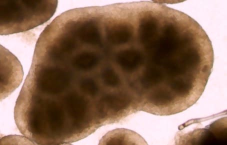
iPSC-derived Human Intestinal Organoids Cultured using Recombinant Human EGF Protein.
iPSC-derived human intestinal organoids were cultured using Cultrex™ UltiMatrix RGF Basement Membrane Extract (BME001-05) and intestinal organoid culture medium, which includes Recombinant Human EGF (Catalog # 236-EG), Recombinant Human Noggin (6057-NG), Recombinant Human R-Spondin 1 (4645-RS), and Recombinant Human Wnt-3a (5036-WN), along with the other reagents listed in the intestinal organoid culture medium recipe in the human intestinal organoid culture protocol. (A) Human intestinal organoids were stained using a Rat Anti-Human/Mouse/Rat Vimentin Monoclonal Antibody (MAB2105; green) and a Goat Anti-Human/Mouse Desmin Antigen Affinity-purified Polyclonal Antibody (AF3844; red) to visualize myofibroblast cells and counterstained with DAPI (5748; blue). (B) Human intestinal organoids were stained using a Goat Anti-Human/Mouse E-Cadherin Antigen Affinity-purified Polyclonal Antibody (AF748; green) and a Mouse Anti-Human MUC2 Monoclonal Antibody (Novus Biologicals, Catalog # NBP2-44431; red) and counterstained with DAPI (5748; blue).
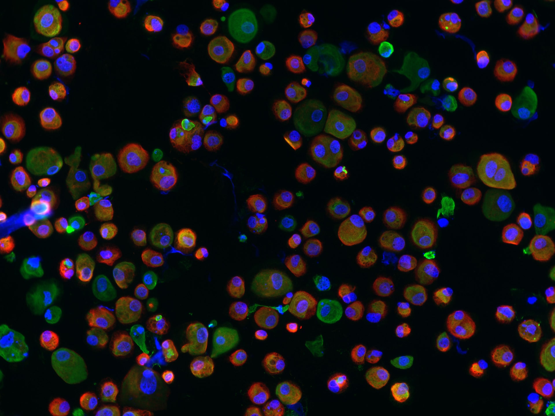
Adult Stem Cell-derived Human Descending Colon Organoids Cultured using Recombinant Human EGF Protein.
Adult stem cells isolated from human descending colon were embedded in Cultrex UltiMatrix RGF Basement Membrane Extract (BME001-05) and cultured for 30 days in intestinal organoid culture medium, which includes Recombinant Human EGF (Catalog # 236-EG), Recombinant Human Noggin (6057-NG), Recombinant Human R-Spondin 1 (4645-RS), and Recombinant Human Wnt-3a (5036-WN), along with the other reagents listed in the intestinal organoid culture medium recipe in the human intestinal organoid culture protocol. (A) Organoids were fixed and stained with a Mouse Anti-Human MUC2 Monoclonal Antibody (Novus Biologicals; Catalog # NBP2-44431; green) to visualize intestinal goblet cells and counterstained with a Goat Anti-Human/Mouse E-Cadherin Antigen Affinity-purified Polyclonal Antibody (AF748; red) and DAPI (5748; blue). The image shown was taken at 10x magnification. (B) Organoids were fixed and stained with a Mouse Anti-Human Chromogranin A Monoclonal Antibody (MAB90981; green) to visualize enteroendocrine cells and counterstained with a Goat Anti-Human/Mouse E-Cadherin Antigen Affinity-purified Polyclonal Antibody (AF748; red) and DAPI (5748; blue). The image shown was taken at 20x magnification.
Culture and Characterization of Mouse Oligodendrocytes.
D3 mouse embryonic stem cells were expanded in KO-ES Media supplemented with Bovine Fibronectin Protein (1030-FN) to support cell attachment and spreading, the ITS and N-2 Plus Media Supplements (AR013 and AR003), and a panel of growth factors for effective oligodendrocyte differentiation, including Recombinant Human FGF-basic, Recombinant Human EGF (Catalog # 236-EG), and Recombinant Human PDGF-AA (221-AA). Oligodendrocytes were detected using a Mouse Anti-Human/Mouse/Rat/Chicken Oligodendrocyte Marker O4 Monoclonal Antibody (MAB1326). The cells were stained with the NorthernLights™-557 Affinity-purified Goat Anti-Mouse IgM Secondary Antibody (NL019; red). The nuclei were counterstained with DAPI (5748;blue).Formulation, Preparation, and Storage
236-EG
| Formulation | Lyophilized from a 0.2 μm filtered solution in PBS. |
| Reconstitution | Reconstitute at 500 μg/mL in sterile PBS. |
| Shipping | The product is shipped at ambient temperature. Upon receipt, store it immediately at the temperature recommended below. |
| Stability & Storage | Use a manual defrost freezer and avoid repeated freeze-thaw cycles.
|
Calculators
Background: EGF
References
- Harris, R.C. et al. (2003) Exp. Cell Res. 284:2.
- Carpenter, G. and Cohen, S. (1990) J. Biol. Chem. 265:7709.
- Bell, G.I. et al. (1986) Nucl. Acids Res. 14:8427.
- Carpenter, G. and Zendegui, J.G. (1986) Exp. Cell Res. 164:1.
- Jorissen, R.N. et al. (2003) Exp. Cell Res. 284:31.
- Gamett, D.C. et al. (1997) J. Biol. Chem. 272:12052.
- Qian, X. et al. (1994) Proc. Natl. Acad. Sci. 91:1500.
- Qian, X. et al. (1999) J. Biol. Chem. 274:574.
Long Name
Alternate Names
Gene Symbol
UniProt
Additional EGF Products
Product Documents for Recombinant Human EGF Protein, CF
Certificate of Analysis
To download a Certificate of Analysis, please enter a lot or batch number in the search box below.
Note: Certificate of Analysis not available for kit components.
Product Specific Notices for Recombinant Human EGF Protein, CF
For research use only
Related Research Areas
Citations for Recombinant Human EGF Protein, CF
 Powered by Bioz
Customer Reviews for Recombinant Human EGF Protein, CF (32)
Have you used Recombinant Human EGF Protein, CF?
Submit a review and receive an Amazon gift card!
$25/€18/£15/$25CAN/¥2500 Yen for a review with an image
$10/€7/£6/$10CAN/¥1110 Yen for a review without an image
Submit a review
Customer Images
-
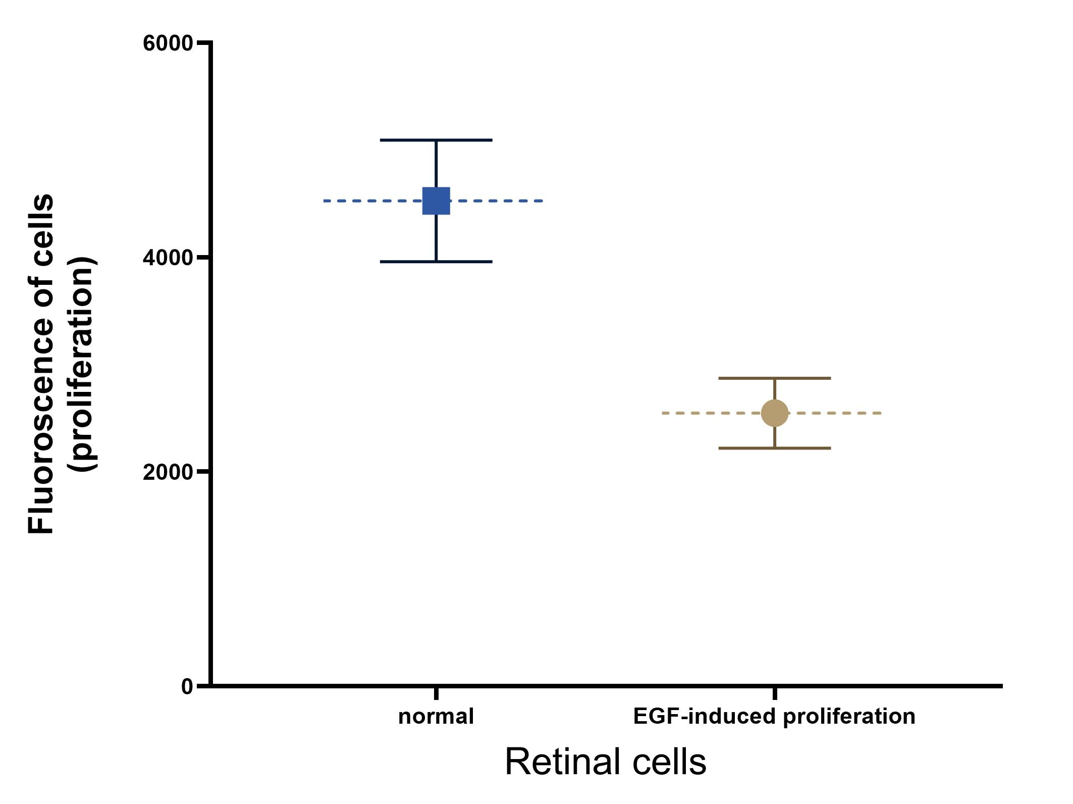
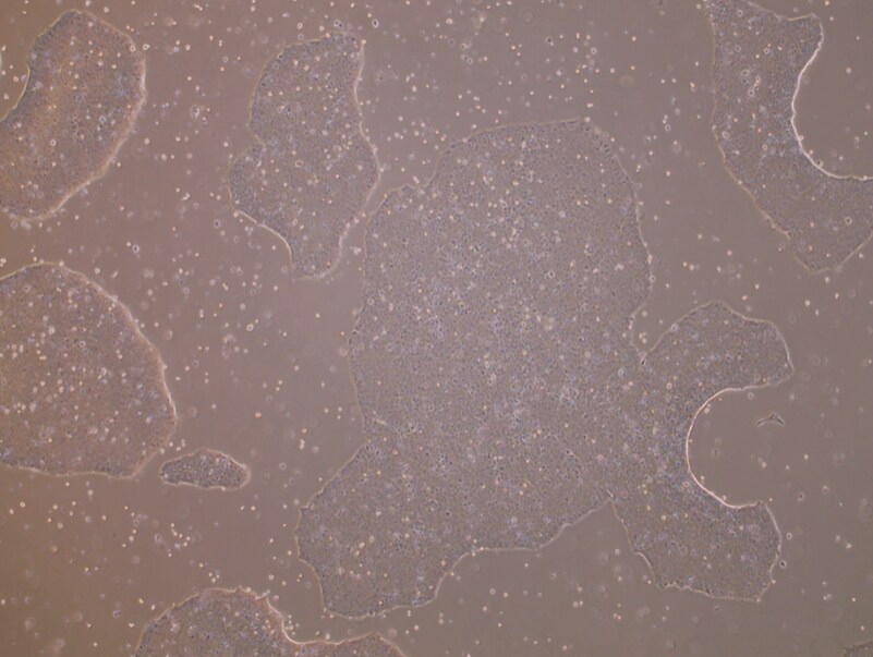
Application: Cell ProliferationVerified Customer | Posted 03/03/2026ARPE-19 proliferation

-
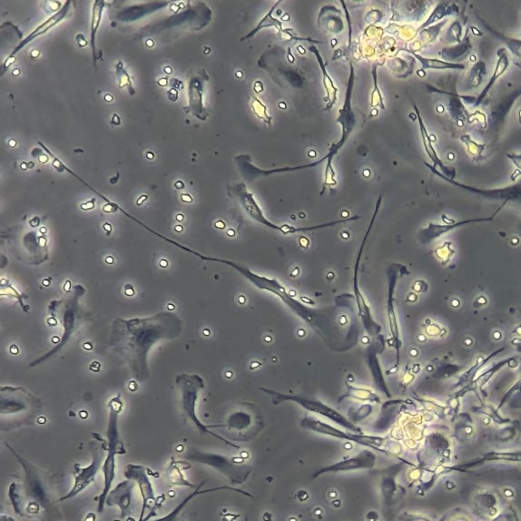
Application: In vitro bioactivity in cell cultureVerified Customer | Posted 07/15/2025
-
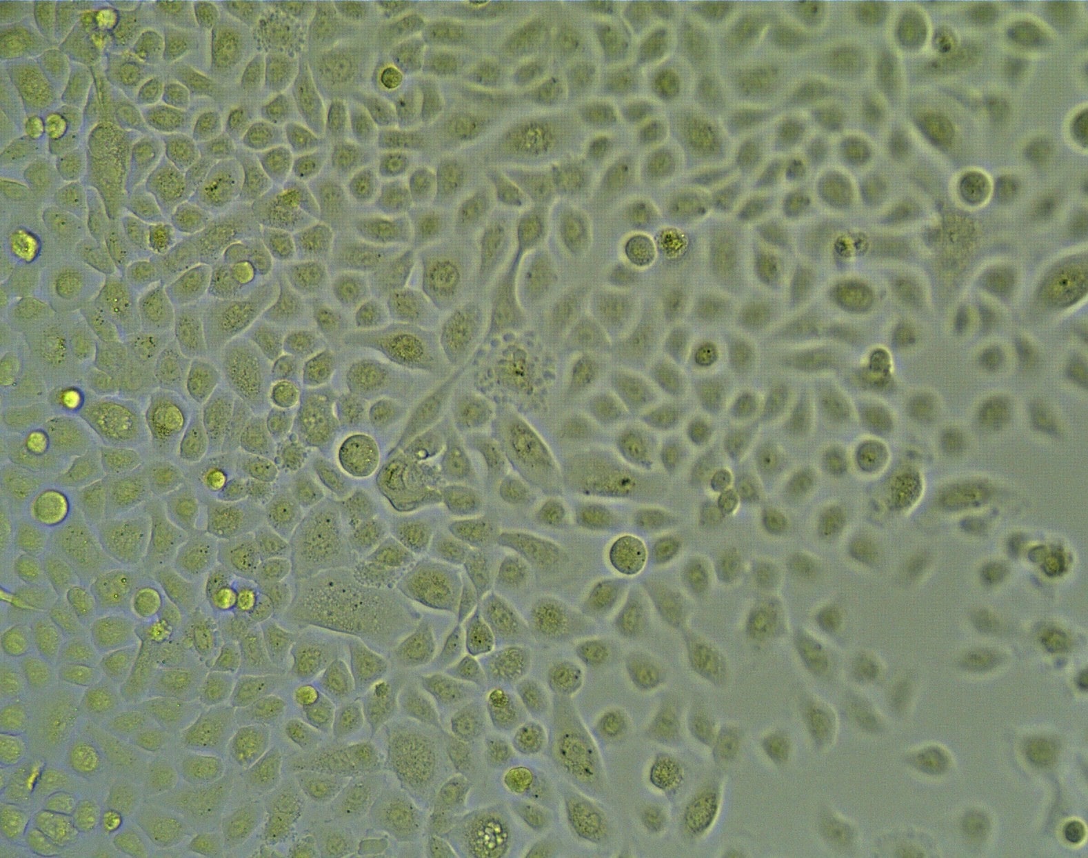
Application: In vitro bioactivity in cell cultureVerified Customer | Posted 07/06/2025
-
Application: Supplement in growth mediaVerified Customer | Posted 10/11/2023Used as supplement in growth media for primary human lung epithelial cells.
-
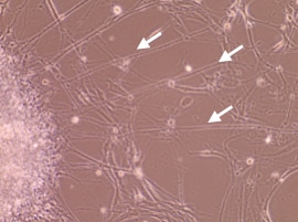
Application: In vivo studyVerified Customer | Posted 05/20/2023EGF was used to promote the growth of axons.
-
Application: Cell ProliferationVerified Customer | Posted 06/18/2022
-
Application: In vitro bioactivity in cell cultureVerified Customer | Posted 05/04/2022
-
Application: Stem/Immune cell maintenance or differentiationVerified Customer | Posted 01/01/2022EGF was used to test if it can promote the growth of ipsc
-
Application: Cell ProliferationVerified Customer | Posted 09/23/2021It's better to store it in -20C freezer.
-
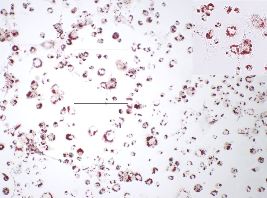
Application: Cell ProliferationVerified Customer | Posted 08/17/2021EGF promote adipogenesis
-
Application: Cell ProliferationVerified Customer | Posted 07/21/20210.2ng/ml EGF can promote the proliferation of limbal stem cells.
-
Application: Cell CultureVerified Customer | Posted 01/02/2021
-
Application: Cell ProliferationVerified Customer | Posted 12/28/2020
-
Application: In vitro bioactivity in cell cultureVerified Customer | Posted 07/02/2020
-
Application: In vitro bioactivity in cell cultureVerified Customer | Posted 06/26/2020
-
Application: In vitro bioactivity in cell cultureVerified Customer | Posted 06/06/2020
-
Application: Apoptosis assayVerified Customer | Posted 04/17/2020
-
Application: Stem/Immune cell maintenance or differentiationVerified Customer | Posted 01/09/2020
-
Application: In vitro bioactivity in cell cultureVerified Customer | Posted 08/02/2019
-
Application: Cell ProliferationVerified Customer | Posted 05/07/2019
-
Application: Binding assay/Protein-protein interactionVerified Customer | Posted 04/05/2019
-
Application: In vitro bioactivity in cell cultureVerified Customer | Posted 09/10/2018
-
Application: Immunoassay StandardVerified Customer | Posted 02/26/2018
-
Application: In vitro bioactivity in cell cultureVerified Customer | Posted 02/22/2018Human lung cancer cells activated by 10ng /ml of EGF
-
Application: Media additive for protein or antibody productionVerified Customer | Posted 12/06/2017
-
Application: Cell ProliferationVerified Customer | Posted 10/21/2017
-
Application: In vitro bioactivity in cell cultureVerified Customer | Posted 07/20/2017
-
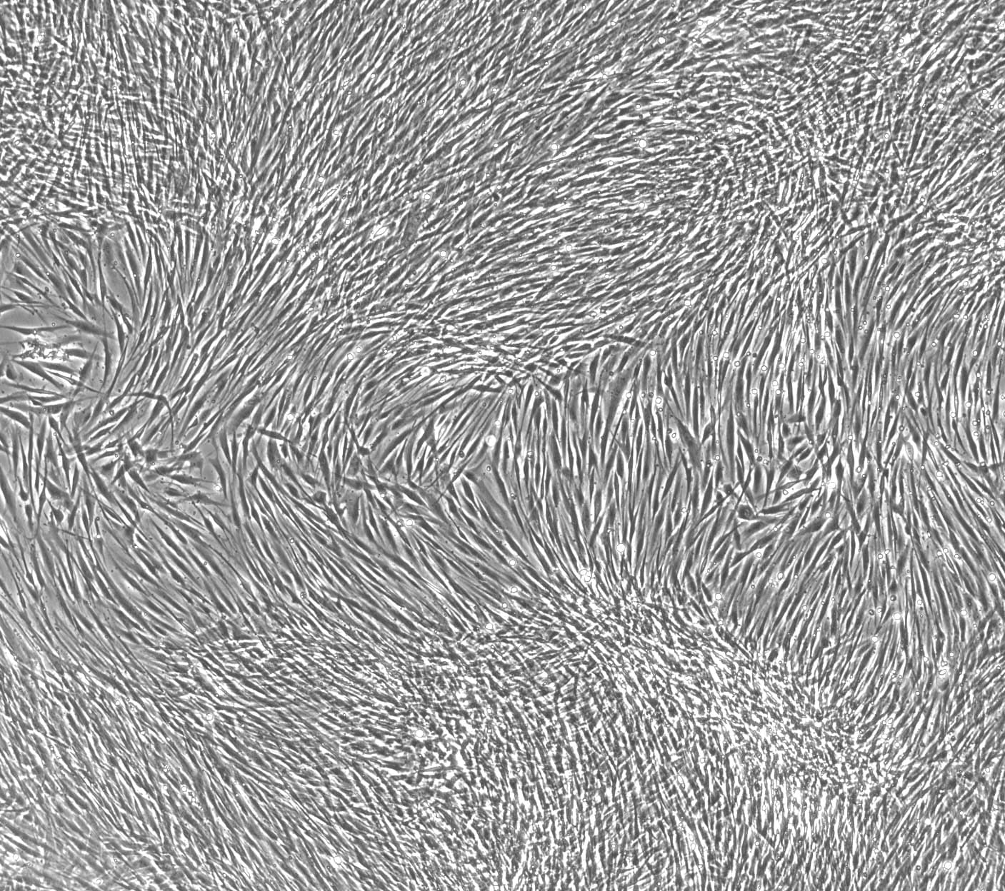
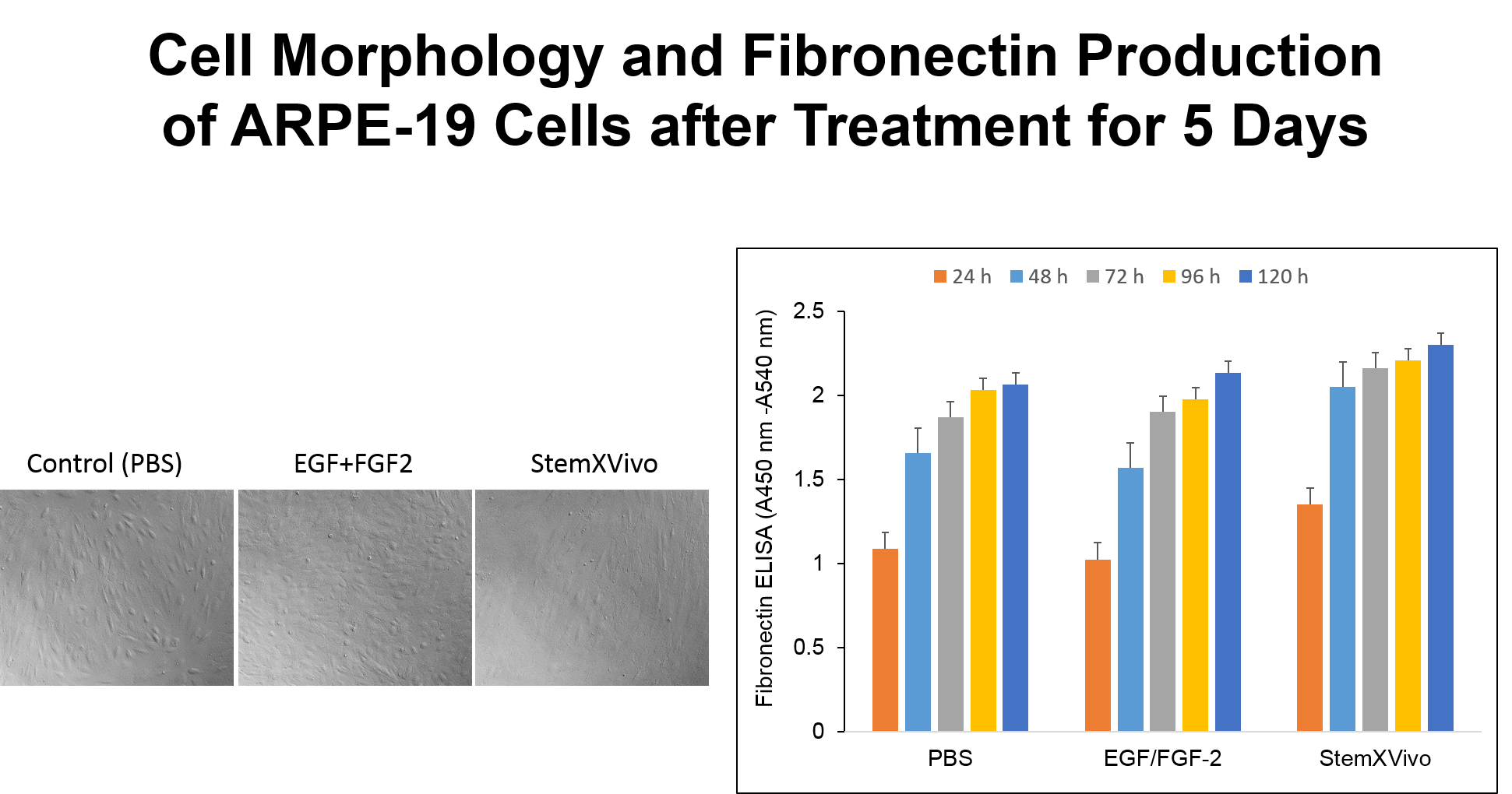
Application: Induction of fibronectin productionVerified Customer | Posted 04/12/2017To induce proliferation and EMT in ARPE-19 cells
-
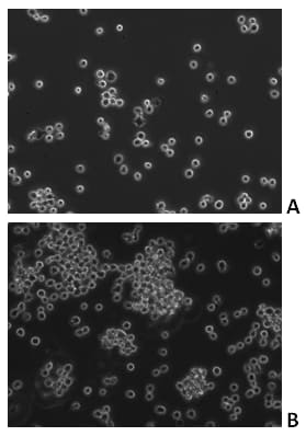
Application: Stem/Immune cell maintenance or differentiationVerified Customer | Posted 03/31/2017Growth of MDA-MB-231 spheroids in serum-free condition in the absence (A) or presence (B) of Recombinant Human EGF (#236-EG) after 72 hours. Medium: RPMI + 2% B-27 supplement + 1% L-glutamine + 1% Pen/Strep.
-
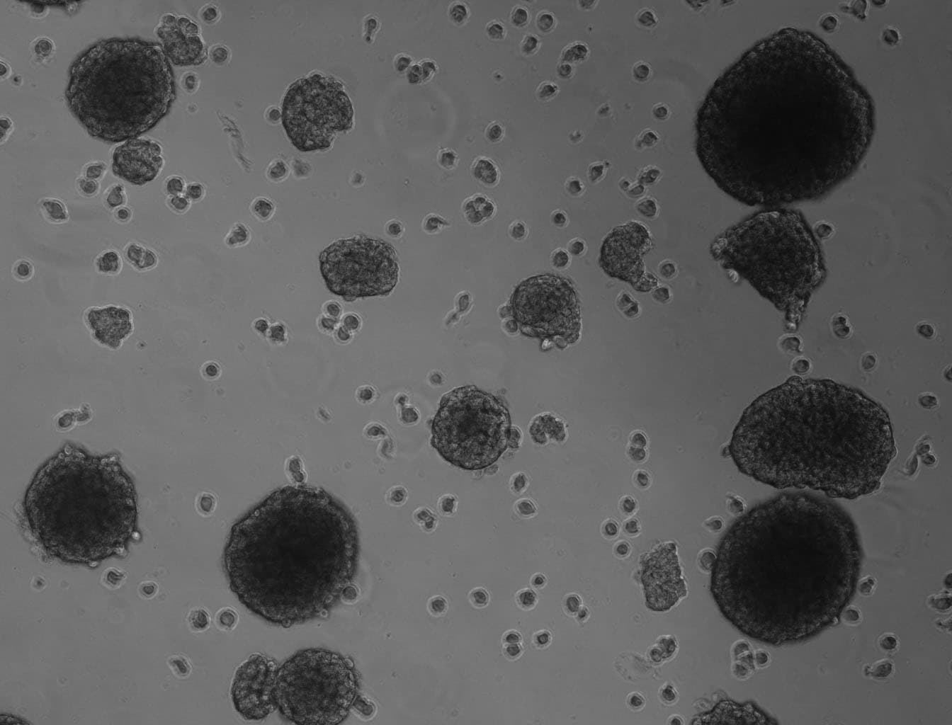
Application: Stem/Immune cell maintenance or differentiationVerified Customer | Posted 08/17/2016Recombinant protein to induced sphere formation on olfactory stem cells
-
Application: Manufacturing In vitro ProductsVerified Customer | Posted 07/21/2016
-
Application: Cell migration/motilityVerified Customer | Posted 05/18/2016
There are no reviews that match your criteria.
FAQs for Recombinant Human EGF Protein, CF
-
Q: The product literature for recombinant human EGF indicates to reconstitute at 500 μg/mL. Can I reconstitute to a lower concentration?
A: For recombinant human EGF, stability and activity testing has been performed after reconstitution to 500 μg/mL. We cannot guarantee performance of the protein after reconstitution to a lower concentration. We would recommend reconstituting and storing at 500 μg/mL in single-use aliquots, diluting to the working concentration just prior to use.


