Human Fibronectin Antibody (960642)
R&D Systems | Catalog # MAB19182

Key Product Details
Validated by
Biological Validation
Species Reactivity
Validated:
Human
Cited:
Human
Applications
Validated:
Multiplex Immunofluorescence, Immunohistochemistry, Western Blot, Immunocytochemistry, COMET
Cited:
Western Blot, Immunocytochemistry/ Immunofluorescence
Label
Unconjugated
Antibody Source
Monoclonal Mouse IgG1 Clone # 960642
Loading...
Product Specifications
Immunogen
E.coli-derived recombinant human Fibronectin
Asn631-Pro705
Accession # P02751
Asn631-Pro705
Accession # P02751
Specificity
Detects human Fibronectin in direct ELISAs and Western blots.
Clonality
Monoclonal
Host
Mouse
Isotype
IgG1
Scientific Data Images for Human Fibronectin Antibody (960642)
Detection of Fibronectin in Human Breast Tumor via seqIF™ staining on COMET™
Fibronectin was detected in immersion fixed paraffin-embedded sections of human Breast Tumor using Mouse Anti-Human Fibronectin Monoclonal Antibody (Catalog # MAB19182) at 15ug/mL at 37 ° Celsius for 4 minutes. Before incubation with the primary antibody, tissue underwent an all-in-one dewaxing and antigen retrieval preprocessing using PreTreatment Module (PT Module) and Dewax and HIER Buffer H (pH 9; Epredia Catalog # TA-999-DHBH). Tissue was stained using the Alexa Fluor™ 555 Goat anti-Mouse IgG Secondary Antibody at 1:100 at 37 ° Celsius for 2 minutes. (Yellow; Lunaphore Catalog # DR555MS) and counterstained with DAPI (blue; Lunaphore Catalog # DR100). Specific staining was localized to the cytoplasm. Protocol available in COMET™ Panel Builder.
Detection of Human Fibronectin by Western Blot.
Western blot shows lysates of HepG2 human hepatocellular carcinoma cell line. PVDF membrane was probed with 2 µg/mL of Mouse Anti-Human Fibronectin Monoclonal Antibody (Catalog # MAB19182) followed by HRP-conjugated Anti-Mouse IgG Secondary Antibody (HAF018). A specific band was detected for Fibronectin at approximately 300 kDa (as indicated). This experiment was conducted under reducing conditions and using Western Blot Buffer Group 1.
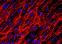
Fibronectin in HepG2 Human Cell Line.
Fibronectin was detected in immersion fixed HepG2 human hepatocellular carcinoma cell line treated with monensin using Mouse Anti-Human Fibronectin Monoclonal Antibody (Catalog # MAB19182) at 3 µg/mL for 3 hours at room temperature. Cells were stained using the NorthernLights™ 557-conjugated Anti-Mouse IgG Secondary Antibody (red; Catalog # NL007) and counterstained with DAPI (blue). Specific staining was localized to cytoplasm (punctate). View our protocol for Fluorescent ICC Staining of Cells on Coverslips.
Fibronectin in Human Liver Cancer Tissue.
Fibronectin was detected in immersion fixed paraffin-embedded sections of human liver cancer tissue using Mouse Anti-Human Fibronectin Monoclonal Antibody (Catalog # MAB19182) at 5 µg/mL for 1 hour at room temperature followed by incubation with the Anti-Mouse IgG VisUCyte™ HRP Polymer Antibody (Catalog # VC001). Tissue was stained using DAB (brown) and counterstained with hematoxylin (blue). Specific staining was localized to cytoplasm and plasma membrane in tumor cells. View our protocol for IHC Staining with VisUCyte HRP Polymer Detection Reagents.
Detection of Fibronectin by Western Blot
KPA treatment decreases activation of human hepatic stellate LX-2 cells. Representative Western blots showing expression of endogenous (A) KISS1 protein (n = 5 biological replicates) and (B) KISS1R protein expression (n = 4 biological replicates). MDA-MB-231, SKBR3, and KISS1R-SKBR3 were used as reference for expression (n = 4). (C) Secreted kisspeptin protein in culture media measured by ELISA (N-4 biological replicates). (D–G) Changes in fibrogenic gene expression in response to KPA (3 nM, 48 h) +/− TGFb (5 ng/mL, 48 h). (n = 4–6 biological replicates) (H) Western blot analysis and (I,J) densitometric analyses of blots, showing changes in fibrogenic protein in response to KPA (3 nM, 72 h) +/− TGFb (5 ng/mL, 72 h). (n = 4 biological replicates) Results are expressed as mean +/− S.E.M. * p < 0.05 vs. control. ** p < 0.01 vs. control. *** p < 0.001 vs. control. **** p < 0.0001 vs. control. One-way ANOVA was conducted, followed by multiple comparison test. Image collected and cropped by CiteAb from the following open publication (https://pubmed.ncbi.nlm.nih.gov/39404414), licensed under a CC-BY license. Not internally tested by R&D Systems.Applications for Human Fibronectin Antibody (960642)
Application
Recommended Usage
COMET
Optimal dilutions of this antibody should be experimentally determined.
Immunocytochemistry
3-25 µg/mL
Sample: Immersion fixed HepG2 human hepatocellular carcinoma cell line treated with monensin
Sample: Immersion fixed HepG2 human hepatocellular carcinoma cell line treated with monensin
Immunohistochemistry
5-25 µg/mL
Sample: Immersion fixed paraffin-embedded sections of human liver cancer tissue
Sample: Immersion fixed paraffin-embedded sections of human liver cancer tissue
Multiplex Immunofluorescence
15 µg/mL
Sample: Immersion fixed paraffin-embedded sections of human Breast Tumor
Sample: Immersion fixed paraffin-embedded sections of human Breast Tumor
Western Blot
2 µg/mL
Sample: HepG2 human hepatocellular carcinoma cell line
Sample: HepG2 human hepatocellular carcinoma cell line
Reviewed Applications
Read 1 review rated 5 using MAB19182 in the following applications:
Formulation, Preparation, and Storage
Purification
Protein A or G purified from hybridoma culture supernatant
Reconstitution
Reconstitute at 0.5 mg/mL in sterile PBS. For liquid material, refer to CoA for concentration.
Loading...
Formulation
Lyophilized from a 0.2 μm filtered solution in PBS with Trehalose. See Certificate of Analysis for details.
*Small pack size (-SP) is supplied either lyophilized or as a 0.2 µm filtered solution in PBS.
*Small pack size (-SP) is supplied either lyophilized or as a 0.2 µm filtered solution in PBS.
Shipping
Lyophilized product is shipped at ambient temperature. Liquid small pack size (-SP) is shipped with polar packs. Upon receipt, store immediately at the temperature recommended below.
Stability & Storage
Use a manual defrost freezer and avoid repeated freeze-thaw cycles.
- 12 months from date of receipt, -20 to -70 °C as supplied.
- 1 month, 2 to 8 °C under sterile conditions after reconstitution.
- 6 months, -20 to -70 °C under sterile conditions after reconstitution.
Calculators
Background: Fibronectin
Alternate Names
CIG, ED-B, FINC, FN1, FNZ, GFND2, LETS, SMDCF
Gene Symbol
FN1
UniProt
Additional Fibronectin Products
Product Documents for Human Fibronectin Antibody (960642)
Certificate of Analysis
To download a Certificate of Analysis, please enter a lot or batch number in the search box below.
Note: Certificate of Analysis not available for kit components.
Product Specific Notices for Human Fibronectin Antibody (960642)
For research use only
Citations for Human Fibronectin Antibody (960642)
 Powered by Bioz
Customer Reviews for Human Fibronectin Antibody (960642) (1)
5 out of 5
1 Customer Rating
Have you used Human Fibronectin Antibody (960642)?
Submit a review and receive an Amazon gift card!
$25/€18/£15/$25CAN/¥2500 Yen for a review with an image
$10/€7/£6/$10CAN/¥1110 Yen for a review without an image
Submit a review
Customer Images
Showing
1
-
1 of
1 review
Showing All
Filter By:
-
Application: Immunocytochemistry/ImmunofluorescenceSample Tested: fibroblastsSpecies: HumanVerified Customer | Posted 10/24/2021

There are no reviews that match your criteria.
Protocols
Find general support by application which include: protocols, troubleshooting, illustrated assays, videos and webinars.
- Antigen Retrieval Protocol (PIER)
- Antigen Retrieval for Frozen Sections Protocol
- Appropriate Fixation of IHC/ICC Samples
- Cellular Response to Hypoxia Protocols
- Chromogenic IHC Staining of Formalin-Fixed Paraffin-Embedded (FFPE) Tissue Protocol
- Chromogenic Immunohistochemistry Staining of Frozen Tissue
- ClariTSA™ Fluorophore Kits
- Detection & Visualization of Antibody Binding
- Fluorescent IHC Staining of Frozen Tissue Protocol
- Graphic Protocol for Heat-induced Epitope Retrieval
- Graphic Protocol for the Preparation and Fluorescent IHC Staining of Frozen Tissue Sections
- Graphic Protocol for the Preparation and Fluorescent IHC Staining of Paraffin-embedded Tissue Sections
- Graphic Protocol for the Preparation of Gelatin-coated Slides for Histological Tissue Sections
- ICC Cell Smear Protocol for Suspension Cells
- ICC Immunocytochemistry Protocol Videos
- ICC for Adherent Cells
- IHC Sample Preparation (Frozen sections vs Paraffin)
- Immunocytochemistry (ICC) Protocol
- Immunocytochemistry Troubleshooting
- Immunofluorescence of Organoids Embedded in Cultrex Basement Membrane Extract
- Immunofluorescent IHC Staining of Formalin-Fixed Paraffin-Embedded (FFPE) Tissue Protocol
- Immunohistochemistry (IHC) and Immunocytochemistry (ICC) Protocols
- Immunohistochemistry Frozen Troubleshooting
- Immunohistochemistry Paraffin Troubleshooting
- Preparing Samples for IHC/ICC Experiments
- Preventing Non-Specific Staining (Non-Specific Binding)
- Primary Antibody Selection & Optimization
- Protocol for Heat-Induced Epitope Retrieval (HIER)
- Protocol for Making a 4% Formaldehyde Solution in PBS
- Protocol for VisUCyte™ HRP Polymer Detection Reagent
- Protocol for the Fluorescent ICC Staining of Cell Smears - Graphic
- Protocol for the Fluorescent ICC Staining of Cultured Cells on Coverslips - Graphic
- Protocol for the Preparation & Fixation of Cells on Coverslips
- Protocol for the Preparation and Chromogenic IHC Staining of Frozen Tissue Sections
- Protocol for the Preparation and Chromogenic IHC Staining of Frozen Tissue Sections - Graphic
- Protocol for the Preparation and Chromogenic IHC Staining of Paraffin-embedded Tissue Sections
- Protocol for the Preparation and Chromogenic IHC Staining of Paraffin-embedded Tissue Sections - Graphic
- Protocol for the Preparation and Fluorescent ICC Staining of Cells on Coverslips
- Protocol for the Preparation and Fluorescent ICC Staining of Non-adherent Cells
- Protocol for the Preparation and Fluorescent ICC Staining of Stem Cells on Coverslips
- Protocol for the Preparation and Fluorescent IHC Staining of Frozen Tissue Sections
- Protocol for the Preparation and Fluorescent IHC Staining of Paraffin-embedded Tissue Sections
- Protocol for the Preparation of Gelatin-coated Slides for Histological Tissue Sections
- Protocol for the Preparation of a Cell Smear for Non-adherent Cell ICC - Graphic
- R&D Systems Quality Control Western Blot Protocol
- TUNEL and Active Caspase-3 Detection by IHC/ICC Protocol
- The Importance of IHC/ICC Controls
- Troubleshooting Guide: Immunohistochemistry
- Troubleshooting Guide: Western Blot Figures
- Western Blot Conditions
- Western Blot Protocol
- Western Blot Protocol for Cell Lysates
- Western Blot Troubleshooting
- Western Blot Troubleshooting Guide
- View all Protocols, Troubleshooting, Illustrated assays and Webinars


