
Key Product Details
Species Reactivity
Validated:
Human
Cited:
Human
Applications
Validated:
Immunohistochemistry, Western Blot, Flow Cytometry, CyTOF-ready
Cited:
Flow Cytometry
Label
Unconjugated
Antibody Source
Polyclonal Goat IgG
Loading...
Product Specifications
Immunogen
Mouse myeloma cell line NS0-derived recombinant human Frizzled-6
His19-Val153
Accession # O60353
His19-Val153
Accession # O60353
Specificity
Detects human Frizzled-6 in direct ELISAs and Western blots.
Clonality
Polyclonal
Host
Goat
Isotype
IgG
Scientific Data Images for Human Frizzled-6 Antibody
Frizzled‑6 in Human Kidney Cancer Tissue.
Frizzled-6 was detected in immersion fixed paraffin-embedded sections of human kidney cancer tissue using Goat Anti-Human Frizzled-6 Antigen Affinity-purified Polyclonal Antibody (Catalog # AF3149) at 10 µg/mL overnight at 4 °C. Before incubation with the primary antibody, tissue was subjected to heat-induced epitope retrieval using Antigen Retrieval Reagent-Basic (Catalog # CTS013). Tissue was stained using the Anti-Goat HRP-DAB Cell & Tissue Staining Kit (brown; (CTS008) and counterstained with hematoxylin (blue). Specific staining was localized to cell membranes. View our protocol for Chromogenic IHC Staining of Paraffin-embedded Tissue Sections.
Detection of Frizzled-6 by Flow Cytometry
miR-20b and miR-125b augment Wnt signalling through targeting FZD6. (h) sphere formation of MES 83 expressing control shRNA or shRNAs of FZD6, with or without re-expression of FZD6, or a control (right). Image collected and cropped by CiteAb from the following open publication (https://pubmed.ncbi.nlm.nih.gov/27698350), licensed under a CC-BY license. Not internally tested by R&D Systems.
Detection of Frizzled-6 by Western Blot
miR-20b and miR-125b augment Wnt signalling through targeting FZD6. (d) IB for FZD6 and APC in miR-transduced MES 83 and 1123, or miRZIP-transduced PN 157 and 84. Image collected and cropped by CiteAb from the following open publication (https://pubmed.ncbi.nlm.nih.gov/27698350), licensed under a CC-BY license. Not internally tested by R&D Systems.
Detection of Frizzled-6 by Western Blot
FZD6 mediates induction of the MES-associated phenotype and suppression of the Wnt signalling through TAK1.(a) IB analysis on PN 23 and 17 spheres with or without FZD6 overexpression using indicated antibodies. (b) qRT–PCR analysis of PN-associated genes OLIG2 and SOX2, and MES-associated genes ALDH1A3 and CD44 in PN 23 spheres with or without FZD6 overexpression. (c) FZD6 expression promoted PN 23 spheres proliferation and self-renewal. (d) Ectopic expression of FZD6 promoted tumour growth of PN 23 spheres in GBM xenografts in the brain (n=5). Scale bar, 1.0 mm. (e) TOPFlash/FOPFlash assays on PN 23 with overexpressing FZD6 or control, with or without CaMKII inhibitor KN93 (5 μM), TAK1 inhibitor 5Z-7-oxo (5 μM), or a shRNA-NLK. (f,g) qRT–PCR analyses of the relative expression of miR-20b and miR-125b (f), and Wnt signalling target gene LEF1 (g) in PN 23 overexpressing FZD6, or a vector control, with or without CaMKII inhibitor KN93 (5 μM), TAK1 inhibitor 5Z-7-oxo (5 μM), or a shRNA-NLK. (h) sphere formation assays for PN 23 and 17 spheres with or without overexpression of FZD1, FZD3, and FZD5, respectively. (i) Summary of data shown in Supplementary Fig. 10. An overview of transmembrane receptors and Wnt ligands that affect FZD6 overexpression-induced phenotypes. Data were normalized to the control. *Relative inhibitory effects on indicated protein expression or cellular properties. Error bars (s.d.) represent the data of triplicate samples for each cell line or tumours from five individual mice in each group. *P<0.05, **P<0.01, paired two-way Student's t-test. Data are representative from three independent experiments with similar results. Image collected and cropped by CiteAb from the following open publication (https://pubmed.ncbi.nlm.nih.gov/27698350), licensed under a CC-BY license. Not internally tested by R&D Systems.Applications for Human Frizzled-6 Antibody
Application
Recommended Usage
CyTOF-ready
Ready to be labeled using established conjugation methods. No BSA or other carrier proteins that could interfere with conjugation.
Flow Cytometry
2.5 µg/106 cells
Sample: HEK293 human embryonic kidney cell line
Sample: HEK293 human embryonic kidney cell line
Immunohistochemistry
5-15 µg/mL
Sample: Immersion fixed paraffin-embedded sections of human kidney cancer tissue
Sample: Immersion fixed paraffin-embedded sections of human kidney cancer tissue
Western Blot
0.1 µg/mL
Sample: Recombinant Human Frizzled‑6
Sample: Recombinant Human Frizzled‑6
Reviewed Applications
Read 5 reviews rated 4.8 using AF3149 in the following applications:
Flow Cytometry Panel Builder
Bio-Techne Knows Flow Cytometry
Save time and reduce costly mistakes by quickly finding compatible reagents using the Panel Builder Tool.
Advanced Features
- Spectra Viewer - Custom analysis of spectra from multiple fluorochromes
- Spillover Popups - Visualize the spectra of individual fluorochromes
- Antigen Density Selector - Match fluorochrome brightness with antigen density

Formulation, Preparation, and Storage
Purification
Antigen Affinity-purified
Reconstitution
Reconstitute at 0.2 mg/mL in sterile PBS. For liquid material, refer to CoA for concentration.
Loading...
Formulation
Lyophilized from a 0.2 μm filtered solution in PBS with Trehalose. *Small pack size (SP) is supplied either lyophilized or as a 0.2 µm filtered solution in PBS.
Shipping
Lyophilized product is shipped at ambient temperature. Liquid small pack size (-SP) is shipped with polar packs. Upon receipt, store immediately at the temperature recommended below.
Stability & Storage
Use a manual defrost freezer and avoid repeated freeze-thaw cycles.
- 12 months from date of receipt, -20 to -70 °C as supplied.
- 1 month, 2 to 8 °C under sterile conditions after reconstitution.
- 6 months, -20 to -70 °C under sterile conditions after reconstitution.
Calculators
Background: Frizzled-6
Alternate Names
Frizzled6, FZD6
Gene Symbol
FZD6
UniProt
Additional Frizzled-6 Products
Product Documents for Human Frizzled-6 Antibody
Certificate of Analysis
To download a Certificate of Analysis, please enter a lot or batch number in the search box below.
Note: Certificate of Analysis not available for kit components.
Product Specific Notices for Human Frizzled-6 Antibody
For research use only
Related Research Areas
Citations for Human Frizzled-6 Antibody
 Powered by Bioz
Customer Reviews for Human Frizzled-6 Antibody (5)
4.8 out of 5
5 Customer Ratings
Have you used Human Frizzled-6 Antibody?
Submit a review and receive an Amazon gift card!
$25/€18/£15/$25CAN/¥2500 Yen for a review with an image
$10/€7/£6/$10CAN/¥1110 Yen for a review without an image
Submit a review
Customer Images

Showing
1
-
5 of
5 reviews
Showing All
Filter By:
-
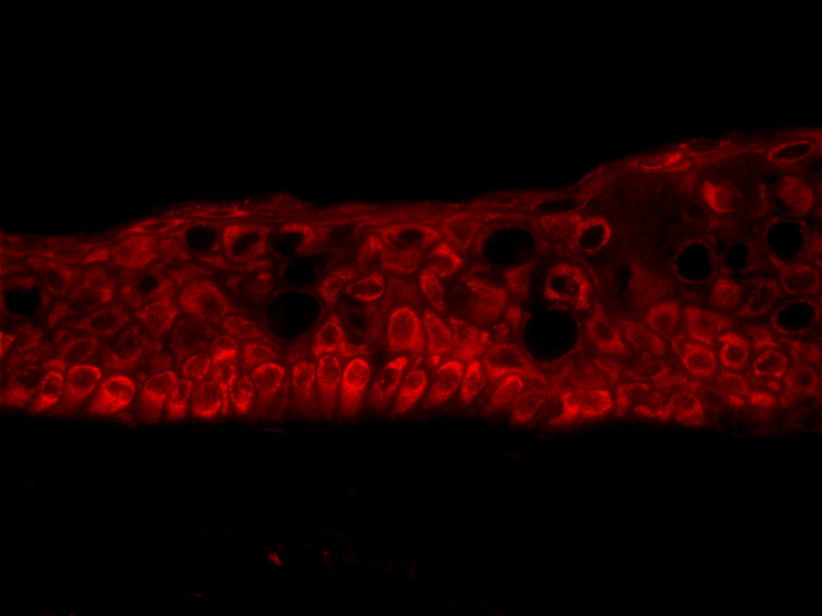
Application: Immunocytochemistry/ImmunofluorescenceSample Tested: corneal tissueSpecies: HumanVerified Customer | Posted 03/21/20231% formalin fixed 0.5% Triton-X100 permeabilized 5% BSA blocking

-
Application: Immunocytochemistry/ImmunofluorescenceSample Tested: MDA-MBA-231 human breast cancer cell lineSpecies: HumanVerified Customer | Posted 10/26/2015Specificity: Specific<br />Sensitivity: Sensitive<br />Buffer: PBS with 2%BSA<br />Dilution: 1:250
-
Application: Immunocytochemistry/ImmunofluorescenceSample Tested: SUM-159PT human breast cancer cell lineSpecies: HumanVerified Customer | Posted 10/26/20154% PFA fixation, Secondary Ab: Alexa488-donkey-anti-goat 1/500. <br />Specificity: Specific<br />Sensitivity: Sensitive<br />Buffer: PBS/Tween<br />Dilution: 1/250
-
Application: Immunocytochemistry/ImmunofluorescenceSample Tested: MDA-MB-231 human breast cancer cell lineSpecies: HumanVerified Customer | Posted 10/26/2015Cells were fixed in 4% PFA. Secondary Ab: Alexa488-donkey-anti-goat 1/500. <br />Specificity: Specific<br />Sensitivity: Sensitive<br />Buffer: PBS/Tween<br />Dilution: 1/250
-
Application: Flow CytometrySample Tested: See PMID 24274766Species: HumanVerified Customer | Posted 01/07/2015
There are no reviews that match your criteria.
Protocols
Find general support by application which include: protocols, troubleshooting, illustrated assays, videos and webinars.
- 7-Amino Actinomycin D (7-AAD) Cell Viability Flow Cytometry Protocol
- Antigen Retrieval Protocol (PIER)
- Antigen Retrieval for Frozen Sections Protocol
- Appropriate Fixation of IHC/ICC Samples
- Cellular Response to Hypoxia Protocols
- Chromogenic IHC Staining of Formalin-Fixed Paraffin-Embedded (FFPE) Tissue Protocol
- Chromogenic Immunohistochemistry Staining of Frozen Tissue
- ClariTSA™ Fluorophore Kits
- Detection & Visualization of Antibody Binding
- Extracellular Membrane Flow Cytometry Protocol
- Flow Cytometry Protocol for Cell Surface Markers
- Flow Cytometry Protocol for Staining Membrane Associated Proteins
- Flow Cytometry Staining Protocols
- Flow Cytometry Troubleshooting Guide
- Fluorescent IHC Staining of Frozen Tissue Protocol
- Graphic Protocol for Heat-induced Epitope Retrieval
- Graphic Protocol for the Preparation and Fluorescent IHC Staining of Frozen Tissue Sections
- Graphic Protocol for the Preparation and Fluorescent IHC Staining of Paraffin-embedded Tissue Sections
- Graphic Protocol for the Preparation of Gelatin-coated Slides for Histological Tissue Sections
- IHC Sample Preparation (Frozen sections vs Paraffin)
- Immunofluorescent IHC Staining of Formalin-Fixed Paraffin-Embedded (FFPE) Tissue Protocol
- Immunohistochemistry (IHC) and Immunocytochemistry (ICC) Protocols
- Immunohistochemistry Frozen Troubleshooting
- Immunohistochemistry Paraffin Troubleshooting
- Intracellular Flow Cytometry Protocol Using Alcohol (Methanol)
- Intracellular Flow Cytometry Protocol Using Detergents
- Intracellular Nuclear Staining Flow Cytometry Protocol Using Detergents
- Intracellular Staining Flow Cytometry Protocol Using Alcohol Permeabilization
- Intracellular Staining Flow Cytometry Protocol Using Detergents to Permeabilize Cells
- Preparing Samples for IHC/ICC Experiments
- Preventing Non-Specific Staining (Non-Specific Binding)
- Primary Antibody Selection & Optimization
- Propidium Iodide Cell Viability Flow Cytometry Protocol
- Protocol for Heat-Induced Epitope Retrieval (HIER)
- Protocol for Liperfluo
- Protocol for Making a 4% Formaldehyde Solution in PBS
- Protocol for VisUCyte™ HRP Polymer Detection Reagent
- Protocol for the Characterization of Human Th22 Cells
- Protocol for the Characterization of Human Th9 Cells
- Protocol for the Preparation & Fixation of Cells on Coverslips
- Protocol for the Preparation and Chromogenic IHC Staining of Frozen Tissue Sections
- Protocol for the Preparation and Chromogenic IHC Staining of Frozen Tissue Sections - Graphic
- Protocol for the Preparation and Chromogenic IHC Staining of Paraffin-embedded Tissue Sections
- Protocol for the Preparation and Chromogenic IHC Staining of Paraffin-embedded Tissue Sections - Graphic
- Protocol for the Preparation and Fluorescent IHC Staining of Frozen Tissue Sections
- Protocol for the Preparation and Fluorescent IHC Staining of Paraffin-embedded Tissue Sections
- Protocol for the Preparation of Gelatin-coated Slides for Histological Tissue Sections
- Protocol: Annexin V and PI Staining by Flow Cytometry
- Protocol: Annexin V and PI Staining for Apoptosis by Flow Cytometry
- R&D Systems Quality Control Western Blot Protocol
- TUNEL and Active Caspase-3 Detection by IHC/ICC Protocol
- The Importance of IHC/ICC Controls
- Troubleshooting Guide: Fluorokine Flow Cytometry Kits
- Troubleshooting Guide: Immunohistochemistry
- Troubleshooting Guide: Western Blot Figures
- Western Blot Conditions
- Western Blot Protocol
- Western Blot Protocol for Cell Lysates
- Western Blot Troubleshooting
- Western Blot Troubleshooting Guide
- View all Protocols, Troubleshooting, Illustrated assays and Webinars
Loading...
Associated Pathways

