Histone Deacetylase 4 (HDAC4; also HD4) is a founding member of the class IIa subfamily, histone deacetylase family of transcriptional regulators. Although its predicted MW is 119 kDa, it runs anomalously at 140‑150 kDa in SDS‑PAGE which may be due to extensive phosphorylation plus SUMOylation. It has an N-terminal region that interacts with transcription factors and corepressors, and a C-terminal domain that shows deacetylase activity, thus repressing gene transcription. HDAC4 is found in osteoblasts, cardiac and skeletal muscle cells, and neurons. Human HDAC4 is 1084 amino acids (aa) in length. It contains one coiled-coil region (aa 67‑177), a SUMOylation site at Lys559, a histone deacetylation domain (aa 665‑993) and one NES (aa 1051‑1084). Caspase cleavage after Asp289 generates bioactive 97 and 34 kDa fragments. There is one potential splice variant that shows an alternative start site at Met118 coupled to a five aa insertion after Thr431. Over aa 1‑68, human HDAC4 shares 96% aa identity with mouse HDAC4.

Key Product Details
Species Reactivity
Human
Applications
Immunohistochemistry
Label
Unconjugated
Antibody Source
Monoclonal Mouse IgG2A Clone # 713802
Loading...
Product Specifications
Immunogen
E. coli-derived recombinant human Histone Deacetylase 4/HDAC4
Met1-Glu68
Accession # P56524
Met1-Glu68
Accession # P56524
Specificity
Detects human Histone Deacetylase 4/HDAC4 in direct ELISAs.
Clonality
Monoclonal
Host
Mouse
Isotype
IgG2A
Scientific Data Images for Human Histone Deacetylase 4/HDAC4 Antibody
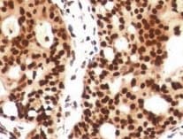
Histone Deacetylase 4/HDAC4 in Human Testis.
Histone Deacetylase 4/HDAC4 was detected in immersion fixed paraffin-embedded sections of human testis using Mouse Anti-Human Histone Deacetylase 4/HDAC4 Monoclonal Antibody (Catalog # MAB6205) at 5 µg/mL for 1 hour at room temperature followed by incubation with the Anti-Mouse IgG VisUCyte™ HRP Polymer Antibody (Catalog # VC001). Tissue was stained using DAB (brown) and counterstained with hematoxylin (blue). Specific staining was localized to cytoplasm and nuclei of sperm cells. View our protocol for IHC Staining with VisUCyte HRP Polymer Detection Reagents.Applications for Human Histone Deacetylase 4/HDAC4 Antibody
Application
Recommended Usage
Immunohistochemistry
5-25 µg/mL
Sample: Immersion fixed paraffin-embedded sections of human testis
Sample: Immersion fixed paraffin-embedded sections of human testis
Reviewed Applications
Read 1 review rated 5 using MAB6205 in the following applications:
Formulation, Preparation, and Storage
Purification
Protein A or G purified from hybridoma culture supernatant
Reconstitution
Reconstitute at 0.5 mg/mL in sterile PBS. For liquid material, refer to CoA for concentration.
Loading...
Formulation
Lyophilized from a 0.2 μm filtered solution in PBS with Trehalose. *Small pack size (SP) is supplied either lyophilized or as a 0.2 µm filtered solution in PBS.
Shipping
Lyophilized product is shipped at ambient temperature. Liquid small pack size (-SP) is shipped with polar packs. Upon receipt, store immediately at the temperature recommended below.
Stability & Storage
Use a manual defrost freezer and avoid repeated freeze-thaw cycles.
- 12 months from date of receipt, -20 to -70 °C as supplied.
- 1 month, 2 to 8 °C under sterile conditions after reconstitution.
- 6 months, -20 to -70 °C under sterile conditions after reconstitution.
Calculators
Background: Histone Deacetylase 4/HDAC4
Long Name
Histone Deacetylase 4
Alternate Names
HDAC4
Gene Symbol
HDAC4
UniProt
Additional Histone Deacetylase 4/HDAC4 Products
Product Documents for Human Histone Deacetylase 4/HDAC4 Antibody
Certificate of Analysis
To download a Certificate of Analysis, please enter a lot or batch number in the search box below.
Note: Certificate of Analysis not available for kit components.
Product Specific Notices for Human Histone Deacetylase 4/HDAC4 Antibody
For research use only
Related Research Areas
Customer Reviews for Human Histone Deacetylase 4/HDAC4 Antibody (1)
5 out of 5
1 Customer Rating
Have you used Human Histone Deacetylase 4/HDAC4 Antibody?
Submit a review and receive an Amazon gift card!
$25/€18/£15/$25CAN/¥2500 Yen for a review with an image
$10/€7/£6/$10CAN/¥1110 Yen for a review without an image
Submit a review
Customer Images
Showing
1
-
1 of
1 review
Showing All
Filter By:
-
Application: ImmunohistochemistrySample Tested: Breast cancer tissueSpecies: HumanVerified Customer | Posted 02/23/2022

There are no reviews that match your criteria.
Protocols
Find general support by application which include: protocols, troubleshooting, illustrated assays, videos and webinars.
- Antigen Retrieval Protocol (PIER)
- Antigen Retrieval for Frozen Sections Protocol
- Appropriate Fixation of IHC/ICC Samples
- Cellular Response to Hypoxia Protocols
- Chromogenic IHC Staining of Formalin-Fixed Paraffin-Embedded (FFPE) Tissue Protocol
- Chromogenic Immunohistochemistry Staining of Frozen Tissue
- ClariTSA™ Fluorophore Kits
- Detection & Visualization of Antibody Binding
- Fluorescent IHC Staining of Frozen Tissue Protocol
- Graphic Protocol for Heat-induced Epitope Retrieval
- Graphic Protocol for the Preparation and Fluorescent IHC Staining of Frozen Tissue Sections
- Graphic Protocol for the Preparation and Fluorescent IHC Staining of Paraffin-embedded Tissue Sections
- Graphic Protocol for the Preparation of Gelatin-coated Slides for Histological Tissue Sections
- IHC Sample Preparation (Frozen sections vs Paraffin)
- Immunofluorescent IHC Staining of Formalin-Fixed Paraffin-Embedded (FFPE) Tissue Protocol
- Immunohistochemistry (IHC) and Immunocytochemistry (ICC) Protocols
- Immunohistochemistry Frozen Troubleshooting
- Immunohistochemistry Paraffin Troubleshooting
- Preparing Samples for IHC/ICC Experiments
- Preventing Non-Specific Staining (Non-Specific Binding)
- Primary Antibody Selection & Optimization
- Protocol for Heat-Induced Epitope Retrieval (HIER)
- Protocol for Making a 4% Formaldehyde Solution in PBS
- Protocol for VisUCyte™ HRP Polymer Detection Reagent
- Protocol for the Preparation & Fixation of Cells on Coverslips
- Protocol for the Preparation and Chromogenic IHC Staining of Frozen Tissue Sections
- Protocol for the Preparation and Chromogenic IHC Staining of Frozen Tissue Sections - Graphic
- Protocol for the Preparation and Chromogenic IHC Staining of Paraffin-embedded Tissue Sections
- Protocol for the Preparation and Chromogenic IHC Staining of Paraffin-embedded Tissue Sections - Graphic
- Protocol for the Preparation and Fluorescent IHC Staining of Frozen Tissue Sections
- Protocol for the Preparation and Fluorescent IHC Staining of Paraffin-embedded Tissue Sections
- Protocol for the Preparation of Gelatin-coated Slides for Histological Tissue Sections
- TUNEL and Active Caspase-3 Detection by IHC/ICC Protocol
- The Importance of IHC/ICC Controls
- Troubleshooting Guide: Immunohistochemistry
- View all Protocols, Troubleshooting, Illustrated assays and Webinars
Loading...