
Key Product Details
Species Reactivity
Validated:
Human
Cited:
Human, Feline, Primate - Macaca mulatta (Rhesus Macaque)
Applications
Validated:
Immunohistochemistry, Western Blot, Neutralization
Cited:
Immunohistochemistry-Paraffin, Immunohistochemistry-Frozen, Western Blot, Neutralization, Immunocytochemistry
Label
Unconjugated
Antibody Source
Polyclonal Goat IgG
Loading...
Product Specifications
Immunogen
E. coli-derived recombinant human IL-15
Asn49-Ser162
Accession # P40933
Asn49-Ser162
Accession # P40933
Specificity
Detects human IL-15 in direct ELISAs and Western blots. In direct ELISAs, less than 1% cross-reactivity with recombinant mouse IL‑15 is observed.
Clonality
Polyclonal
Host
Goat
Isotype
IgG
Endotoxin Level
<0.10 EU per 1 μg of the antibody by the LAL method.
Scientific Data Images for Human IL-15 Antibody
IL‑15 in Human Tonsil.
IL‑15 was detected in immersion fixed paraffin-embedded sections of human tonsil using Goat Anti-Human IL‑15 Antigen Affinity-purified Polyclonal Antibody (Catalog # AF315) at 10 µg/mL overnight at 4 °C. Before incubation with the primary antibody tissue was subjected to heat-induced epitope retrieval using Antigen Retrieval Reagent-Basic (Catalog # CTS013). Tissue was stained using the Anti-Goat HRP-DAB Cell & Tissue Staining Kit (brown; Catalog # CTS008) and counterstained with hematoxylin (blue). View our protocol for Chromogenic IHC Staining of Paraffin-embedded Tissue Sections.
Cell Proliferation Induced by IL‑15 and Neutralization by Human IL‑15 Antibody.
Recombinant Human IL-15 (Catalog # 247-IL) stimulates proliferation in the MO7e human megakaryocytic leukemic cell line in a dose-dependent manner (orange line). Proliferation elicited by Recombinant Human IL-15 (10 ) is neutralized (green line) by increasing concen-trations of Goat Anti-Human IL-15 Antigen Affinity-purified Polyclonal Antibody (Catalog # AF315). The ND50 is typically 0.5-1.5 µg/mL.Applications for Human IL-15 Antibody
Application
Recommended Usage
Immunohistochemistry
5-15 µg/mL
Sample: Immersion fixed paraffin-embedded sections of human tonsil subjected to Antigen Retrieval Reagent-Basic (Catalog # CTS013)
Sample: Immersion fixed paraffin-embedded sections of human tonsil subjected to Antigen Retrieval Reagent-Basic (Catalog # CTS013)
Western Blot
0.1 µg/mL
Sample: Recombinant Human IL‑15 (Catalog # 247-IL)
Sample: Recombinant Human IL‑15 (Catalog # 247-IL)
Neutralization
Measured by its ability to neutralize IL‑15-induced proliferation in the MO7e human megakaryocytic leukemic cell line [Avanzi, G. et al. (1988) Br. J. Haematol. 69:359]. The Neutralization Dose (ND50) is typically 0.5-1.5 µg/mL in the presence of 10 ng/mL of Recombinant Human IL‑15.
Reviewed Applications
Read 1 review rated 5 using AF315 in the following applications:
Formulation, Preparation, and Storage
Purification
Antigen Affinity-purified
Reconstitution
Reconstitute at 0.2 mg/mL in sterile PBS. For liquid material, refer to CoA for concentration.
Loading...
Formulation
Lyophilized from a 0.2 μm filtered solution in PBS with Trehalose. *Small pack size (SP) is supplied either lyophilized or as a 0.2 µm filtered solution in PBS.
Shipping
Lyophilized product is shipped at ambient temperature. Liquid small pack size (-SP) is shipped with polar packs. Upon receipt, store immediately at the temperature recommended below.
Stability & Storage
Use a manual defrost freezer and avoid repeated freeze-thaw cycles.
- 12 months from date of receipt, -20 to -70 °C as supplied.
- 1 month, 2 to 8 °C under sterile conditions after reconstitution.
- 6 months, -20 to -70 °C under sterile conditions after reconstitution.
Calculators
Background: IL-15
References
- Grabstein, K. et al. (1994) Science 264:965.
- Budagian, V. et al. (2006) Cytokine Growth Factor Rev. 17:259.
- Ma, A. et al. (2006) Annu. Rev. Immunol. 24:657.
- Tagaya, Y. et al. (1997) Proc. Natl. Acad. Sci. USA 94:14444.
- Giri, J.G. et al. (1995) EMBO 14:3654.
- Giri, J. et al. (1994) EMBO J. 13:2822.
- Duitman, E.H. et al. (2008) Mol. Cell. Biol. 28:4851.
- Dubois, S. et al. (2002) Immunity 17:537.
- Stonier, S.W. and K.S. Schluns (2010) Immunol. Lett. 127:85.
- Burkett, P.R. et al. (2004) J. Exp. Med. 200:825.
- Budagian, V. et al. (2004) J. Biol. Chem. 279:40368.
- Mortier, E. et al. (2004) J. Immunol. 173:1681.
- Bulanova, E. et al. (2007) J. Biol. Chem. 282:13167.
- Budagian, V. et al. (2004) J. Biol. Chem. 279:42192.
- Neely, G.G. et al. (2004) J. Immunol. 172:4225.
Long Name
Interleukin 15
Alternate Names
IL15
Entrez Gene IDs
Gene Symbol
IL15
UniProt
Additional IL-15 Products
Product Documents for Human IL-15 Antibody
Certificate of Analysis
To download a Certificate of Analysis, please enter a lot or batch number in the search box below.
Note: Certificate of Analysis not available for kit components.
Product Specific Notices for Human IL-15 Antibody
For research use only
Citations for Human IL-15 Antibody
 Powered by Bioz
Customer Reviews for Human IL-15 Antibody (1)
5 out of 5
1 Customer Rating
Have you used Human IL-15 Antibody?
Submit a review and receive an Amazon gift card!
$25/€18/£15/$25CAN/¥2500 Yen for a review with an image
$10/€7/£6/$10CAN/¥1110 Yen for a review without an image
Submit a review
Customer Images
Showing
1
-
1 of
1 review
Showing All
Filter By:
-
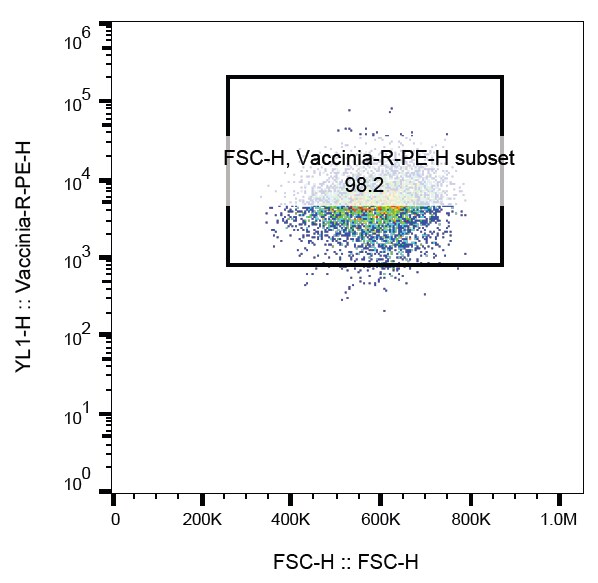
Application: Flow CytometrySample Tested: U-87 MG human glioblastoma/astrocytoma cell lineSpecies: HumanVerified Customer | Posted 05/01/2024

There are no reviews that match your criteria.
Protocols
Find general support by application which include: protocols, troubleshooting, illustrated assays, videos and webinars.
- Antigen Retrieval Protocol (PIER)
- Antigen Retrieval for Frozen Sections Protocol
- Appropriate Fixation of IHC/ICC Samples
- Cellular Response to Hypoxia Protocols
- Chromogenic IHC Staining of Formalin-Fixed Paraffin-Embedded (FFPE) Tissue Protocol
- Chromogenic Immunohistochemistry Staining of Frozen Tissue
- ClariTSA™ Fluorophore Kits
- Detection & Visualization of Antibody Binding
- Fluorescent IHC Staining of Frozen Tissue Protocol
- Graphic Protocol for Heat-induced Epitope Retrieval
- Graphic Protocol for the Preparation and Fluorescent IHC Staining of Frozen Tissue Sections
- Graphic Protocol for the Preparation and Fluorescent IHC Staining of Paraffin-embedded Tissue Sections
- Graphic Protocol for the Preparation of Gelatin-coated Slides for Histological Tissue Sections
- IHC Sample Preparation (Frozen sections vs Paraffin)
- Immunofluorescent IHC Staining of Formalin-Fixed Paraffin-Embedded (FFPE) Tissue Protocol
- Immunohistochemistry (IHC) and Immunocytochemistry (ICC) Protocols
- Immunohistochemistry Frozen Troubleshooting
- Immunohistochemistry Paraffin Troubleshooting
- Preparing Samples for IHC/ICC Experiments
- Preventing Non-Specific Staining (Non-Specific Binding)
- Primary Antibody Selection & Optimization
- Protocol for Heat-Induced Epitope Retrieval (HIER)
- Protocol for Making a 4% Formaldehyde Solution in PBS
- Protocol for VisUCyte™ HRP Polymer Detection Reagent
- Protocol for the Preparation & Fixation of Cells on Coverslips
- Protocol for the Preparation and Chromogenic IHC Staining of Frozen Tissue Sections
- Protocol for the Preparation and Chromogenic IHC Staining of Frozen Tissue Sections - Graphic
- Protocol for the Preparation and Chromogenic IHC Staining of Paraffin-embedded Tissue Sections
- Protocol for the Preparation and Chromogenic IHC Staining of Paraffin-embedded Tissue Sections - Graphic
- Protocol for the Preparation and Fluorescent IHC Staining of Frozen Tissue Sections
- Protocol for the Preparation and Fluorescent IHC Staining of Paraffin-embedded Tissue Sections
- Protocol for the Preparation of Gelatin-coated Slides for Histological Tissue Sections
- R&D Systems Quality Control Western Blot Protocol
- TUNEL and Active Caspase-3 Detection by IHC/ICC Protocol
- The Importance of IHC/ICC Controls
- Troubleshooting Guide: Immunohistochemistry
- Troubleshooting Guide: Western Blot Figures
- Western Blot Conditions
- Western Blot Protocol
- Western Blot Protocol for Cell Lysates
- Western Blot Troubleshooting
- Western Blot Troubleshooting Guide
- View all Protocols, Troubleshooting, Illustrated assays and Webinars


