Functional IL-2 receptors can exist in two affinity states on cell surfaces, the high affinity complex consisting of heterotrimers of the alpha, beta, and gamma chains and the intermediate affinity complex comprising heterodimers of the beta and gamma chains. Individual beta chains and alpha chains exhibit low affinity IL-2 binding, and the gamma chain alone does not bind IL-2. In addition to their involvement in IL-2 mediated signal transduction, both the beta chain and gamma chain have been shown to be required for IL-15 mediated signaling. IL-2 R beta is a member of the cytokine receptor superfamily. Human IL-2 R beta cDNA encodes a 551 amino acid (aa) precursor Type I membrane protein with a 26 aa signal peptide, a 214 aa extracellular region, a 25 aa transmembrane region and a 286 aa cytoplasmic domain. A soluble IL-2 R beta has been identified in the culture supernatants of a human lymphoid cell line, YT, that displays IL-2 R beta. Soluble IL-2 R beta binds IL-2 with low affinity and is not an effective IL-2 antagonist on cells displaying the high or intermediate affinity IL-2 signaling receptors. Nevertheless, soluble IL-2 R beta binds IL-15 with sufficient affinity to neutralize IL-15 biological activities.
Human IL-2 R beta Antibody (27302)
R&D Systems | Catalog # MAB224

Key Product Details
Species Reactivity
Validated:
Human
Cited:
Human, Primate - Macaca mulatta (Rhesus Macaque)
Applications
Validated:
Immunohistochemistry, Neutralization, Flow Cytometry, CyTOF-ready
Cited:
Neutralization, Flow Cytometry, ELISA Development, Protein Array
Label
Unconjugated
Antibody Source
Monoclonal Mouse IgG1 Clone # 27302
Loading...
Product Specifications
Immunogen
S. frugiperda insect ovarian cell line Sf 21-derived recombinant human IL‑2 R beta
Ala27-Asp239
Accession # NP_000869
Ala27-Asp239
Accession # NP_000869
Specificity
Detects human IL-2 R beta in direct ELISAs. In direct ELISAs, no cross-reactivity with recombinant human (rh) IL‑2 R alpha, rh gamma c, rhIL‑4 R, or rhIL‑6 R is observed.
Clonality
Monoclonal
Host
Mouse
Isotype
IgG1
Endotoxin Level
<0.10 EU per 1 μg of the antibody by the LAL method.
Scientific Data Images for Human IL-2 R beta Antibody (27302)
IL‑2 R beta in Human Lymph Node.
IL‑2 R beta was detected in immersion fixed frozen sections of human lymph node using 15 µg/mL Mouse Anti-Human IL‑2 R beta Monoclonal Antibody (Catalog # MAB224) overnight at 4 °C. Tissue was stained (red) and counterstained with hematoxylin (blue). View our protocol for Chromogenic IHC Staining of Frozen Tissue Sections.
Cell Proliferation Induced by IL‑2 and Neutralization by Human IL‑2 R beta Antibody.
Recombinant Human IL-2 (Catalog # 202-IL) stimulates proliferation in the MO7e human megakaryocytic leukemic cell line in a dose-dependent manner (orange line). Proliferation elicited by Recombinant Human IL-2 (30 ng/mL) is neutralized (green line) by increasing concentrations of Mouse Anti-Human IL-2 R beta Monoclonal Antibody (Catalog # MAB224). The ND50 is typically 0.05-0.15 µg/mL.Applications for Human IL-2 R beta Antibody (27302)
Application
Recommended Usage
CyTOF-ready
Ready to be labeled using established conjugation methods. No BSA or other carrier proteins that could interfere with conjugation.
Flow Cytometry
2.5 µg/106 cells
Sample: Human whole blood CD56+ natural killer cells
Sample: Human whole blood CD56+ natural killer cells
Immunohistochemistry
8-25 µg/mL
Sample: Immersion fixed frozen sections of human lymph node
Sample: Immersion fixed frozen sections of human lymph node
Neutralization
Measured by its ability to neutralize IL‑2-induced proliferation in the MO7e human megakaryocytic leukemic cell line. Avanzi, G. et al. (1988) Br. J. Haematol. 69:359. The Neutralization Dose (ND50) is typically 0.05-0.15 µg/mL in the presence of 30 ng/mL Recombinant Human IL‑2.
Reviewed Applications
Read 1 review rated 4 using MAB224 in the following applications:
Flow Cytometry Panel Builder
Bio-Techne Knows Flow Cytometry
Save time and reduce costly mistakes by quickly finding compatible reagents using the Panel Builder Tool.
Advanced Features
- Spectra Viewer - Custom analysis of spectra from multiple fluorochromes
- Spillover Popups - Visualize the spectra of individual fluorochromes
- Antigen Density Selector - Match fluorochrome brightness with antigen density

Formulation, Preparation, and Storage
Purification
Protein A or G purified from ascites
Reconstitution
Reconstitute at 0.5 mg/mL in sterile PBS. For liquid material, refer to CoA for concentration.
Loading...
Formulation
Lyophilized from a 0.2 μm filtered solution in PBS with Trehalose. *Small pack size (SP) is supplied either lyophilized or as a 0.2 µm filtered solution in PBS.
Shipping
Lyophilized product is shipped at ambient temperature. Liquid small pack size (-SP) is shipped with polar packs. Upon receipt, store immediately at the temperature recommended below.
Stability & Storage
Use a manual defrost freezer and avoid repeated freeze-thaw cycles.
- 12 months from date of receipt, -20 to -70 °C as supplied.
- 1 month, 2 to 8 °C under sterile conditions after reconstitution.
- 6 months, -20 to -70 °C under sterile conditions after reconstitution.
Calculators
Background: IL-2 R beta
Long Name
Interleukin 2 Receptor beta
Alternate Names
CD122, IL-15 R beta, IL-2Rb, IL2R beta, IL2RB
Gene Symbol
IL2RB
UniProt
Additional IL-2 R beta Products
Product Documents for Human IL-2 R beta Antibody (27302)
Certificate of Analysis
To download a Certificate of Analysis, please enter a lot or batch number in the search box below.
Note: Certificate of Analysis not available for kit components.
Product Specific Notices for Human IL-2 R beta Antibody (27302)
For research use only
Citations for Human IL-2 R beta Antibody (27302)
 Powered by Bioz
Customer Reviews for Human IL-2 R beta Antibody (27302) (1)
4 out of 5
1 Customer Rating
Have you used Human IL-2 R beta Antibody (27302)?
Submit a review and receive an Amazon gift card!
$25/€18/£15/$25CAN/¥2500 Yen for a review with an image
$10/€7/£6/$10CAN/¥1110 Yen for a review without an image
Submit a review
Customer Images
Showing
1
-
1 of
1 review
Showing All
Filter By:
-
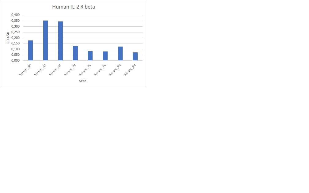
Application: ELISASample Tested: Serum and PlasmaSpecies: HumanVerified Customer | Posted 02/13/2023Working good for the detection of Human IL2 R beta in the serum or plasma samples.

There are no reviews that match your criteria.
Protocols
Find general support by application which include: protocols, troubleshooting, illustrated assays, videos and webinars.
- 7-Amino Actinomycin D (7-AAD) Cell Viability Flow Cytometry Protocol
- Antigen Retrieval Protocol (PIER)
- Antigen Retrieval for Frozen Sections Protocol
- Appropriate Fixation of IHC/ICC Samples
- Cellular Response to Hypoxia Protocols
- Chromogenic IHC Staining of Formalin-Fixed Paraffin-Embedded (FFPE) Tissue Protocol
- Chromogenic Immunohistochemistry Staining of Frozen Tissue
- ClariTSA™ Fluorophore Kits
- Detection & Visualization of Antibody Binding
- Extracellular Membrane Flow Cytometry Protocol
- Flow Cytometry Protocol for Cell Surface Markers
- Flow Cytometry Protocol for Staining Membrane Associated Proteins
- Flow Cytometry Staining Protocols
- Flow Cytometry Troubleshooting Guide
- Fluorescent IHC Staining of Frozen Tissue Protocol
- Graphic Protocol for Heat-induced Epitope Retrieval
- Graphic Protocol for the Preparation and Fluorescent IHC Staining of Frozen Tissue Sections
- Graphic Protocol for the Preparation and Fluorescent IHC Staining of Paraffin-embedded Tissue Sections
- Graphic Protocol for the Preparation of Gelatin-coated Slides for Histological Tissue Sections
- IHC Sample Preparation (Frozen sections vs Paraffin)
- Immunofluorescent IHC Staining of Formalin-Fixed Paraffin-Embedded (FFPE) Tissue Protocol
- Immunohistochemistry (IHC) and Immunocytochemistry (ICC) Protocols
- Immunohistochemistry Frozen Troubleshooting
- Immunohistochemistry Paraffin Troubleshooting
- Intracellular Flow Cytometry Protocol Using Alcohol (Methanol)
- Intracellular Flow Cytometry Protocol Using Detergents
- Intracellular Nuclear Staining Flow Cytometry Protocol Using Detergents
- Intracellular Staining Flow Cytometry Protocol Using Alcohol Permeabilization
- Intracellular Staining Flow Cytometry Protocol Using Detergents to Permeabilize Cells
- Preparing Samples for IHC/ICC Experiments
- Preventing Non-Specific Staining (Non-Specific Binding)
- Primary Antibody Selection & Optimization
- Propidium Iodide Cell Viability Flow Cytometry Protocol
- Protocol for Heat-Induced Epitope Retrieval (HIER)
- Protocol for Liperfluo
- Protocol for Making a 4% Formaldehyde Solution in PBS
- Protocol for VisUCyte™ HRP Polymer Detection Reagent
- Protocol for the Characterization of Human Th22 Cells
- Protocol for the Characterization of Human Th9 Cells
- Protocol for the Preparation & Fixation of Cells on Coverslips
- Protocol for the Preparation and Chromogenic IHC Staining of Frozen Tissue Sections
- Protocol for the Preparation and Chromogenic IHC Staining of Frozen Tissue Sections - Graphic
- Protocol for the Preparation and Chromogenic IHC Staining of Paraffin-embedded Tissue Sections
- Protocol for the Preparation and Chromogenic IHC Staining of Paraffin-embedded Tissue Sections - Graphic
- Protocol for the Preparation and Fluorescent IHC Staining of Frozen Tissue Sections
- Protocol for the Preparation and Fluorescent IHC Staining of Paraffin-embedded Tissue Sections
- Protocol for the Preparation of Gelatin-coated Slides for Histological Tissue Sections
- Protocol: Annexin V and PI Staining by Flow Cytometry
- Protocol: Annexin V and PI Staining for Apoptosis by Flow Cytometry
- TUNEL and Active Caspase-3 Detection by IHC/ICC Protocol
- The Importance of IHC/ICC Controls
- Troubleshooting Guide: Fluorokine Flow Cytometry Kits
- Troubleshooting Guide: Immunohistochemistry
- View all Protocols, Troubleshooting, Illustrated assays and Webinars
Loading...
Associated Pathways


Innate Lymphoid Cell Differentiation Pathways
 Jak/STAT Signaling Pathway
 Th2 Differentiation Pathway
