IL-2 R beta is a member of the cytokine receptor superfamily. Human IL-2 R beta cDNA encodes a 551 amino acid residue precursor Type I membrane protein with a 26 residue signal peptide, a 214 residue extracellular region, a 25 residue transmembrane region and a 286 residue cytoplasmic domain. A soluble IL-2 R beta (IL-2 sR beta ) has been identified in the culture supernatants of a human lymphoid cell line, YT, that displays IL-2 R beta. At present, the function of IL-2 sR beta is unclear. Recombinant human IL-2 sR beta binds IL-2 with low affinity and is not an effective IL-2 antagonist on cells displaying the high or intermediate affinity IL-2 signaling receptors. Nevertheless, IL-2 sR beta binds IL-15 with sufficient affinity to neutralize IL-15 biological activities.

Key Product Details
Species Reactivity
Validated:
Human
Cited:
Human
Applications
Validated:
Western Blot, Neutralization, Simple Western
Cited:
Western Blot, Neutralization
Label
Unconjugated
Antibody Source
Polyclonal Goat IgG
Loading...
Product Specifications
Immunogen
S. frugiperda insect ovarian cell line Sf 21-derived recombinant human IL‑2 R beta
Ala27-Asp239
Accession # NP_000869
Ala27-Asp239
Accession # NP_000869
Specificity
Detects human IL‑2 R beta in direct ELISAs and Western blots. In direct ELISAs, less than 5% cross-reactivity withrecombinant human (rh) IL-2 R alpha, recombinant mouse (rm) IL-2 R beta, rhIL-2 R gamma, and rhIL-15 R is observed. Is also able to block the cell surface of human IL-2 R beta mediated bioactivities induced by IL-2. For optimal neutralization of IL-2 biological activity on cells expressing the high affinity IL-2 receptors, the use of anti-IL-2 R alpha in conjunction with anti-IL-2 R beta antibodies is recommended.
Clonality
Polyclonal
Host
Goat
Isotype
IgG
Endotoxin Level
<0.10 EU per 1 μg of the antibody by the LAL method.
Scientific Data Images for Human IL-2 R beta Antibody
Cell Proliferation Induced by IL‑2 and Neutralization by Human IL‑2 R beta Antibody.
Recombinant Human IL-2 (Catalog # 202-IL) stimulates proliferation in the MO7e human megakaryocytic leukemic cell line in a dose-dependent manner (orange line). Proliferation elicited by Recombinant Human IL-2 (20 ng/mL) is neutralized (green line) by increasing concentrations of Goat Anti-Human IL-2 R beta Antigen Affinity-purified Polyclonal Antibody (Catalog # AF-224-NA). The ND50 is typically 10‑30 µg/mL.
Detection of Human IL‑2 R beta by Simple WesternTM.
Simple Western lane view shows lysates of HDLM‑2 human Hodgkin’s lymphoma cell line, loaded at 0.2 mg/mL. A specific band was detected for IL‑2 R beta at approximately 98 kDa (as indicated) using 25 µg/mL of Goat Anti-Human IL‑2 R beta Antigen Affinity-purified Polyclonal Antibody (Catalog # AF-224-NA). This experiment was conducted under reducing conditions and using the 12-230kDa separation system.Applications for Human IL-2 R beta Antibody
Application
Recommended Usage
Simple Western
25 µg/mL
Sample: HDLM‑2 human Hodgkin's lymphoma cell line
Sample: HDLM‑2 human Hodgkin's lymphoma cell line
Western Blot
0.1 µg/mL
Sample: Recombinant Human IL‑2 R beta (Catalog # 224-2B)
Sample: Recombinant Human IL‑2 R beta (Catalog # 224-2B)
Neutralization
Measured by its ability to neutralize IL‑2-induced proliferation in the MO7e human megakaryocytic leukemic cell line. Avanzi, G. et al. (1988) Br. J. Haematol. 69:359. The Neutralization Dose (ND50) is typically 10-30 µg/mL in the presence of 20 ng/mL Recombinant Human IL‑2.
Reviewed Applications
Read 1 review rated 4 using AF-224-NA in the following applications:
Formulation, Preparation, and Storage
Purification
Antigen Affinity-purified
Reconstitution
Reconstitute at 0.2 mg/mL in sterile PBS. For liquid material, refer to CoA for concentration.
Loading...
Formulation
Lyophilized from a 0.2 μm filtered solution in PBS with Trehalose. *Small pack size (SP) is supplied either lyophilized or as a 0.2 µm filtered solution in PBS.
Shipping
Lyophilized product is shipped at ambient temperature. Liquid small pack size (-SP) is shipped with polar packs. Upon receipt, store immediately at the temperature recommended below.
Stability & Storage
Use a manual defrost freezer and avoid repeated freeze-thaw cycles.
- 12 months from date of receipt, -20 to -70 °C as supplied.
- 1 month, 2 to 8 °C under sterile conditions after reconstitution.
- 6 months, -20 to -70 °C under sterile conditions after reconstitution.
Calculators
Background: IL-2 R beta
Long Name
Interleukin 2 Receptor beta
Alternate Names
CD122, IL-15 R beta, IL-2Rb, IL2R beta, IL2RB
Gene Symbol
IL2RB
UniProt
Additional IL-2 R beta Products
Product Documents for Human IL-2 R beta Antibody
Certificate of Analysis
To download a Certificate of Analysis, please enter a lot or batch number in the search box below.
Note: Certificate of Analysis not available for kit components.
Product Specific Notices for Human IL-2 R beta Antibody
For research use only
Citations for Human IL-2 R beta Antibody
 Powered by Bioz
Customer Reviews for Human IL-2 R beta Antibody (1)
4 out of 5
1 Customer Rating
Have you used Human IL-2 R beta Antibody?
Submit a review and receive an Amazon gift card!
$25/€18/£15/$25CAN/¥2500 Yen for a review with an image
$10/€7/£6/$10CAN/¥1110 Yen for a review without an image
Submit a review
Customer Images
Showing
1
-
1 of
1 review
Showing All
Filter By:
-
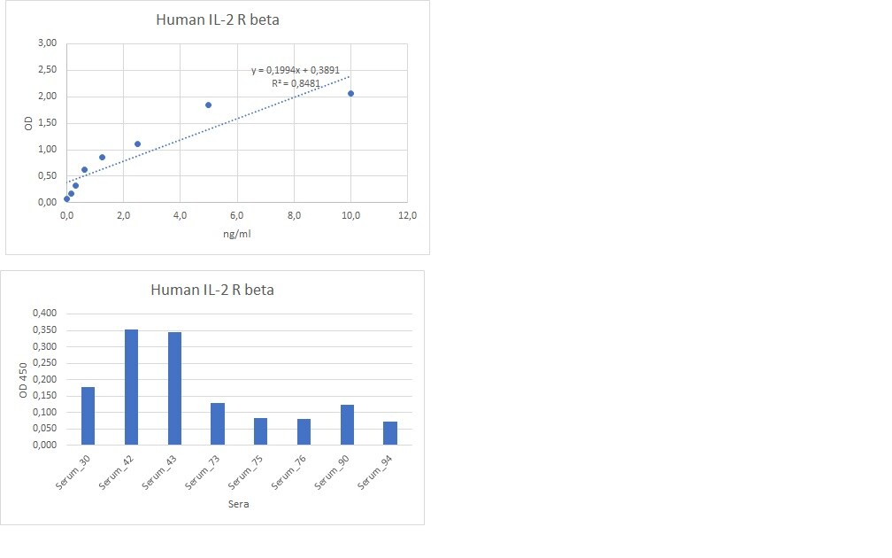
Application: ELISASample Tested: Serum and PlasmaSpecies: HumanVerified Customer | Posted 02/13/2023Working good for the detection of Human IL2 R beta in the serum or plasma samples.

There are no reviews that match your criteria.
Protocols
Find general support by application which include: protocols, troubleshooting, illustrated assays, videos and webinars.
- Cellular Response to Hypoxia Protocols
- R&D Systems Quality Control Western Blot Protocol
- Troubleshooting Guide: Western Blot Figures
- Western Blot Conditions
- Western Blot Protocol
- Western Blot Protocol for Cell Lysates
- Western Blot Troubleshooting
- Western Blot Troubleshooting Guide
- View all Protocols, Troubleshooting, Illustrated assays and Webinars
Loading...
Associated Pathways


Innate Lymphoid Cell Differentiation Pathways
 Jak/STAT Signaling Pathway
 Th2 Differentiation Pathway
