The Insulin Receptor (INS R) and insulin-like growth factor-1 receptor (IGF-I R) constitute a subfamily of receptor tyrosine kinases (1-4). The two receptors share structural similarity as well as overlapping intracellular signaling events, and are believed to have evolved through gene duplication from a common ancestral gene. INS R cDNA encodes a type I transmembrane single chain preproprotein with a putative 27 amino acid residues (aa) signal peptide. The large INS R extracellular domain is organized into two successive homologous globular domains, which are separated by a Cysteine-rich domain, followed by three fibronectin type III domains. The intracellular region contains the kinase domain sandwiched between the juxtamembrane domain used for docking insulin-receptor substrates (IRS), and the carboxy-terminal tail that contains two phosphotyrosine-binding sites. After synthesis, the single chain INS R precursor is glycosylated, dimerized and transported to the Golgi apparatus where it is processed at a furin-cleavage site within the middle fibronectin type III domain to generate the mature disulfide-linked alpha 2 beta 2 tetrameric receptor. The alpha subunit is localized extracellularly and mediates ligand binding while the transmembrane beta subunit contains the cytoplasmic kinase domain and mediates intracellular signaling. As a result of alternative splicing, two INS R isoforms (A and B) that differ by the absence or presence, respectively, of a 12 aa residue sequence in the carboxyl terminus of the alpha subunit exist. Whereas the A isoform is predominantly expressed in fetal tissues and cancer cells, the B isoform is primarily expressed in adult differentiated cells. Both the A and B isoforms bind insulin with high-affinity, but the A isoform has considerably higher affinity for IGF‑I and IGF-II. Ligand binding induces a conformational change of the receptor, resulting in ATP binding, autophosphorylation, and subsequent downstream signaling. INS R signaling is important in metabolic regulation, but may also contribute to cell growth, differentiation and apoptosis. Mutations in the INS R gene have been linked to insulin-resistant diabetes mellitus, noninsulin-dependent diabetes mellitus and leprechaunism, an extremely rare disorder characterized by abnormal resistance to insulin that results in a variety of distinguishing characteristics, including growth delays and abnormalities affecting the endocrine system. INS R is highly conserved between species, rat INS R shares 94% and 97% aa sequence homology with the human and mouse receptor, respectively.

Key Product Details
Species Reactivity
Human
Applications
Flow Cytometry, Immunocytochemistry
Label
Unconjugated
Antibody Source
Monoclonal Mouse IgG2B Clone # 243524
Loading...
Product Specifications
Immunogen
Mouse myeloma cell line NS0-derived recombinant human Insulin R/CD220
His28-Lys944
Accession # NP_001073285
His28-Lys944
Accession # NP_001073285
Specificity
Detects human Insulin R/CD220 in direct ELISAs.
Clonality
Monoclonal
Host
Mouse
Isotype
IgG2B
Scientific Data Images for Human Insulin R/CD220 Antibody (243524)
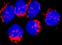
Insulin R/CD220 in HepG2 Human Cell Line.
Insulin R/CD220 was detected in immersion fixed HepG2 human hepatocellular carcinoma cell line (positive staining) and HDLM‑2 human Hodgkin’s lymphoma cell line (negative control) using Mouse Anti-Human Insulin R/CD220 Monoclonal Antibody (Catalog # MAB1544) at 8 µg/mL for 3 hours at room temperature. Cells were stained using the NorthernLights™ 557-conjugated Anti-Mouse IgG Secondary Antibody (red; NL007) and counterstained with DAPI (blue). Specific staining was localized to cell membrane and cytoplasm. Staining was performed using our protocol for Fluorescent ICC Staining of Non-adherent Cells.
Detection of Insulin R/CD220 in PBMC by Flow Cytometry
PBMC were stained with Mouse Anti-Human CD14 PE‑conjugated Monoclonal Antibody (Catalog # FAB3832P) and either (A) Mouse Anti-Human Insulin R/CD220 Monoclonal Antibody (Catalog # MAB1544) or (B) Mouse IgG2B Isotype Control (Catalog # MAB004) followed by Allophycocyanin-conjugated Anti-Mouse IgG Secondary Antibody (Catalog # F0101B). View our protocol for Staining Membrane-associated Proteins.
Detection of Insulin R/CD220 in NS0 cell line transfected with hINSR vs irrelevant transfectant by Flow Cytometry
NSO cell line transfected with hINSR (filled histogram) vs irrelevant transfectant (open histogram) were stained with Mouse Anti-Human Insulin R/CD220 Monoclonal Antibody (Catalog # MAB1544) followed by Allophycocyanin-conjugated Anti-Mouse IgG Secondary Antibody (Catalog # F0101B). View our protocol for Staining Membrane-associated Proteins.Applications for Human Insulin R/CD220 Antibody (243524)
Application
Recommended Usage
Flow Cytometry
2.5 µg/106 cells
Sample: Human peripheral blood monocytes, NS0 cell line transfected with hINSR vs irrelevant transfectant
Sample: Human peripheral blood monocytes, NS0 cell line transfected with hINSR vs irrelevant transfectant
Immunocytochemistry
8-25 µg/mL
Sample: Immersion fixed HepG2 human hepatocellular carcinoma cell line
Sample: Immersion fixed HepG2 human hepatocellular carcinoma cell line
Reviewed Applications
Read 1 review rated 5 using MAB1544 in the following applications:
Flow Cytometry Panel Builder
Bio-Techne Knows Flow Cytometry
Save time and reduce costly mistakes by quickly finding compatible reagents using the Panel Builder Tool.
Advanced Features
- Spectra Viewer - Custom analysis of spectra from multiple fluorochromes
- Spillover Popups - Visualize the spectra of individual fluorochromes
- Antigen Density Selector - Match fluorochrome brightness with antigen density

Formulation, Preparation, and Storage
Purification
Protein A or G purified from hybridoma culture supernatant
Reconstitution
For liquid material, refer to CoA for concentration.
Formulation
Supplied as a 0.2 μm filtered solution in PBS. *Small pack size (SP) is supplied either lyophilized or as a 0.2 µm filtered solution in PBS.
Shipping
Lyophilized product is shipped at ambient temperature. Liquid small pack size (-SP) is shipped with polar packs. Upon receipt, store immediately at the temperature recommended below.
Stability & Storage
Use a manual defrost freezer and avoid repeated freeze-thaw cycles.
- 12 months from date of receipt, -20 to -70 °C, as supplied.
- 1 month, 2 to 8 °C under sterile conditions after opening.
- 6 months, -20 to -70 °C under sterile conditions after opening.
Calculators
Background: Insulin R/CD220
References
- Nakae, J. et al. (2001) Endoc. Rev. 22:818.
- De Meyts, P. and J. Whittaker (2002) Nature Rev. Drug Disc. 1:769.
- Kim, J.J. and D. Accili (2002) Growth Hormone and IGF Res. 12:84.
- Sciacca, L. et al. (2003) Endocrinology 144:2650.
Long Name
Insulin Receptor
Alternate Names
CD220, INSR, InsulinR
Gene Symbol
INSR
UniProt
Additional Insulin R/CD220 Products
Product Documents for Human Insulin R/CD220 Antibody (243524)
Certificate of Analysis
To download a Certificate of Analysis, please enter a lot or batch number in the search box below.
Note: Certificate of Analysis not available for kit components.
Product Specific Notices for Human Insulin R/CD220 Antibody (243524)
For research use only
Related Research Areas
Customer Reviews for Human Insulin R/CD220 Antibody (243524) (1)
5 out of 5
1 Customer Rating
Have you used Human Insulin R/CD220 Antibody (243524)?
Submit a review and receive an Amazon gift card!
$25/€18/£15/$25CAN/¥2500 Yen for a review with an image
$10/€7/£6/$10CAN/¥1110 Yen for a review without an image
Submit a review
Customer Images
Showing
1
-
1 of
1 review
Showing All
Filter By:
-
Application: Immunocytochemistry/ImmunofluorescenceSample Tested: HepG2 human hepatocellular carcinoma cell lineSpecies: HumanVerified Customer | Posted 03/04/2022

There are no reviews that match your criteria.
Protocols
Find general support by application which include: protocols, troubleshooting, illustrated assays, videos and webinars.
- 7-Amino Actinomycin D (7-AAD) Cell Viability Flow Cytometry Protocol
- Appropriate Fixation of IHC/ICC Samples
- Cellular Response to Hypoxia Protocols
- ClariTSA™ Fluorophore Kits
- Detection & Visualization of Antibody Binding
- Extracellular Membrane Flow Cytometry Protocol
- Flow Cytometry Protocol for Cell Surface Markers
- Flow Cytometry Protocol for Staining Membrane Associated Proteins
- Flow Cytometry Staining Protocols
- Flow Cytometry Troubleshooting Guide
- ICC Cell Smear Protocol for Suspension Cells
- ICC Immunocytochemistry Protocol Videos
- ICC for Adherent Cells
- Immunocytochemistry (ICC) Protocol
- Immunocytochemistry Troubleshooting
- Immunofluorescence of Organoids Embedded in Cultrex Basement Membrane Extract
- Immunohistochemistry (IHC) and Immunocytochemistry (ICC) Protocols
- Intracellular Flow Cytometry Protocol Using Alcohol (Methanol)
- Intracellular Flow Cytometry Protocol Using Detergents
- Intracellular Nuclear Staining Flow Cytometry Protocol Using Detergents
- Intracellular Staining Flow Cytometry Protocol Using Alcohol Permeabilization
- Intracellular Staining Flow Cytometry Protocol Using Detergents to Permeabilize Cells
- Preparing Samples for IHC/ICC Experiments
- Preventing Non-Specific Staining (Non-Specific Binding)
- Primary Antibody Selection & Optimization
- Propidium Iodide Cell Viability Flow Cytometry Protocol
- Protocol for Liperfluo
- Protocol for VisUCyte™ HRP Polymer Detection Reagent
- Protocol for the Characterization of Human Th22 Cells
- Protocol for the Characterization of Human Th9 Cells
- Protocol for the Fluorescent ICC Staining of Cell Smears - Graphic
- Protocol for the Fluorescent ICC Staining of Cultured Cells on Coverslips - Graphic
- Protocol for the Preparation and Fluorescent ICC Staining of Cells on Coverslips
- Protocol for the Preparation and Fluorescent ICC Staining of Non-adherent Cells
- Protocol for the Preparation and Fluorescent ICC Staining of Stem Cells on Coverslips
- Protocol for the Preparation of a Cell Smear for Non-adherent Cell ICC - Graphic
- Protocol: Annexin V and PI Staining by Flow Cytometry
- Protocol: Annexin V and PI Staining for Apoptosis by Flow Cytometry
- TUNEL and Active Caspase-3 Detection by IHC/ICC Protocol
- The Importance of IHC/ICC Controls
- Troubleshooting Guide: Fluorokine Flow Cytometry Kits
- View all Protocols, Troubleshooting, Illustrated assays and Webinars
Loading...
Associated Pathways


