Human Integrin beta 1/CD29 Antibody (P5D2)
R&D Systems | Catalog # MAB17781

Key Product Details
Validated by
Species Reactivity
Validated:
Cited:
Applications
Validated:
Cited:
Label
Antibody Source
Product Specifications
Immunogen
Specificity
Clonality
Host
Isotype
Endotoxin Level
Scientific Data Images for Human Integrin beta 1/CD29 Antibody (P5D2)
Detection of Integrin beta 1/CD29 in Human PBMCs by Flow Cytometry.
Human peripheral blood mononuclear cells (PBMCs) were stained with Mouse Anti-Human Integrin beta 1/CD29 Monoclonal Antibody (Catalog # MAB17781, filled histogram) or isotype control antibody (Catalog # MAB002, open histogram), followed by Phycoerythrin-conjugated Anti-Mouse IgG Secondary Antibody (Catalog # F0102B).
Detection of Integrin beta 1/CD29 in A549 human lung carcinoma cell line by Flow Cytometry.
A549 human lung carcinoma cell line was stained with Mouse Anti-Human Integrin beta 1/CD29 Monoclonal Antibody (Catalog # MAB17781, filled histogram) or isotype control antibody (Catalog # MAB002, open histogram), followed by Phycoerythrin-conjugated Anti-Mouse IgG Secondary Antibody (Catalog # F0102B). View our protocol for Staining Membrane-associated Proteins.
Integrin beta 1/CD29 in human PBMCs.
Integrin beta 1/CD29 was detected in immersion fixed human peripheral blood mononuclear cells (PBMCs) using Mouse Anti-Human Integrin beta 1/CD29 Monoclonal Antibody (Catalog # MAB17781) at 25 µg/mL for 3 hours at room temperature. Cells were stained using the NorthernLights™ 557-conjugated Anti-Mouse IgG Secondary Antibody (red; Catalog # NL007) and counterstained with DAPI (blue). Specific staining was localized to plasma membrane. View our protocol for Fluorescent ICC Staining of Cells on Coverslips.
Integrin beta 1/CD29 Specificity is Shown by Flow Cytometry in Knockout Cell Line.
Integrin beta 1/CD29 knockout A549 human lung carcinoma cell line was stained with Mouse Anti-Human Integrin beta 1/CD29 Monoclonal Antibody (Catalog # MAB17781, filled histogram) or isotype control antibody (Catalog # MAB002, open histogram) followed by anti-Mouse IgG PE-conjugated secondary antibody (Catalog# F0102B). No staining in the Integrin beta 1/CD29 knockout A549 cell line was observed. View our protocol for Staining Membrane-associated Proteins.
Detection of Integrin beta 1/CD29 by Flow Cytometry
DC-conditioned medium inhibits mesenchymal stromal cells (MSC) differentiation into adipocytes through osteopontin release. (A) MSCs were cultured for 15 days in adipocyte differentiation medium in the presence of 30% DC-CM or RPMI (control condition, ctrl) and stained with Oil Red O to reveal lipid droplets (original magnification 5×) (left panel). Adipocytes were counted in five random fields from one representative well per group (middle panel) and Oil Red O extracted with isopropanol was measured at optical density 490 (right panel) (mean ± SEM of four independent wells). *p < 0.05 vs. ctrl by Student’s t-test. (B) The mRNA levels of ADIPOQ, FABP4, and PPAR gamma 2 were analyzed by real-time PCR at days 5 and 12 of culture. Data were shown as means ± SEM (n = 3). *p < 0.05 vs. ctrl by Student’s t-test. (C) MSCs were examined for the expression of CD29 and CD44 by flow cytometry (gray area, isotype control; white area, specific antibody). MSCs were induced by adipogenic differentiation medium in control condition, with rhOPN or DC-CM in the presence or the absence of the indicated antibodies. Relative mRNA expression of ADIPOQ and FABP4 was measured by real-time PCR on day 12 of adipogenic induction. RPL13A was used for normalization. Data were shown as means ± SEM (n = 3). *p < 0.05 vs. ctrl; #p < 0.05 vs DC-CM in presence of the isotype control by one-way ANOVA followed by Tukey’s Multiple Comparison Test. Image collected and cropped by CiteAb from the following open publication (https://pubmed.ncbi.nlm.nih.gov/29910810), licensed under a CC-BY license. Not internally tested by R&D Systems.Applications for Human Integrin beta 1/CD29 Antibody (P5D2)
Blockade of Receptor-ligand Interaction
Blaschke, F. et al. (2002) Biochem. Biophys. Res. Commun. 296:890.
CyTOF-ready
Flow Cytometry
Sample: Human peripheral blood mononuclear cells (PBMCs) and A549 human lung carcinoma cell line
Immunocytochemistry
Sample: Immersion fixed human peripheral blood mononuclear cells (PBMCs)
Immunoprecipitation
Lin, Q. et al. (2004) Biochim. Biophys. Acta 1689:175.
Joneckis, C.C. et al. (1993) Blood 82:3548.
Knockout Validated
Reviewed Applications
Read 5 reviews rated 4.6 using MAB17781 in the following applications:
Flow Cytometry Panel Builder
Bio-Techne Knows Flow Cytometry
Save time and reduce costly mistakes by quickly finding compatible reagents using the Panel Builder Tool.
Advanced Features
- Spectra Viewer - Custom analysis of spectra from multiple fluorochromes
- Spillover Popups - Visualize the spectra of individual fluorochromes
- Antigen Density Selector - Match fluorochrome brightness with antigen density

Formulation, Preparation, and Storage
Purification
Reconstitution
Reconstitute at 0.5 mg/mL in sterile PBS. For liquid material, refer to CoA for concentration.
Formulation
Shipping
Stability & Storage
- 12 months from date of receipt, -20 to -70 °C as supplied.
- 1 month, 2 to 8 °C under sterile conditions after reconstitution.
- 6 months, -20 to -70 °C under sterile conditions after reconstitution.
Calculators
Background: Integrin beta 1/CD29
References
- Barkan, D. and A.F. Chambers (2011) Clin. Cancer Res. 17:7219.
- Humphries, M.J. (2000) Biochem. Soc. Trans. 28:311.
Alternate Names
Gene Symbol
Additional Integrin beta 1/CD29 Products
Product Documents for Human Integrin beta 1/CD29 Antibody (P5D2)
Certificate of Analysis
To download a Certificate of Analysis, please enter a lot or batch number in the search box below.
Note: Certificate of Analysis not available for kit components.
Product Specific Notices for Human Integrin beta 1/CD29 Antibody (P5D2)
For research use only
Citations for Human Integrin beta 1/CD29 Antibody (P5D2)
 Powered by Bioz
Customer Reviews for Human Integrin beta 1/CD29 Antibody (P5D2) (5)
Have you used Human Integrin beta 1/CD29 Antibody (P5D2)?
Submit a review and receive an Amazon gift card!
$25/€18/£15/$25CAN/¥2500 Yen for a review with an image
$10/€7/£6/$10CAN/¥1110 Yen for a review without an image
Submit a review
Customer Images


-
Application: Tethering reagent to attach non-adherent cellsSample Tested: Karpas 422 Human B cell non-Hodgkin lymphomaSpecies: HumanVerified Customer | Posted 07/19/2023Cell proliferation was measured by Xcelligence impedance assay.

-
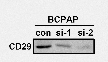
Application: Western BlotSample Tested: Tumor cell lysatesSpecies: HumanVerified Customer | Posted 03/20/2022
-
Application: Western BlotSample Tested: Mucociliary epitheliumSpecies: HumanVerified Customer | Posted 04/02/2020
-
Application: Western BlotSample Tested: Tumor cell lyastesSpecies: HumanVerified Customer | Posted 02/16/2020
-
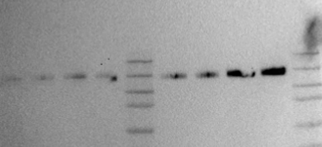
Application: Western BlotSample Tested: blood endothelial cellsSpecies: HumanVerified Customer | Posted 04/24/2018Worked in both reducing and non-reducing conditions Band seen around 130kDa Would use again on a new blot, not a stripped blot as got very similar band to that with alpha 5
There are no reviews that match your criteria.
Protocols
Find general support by application which include: protocols, troubleshooting, illustrated assays, videos and webinars.
- 7-Amino Actinomycin D (7-AAD) Cell Viability Flow Cytometry Protocol
- Appropriate Fixation of IHC/ICC Samples
- Cellular Response to Hypoxia Protocols
- ClariTSA™ Fluorophore Kits
- Detection & Visualization of Antibody Binding
- Extracellular Membrane Flow Cytometry Protocol
- Flow Cytometry Protocol for Cell Surface Markers
- Flow Cytometry Protocol for Staining Membrane Associated Proteins
- Flow Cytometry Staining Protocols
- Flow Cytometry Troubleshooting Guide
- ICC Cell Smear Protocol for Suspension Cells
- ICC Immunocytochemistry Protocol Videos
- ICC for Adherent Cells
- Immunocytochemistry (ICC) Protocol
- Immunocytochemistry Troubleshooting
- Immunofluorescence of Organoids Embedded in Cultrex Basement Membrane Extract
- Immunohistochemistry (IHC) and Immunocytochemistry (ICC) Protocols
- Immunoprecipitation Protocol
- Intracellular Flow Cytometry Protocol Using Alcohol (Methanol)
- Intracellular Flow Cytometry Protocol Using Detergents
- Intracellular Nuclear Staining Flow Cytometry Protocol Using Detergents
- Intracellular Staining Flow Cytometry Protocol Using Alcohol Permeabilization
- Intracellular Staining Flow Cytometry Protocol Using Detergents to Permeabilize Cells
- Preparing Samples for IHC/ICC Experiments
- Preventing Non-Specific Staining (Non-Specific Binding)
- Primary Antibody Selection & Optimization
- Propidium Iodide Cell Viability Flow Cytometry Protocol
- Protocol for Liperfluo
- Protocol for VisUCyte™ HRP Polymer Detection Reagent
- Protocol for the Characterization of Human Th22 Cells
- Protocol for the Characterization of Human Th9 Cells
- Protocol for the Fluorescent ICC Staining of Cell Smears - Graphic
- Protocol for the Fluorescent ICC Staining of Cultured Cells on Coverslips - Graphic
- Protocol for the Preparation and Fluorescent ICC Staining of Cells on Coverslips
- Protocol for the Preparation and Fluorescent ICC Staining of Non-adherent Cells
- Protocol for the Preparation and Fluorescent ICC Staining of Stem Cells on Coverslips
- Protocol for the Preparation of a Cell Smear for Non-adherent Cell ICC - Graphic
- Protocol: Annexin V and PI Staining by Flow Cytometry
- Protocol: Annexin V and PI Staining for Apoptosis by Flow Cytometry
- TUNEL and Active Caspase-3 Detection by IHC/ICC Protocol
- The Importance of IHC/ICC Controls
- Troubleshooting Guide: Fluorokine Flow Cytometry Kits
- View all Protocols, Troubleshooting, Illustrated assays and Webinars
Associated Pathways


