mGluR8 is a Group III metabotropic glutamate receptor. These 7-transmembrane glycoproteins are negatively coupled to adenylate cyclase and expressed presynaptically throughout the brain. mGluR8 shows pronounced expression in the dentate gyrus and CA3 regions of the hippocampus, and is thought to play a role in memory formation. Two splice forms, A and B, differ only in the C-terminal 16 amino acids (aa 893‑909), while splice form C diverges at aa 454 and is truncated prior to the first transmembrane segment. Within the N-terminal extracellular region used as an immunogen (aa 34‑514), human, mouse and rat mGluR8 share 98%‑99% aa identity.

Key Product Details
Species Reactivity
Human
Applications
Immunohistochemistry, Western Blot
Label
Unconjugated
Antibody Source
Monoclonal Mouse IgG2B Clone # 476410
Loading...
Product Specifications
Immunogen
Chinese hamster ovary cell line CHO-derived recombinant human mGluR8
Gln34-Ser514 (predicted)
Accession # O00222
Gln34-Ser514 (predicted)
Accession # O00222
Specificity
Detects human mGluR8 in direct ELISAs and Western blots. In Western blots, no cross-reactivity with recombinant human mGluR1, 2, 3, 4, 5, or 7 is observed.
Clonality
Monoclonal
Host
Mouse
Isotype
IgG2B
Scientific Data Images for Human mGluR8 Antibody (476410)
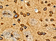
mGluR8 in Human Brain.
mGluR8 was detected in immersion fixed paraffin-embedded sections of human brain (hippocampus) using Mouse Anti-Human mGluR8 Monoclonal Antibody (Catalog # MAB5277) at 25 µg/mL overnight at 4 °C. Tissue was stained using the Anti-Mouse HRP-DAB Cell & Tissue Staining Kit (brown; Catalog # CTS002) and counterstained with hematoxylin (blue). View our protocol for Chromogenic IHC Staining of Paraffin-embedded Tissue Sections.Applications for Human mGluR8 Antibody (476410)
Application
Recommended Usage
Immunohistochemistry
8-25 µg/mL
Sample: Immersion fixed paraffin-embedded sections of human brain (hippocampus)
Sample: Immersion fixed paraffin-embedded sections of human brain (hippocampus)
Western Blot
1 µg/mL
Sample: Human mGluR8
Sample: Human mGluR8
Reviewed Applications
Read 1 review rated 5 using MAB5277 in the following applications:
Formulation, Preparation, and Storage
Purification
Protein A or G purified from hybridoma culture supernatant
Reconstitution
Reconstitute at 0.5 mg/mL in sterile PBS. For liquid material, refer to CoA for concentration.
Loading...
Formulation
Lyophilized from a 0.2 μm filtered solution in PBS with Trehalose. *Small pack size (SP) is supplied either lyophilized or as a 0.2 µm filtered solution in PBS.
Shipping
Lyophilized product is shipped at ambient temperature. Liquid small pack size (-SP) is shipped with polar packs. Upon receipt, store immediately at the temperature recommended below.
Stability & Storage
Use a manual defrost freezer and avoid repeated freeze-thaw cycles.
- 12 months from date of receipt, -20 to -70 °C as supplied.
- 1 month, 2 to 8 °C under sterile conditions after reconstitution.
- 6 months, -20 to -70 °C under sterile conditions after reconstitution.
Calculators
Background: mGluR8
Long Name
Metabotropic Glutamate Receptor 8
Alternate Names
GPRC1H, GRM8
Gene Symbol
GRM8
UniProt
Additional mGluR8 Products
Product Documents for Human mGluR8 Antibody (476410)
Certificate of Analysis
To download a Certificate of Analysis, please enter a lot or batch number in the search box below.
Note: Certificate of Analysis not available for kit components.
Product Specific Notices for Human mGluR8 Antibody (476410)
For research use only
Related Research Areas
Customer Reviews for Human mGluR8 Antibody (476410) (1)
5 out of 5
1 Customer Rating
Have you used Human mGluR8 Antibody (476410)?
Submit a review and receive an Amazon gift card!
$25/€18/£15/$25CAN/¥2500 Yen for a review with an image
$10/€7/£6/$10CAN/¥1110 Yen for a review without an image
Submit a review
Customer Images
Showing
1
-
1 of
1 review
Showing All
Filter By:
-
Application: ImmunohistochemistrySample Tested: Brain tissueSpecies: HumanVerified Customer | Posted 05/16/2022

There are no reviews that match your criteria.
Protocols
Find general support by application which include: protocols, troubleshooting, illustrated assays, videos and webinars.
- Antigen Retrieval Protocol (PIER)
- Antigen Retrieval for Frozen Sections Protocol
- Appropriate Fixation of IHC/ICC Samples
- Cellular Response to Hypoxia Protocols
- Chromogenic IHC Staining of Formalin-Fixed Paraffin-Embedded (FFPE) Tissue Protocol
- Chromogenic Immunohistochemistry Staining of Frozen Tissue
- ClariTSA™ Fluorophore Kits
- Detection & Visualization of Antibody Binding
- Fluorescent IHC Staining of Frozen Tissue Protocol
- Graphic Protocol for Heat-induced Epitope Retrieval
- Graphic Protocol for the Preparation and Fluorescent IHC Staining of Frozen Tissue Sections
- Graphic Protocol for the Preparation and Fluorescent IHC Staining of Paraffin-embedded Tissue Sections
- Graphic Protocol for the Preparation of Gelatin-coated Slides for Histological Tissue Sections
- IHC Sample Preparation (Frozen sections vs Paraffin)
- Immunofluorescent IHC Staining of Formalin-Fixed Paraffin-Embedded (FFPE) Tissue Protocol
- Immunohistochemistry (IHC) and Immunocytochemistry (ICC) Protocols
- Immunohistochemistry Frozen Troubleshooting
- Immunohistochemistry Paraffin Troubleshooting
- Preparing Samples for IHC/ICC Experiments
- Preventing Non-Specific Staining (Non-Specific Binding)
- Primary Antibody Selection & Optimization
- Protocol for Heat-Induced Epitope Retrieval (HIER)
- Protocol for Making a 4% Formaldehyde Solution in PBS
- Protocol for VisUCyte™ HRP Polymer Detection Reagent
- Protocol for the Preparation & Fixation of Cells on Coverslips
- Protocol for the Preparation and Chromogenic IHC Staining of Frozen Tissue Sections
- Protocol for the Preparation and Chromogenic IHC Staining of Frozen Tissue Sections - Graphic
- Protocol for the Preparation and Chromogenic IHC Staining of Paraffin-embedded Tissue Sections
- Protocol for the Preparation and Chromogenic IHC Staining of Paraffin-embedded Tissue Sections - Graphic
- Protocol for the Preparation and Fluorescent IHC Staining of Frozen Tissue Sections
- Protocol for the Preparation and Fluorescent IHC Staining of Paraffin-embedded Tissue Sections
- Protocol for the Preparation of Gelatin-coated Slides for Histological Tissue Sections
- R&D Systems Quality Control Western Blot Protocol
- TUNEL and Active Caspase-3 Detection by IHC/ICC Protocol
- The Importance of IHC/ICC Controls
- Troubleshooting Guide: Immunohistochemistry
- Troubleshooting Guide: Western Blot Figures
- Western Blot Conditions
- Western Blot Protocol
- Western Blot Protocol for Cell Lysates
- Western Blot Troubleshooting
- Western Blot Troubleshooting Guide
- View all Protocols, Troubleshooting, Illustrated assays and Webinars
Loading...
Associated Pathways
