Angiopoietin-2 (Ang-2; also ANGPT2) is a secreted glycoprotein that plays a complex role in angiogenesis and inflammation (1, 2). Mature Ang-2 is 478 amino acids (aa) in length. It contains one coiled-coil domain (aa 166 - 248) that mediates multimerization, and a C-terminal fibrinogen-like domain (aa 275 - 495) that mediates receptor binding. Under reducing conditions, secreted monomeric Ang-2 is 65 - 66 kDa in size. Under nonreducing conditions, both natural and recombinant Ang-2 form 140 kDa dimers, 200 kDa trimers, and 250 - 300 kDa tetramers and pentamers (3 - 6). Alternate splicing generates a short isoform that lacks 52 amino acids (aa) preceding the coiled-coil domain (4). Mature human Ang-2 shares 86% aa sequence identity with mouse and rat Ang-2. Ang-2 is widely expressed during development, but it is restricted postnatally to highly angiogenic tissues such as the placenta, ovaries, and uterus (3). It is particularly abundant in vascular endothelial cells (EC) where it is stored in intracellular Weibel-Palade bodies (1, 3, 7). Both Ang-2 and the related Angiopoietin-1 (Ang-1) are ligands for the receptor tyrosine kinase Tie-2 (2). While Ang-1 is a potent Tie-2 agonist, Ang-2 may act as either a Tie-2 antagonist or agonist, depending upon its state of multimerization. The higher the order of oligomer, the more effective Ang-2 becomes as a Tie-2 agonist (3, 8 - 11). The short isoform appears to block the binding of either Ang-1 or full-length Ang-2 to Tie-2 (4). Ang-2 functions as a pro-angiogenic factor, although it can also induce EC death and vessel regression (12, 13). Upon its release from quiescent EC, it regulates vascular remodeling by promoting EC survival, proliferation, and migration and destabilizing the interaction between EC and perivascular cells (8, 13, 14). Ang-2 is required for postnatal vascular remodeling, and it cooperates with Ang-1 during lymphatic vessel development (7, 15). It mediates the upregulation of ICAM-1 and VCAM-1 on EC, which facilitates the adhesion of leukocytes during inflammation (16). Ang-2 is upregulated in both the endothelium and tumor cells of several cancers as well as in ischemic tissue (17 - 20). Its direct interaction with Integrins promotes tumor cell invasion (21, 22). Ang-2 also promotes the neuronal differentiation and migration of subventricular zone progenitor cells (20).

Key Product Details
Species Reactivity
Validated:
Human, Mouse
Cited:
Human, Mouse
Applications
Validated:
Immunohistochemistry, ELISA Capture (Matched Antibody Pair)
Cited:
Immunohistochemistry, Immunohistochemistry-Paraffin, Western Blot, Immunocytochemistry, ELISA Capture
Label
Unconjugated
Antibody Source
Monoclonal Mouse IgG2A Clone # 85816
Loading...
Product Specifications
Immunogen
Mouse myeloma cell line NS0-derived recombinant human Angiopoietin-2
Asp68-Phe496
Accession # O15123
Asp68-Phe496
Accession # O15123
Specificity
Detects human and mouse Angiopoietin-2 in ELISAs. In ELISAs, no cross-reactivity or interference with recombinant human (rh) Ang‑1, rhAng-4, rhAng-X, recombinant mouse (rm) Ang-3, rmANGPTL-3, rhTie-1, rhTie-2, and rmTie-2 is observed.
Clonality
Monoclonal
Host
Mouse
Isotype
IgG2A
Scientific Data Images for Angiopoietin-2 Antibody (85816)
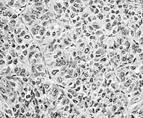
Angiopoietin‑2 in Human Hepatocellular Carcinoma Tissue.
Angiopoietin-2 was detected in immersion fixed paraffin-embedded sections of human hepatocellular carcinoma tissue (right panel; positive stain) using Mouse Anti-Human/Mouse Angiopoietin-2 Monoclonal Antibody (Catalog # MAB098) at 5 µg/mL for 1 hour at room temperature followed by incubation with the Anti-Mouse IgG VisUCyte™ HRP Polymer Antibody (Catalog # VC001). Before incubation with the primary antibody, tissue was subjected to heat-induced epitope retrieval using Antigen Retrieval Reagent-Basic (Catalog # CTS013). Tissue was stained using DAB (brown) and counterstained with hematoxylin (blue). Specific staining was localized to secreted and plasma membrane. View our protocol for IHC Staining with VisUCyte HRP Polymer Detection Reagents.
Angiopoietin-2 in Mouse Lymph Node Tissue.
Angiopoietin-2 was detected in perfusion fixed frozen sections of mouse lymph node tissue using Mouse Anti-Human/Mouse Angiopoietin-2 Monoclonal Antibody (Catalog # MAB098) at 1 µg/mL overnight at 4 °C. Before incubation with the primary antibody, tissue was subjected to heat-induced epitope retrieval using Antigen Retrieval Reagent-Basic (Catalog # CTS013). Tissue was stained using the Anti-Mouse IgG VisUCyte™ HRP Polymer Antibody (brown; Catalog # VC001) and counterstained with hematoxylin (blue). Specific staining was localized to cell membrane. View our protocol for IHC Staining with VisUCyte HRP Polymer Detection Reagents.
Angiopoietin-2 in Mouse Colon Tissue.
Angiopoietin-2 was detected in perfusion fixed frozen sections of mouse colon tissue using Mouse Anti-Human/Mouse Angiopoietin-2 Monoclonal Antibody (Catalog # MAB098) at 1 µg/mL overnight at 4 °C. Before incubation with the primary antibody, tissue was subjected to heat-induced epitope retrieval using Antigen Retrieval Reagent-Basic (Catalog # CTS013). Tissue was stained using the Anti-Mouse IgG VisUCyte™ HRP Polymer Antibody (brown; Catalog # VC001) and counterstained with hematoxylin (blue). Specific staining was localized to lymphocytes in colon mucosa. View our protocol for IHC Staining with VisUCyte HRP Polymer Detection Reagents.
Detection of Mouse Angiopoietin-2 by Western Blot
Sirt3 deficiency increases the hypoxic and oxidative state of the kidney and exacerbates ADR-induced oxidative stress.(A) Representative images and quantification of HIF-1 alpha expression in WT and Sirt3-/- mice after 7 weeks receiving saline or ADR (n = 4 mice for all groups except for n = 3 mice in Sirt3-/-+saline). (B) Representative Western Blot and quantification of Angpt-2 in WT and Sirt3-/- mice, treated with saline or ADR, (n = 4 mice for groups). (C) Representative images and quantification of nitrotyrosine (N-tyrosine) staining in WT and Sirt3-/- mice after 7 weeks receiving saline or ADR (n = 4 mice for all groups). (D) Representative Western Blot and quantification of acetylated SOD2 at lysine 68 (SOD2 AcK) in WT and Sirt3-/- mice, treated with saline or ADR (n = 4 mice for all groups). Data represent mean ± SEM and were analyzed by one-way ANOVA followed by Tukey’s multiple comparisons test. °P<0.05, °°P<0.01, and °°°P<0.001 vs WT+saline. *P<0.05, **P<0.01, and ***P<0.001 vs corresponding saline; #P<0.05, and ###P<0.001 vs WT+ADR. Scale bars, 20 μm. Image collected and cropped by CiteAb from the following open publication (https://pubmed.ncbi.nlm.nih.gov/37816025), licensed under a CC-BY license. Not internally tested by R&D Systems.Applications for Angiopoietin-2 Antibody (85816)
Application
Recommended Usage
Immunohistochemistry
1-25 µg/mL
Sample: Immersion fixed paraffin-embedded sections of human hepatocellular carcinoma tissue, perfusion fixed frozen sections of mouse lymph node tissue, and perfusion fixed frozen sections of mouse colon tissue
Sample: Immersion fixed paraffin-embedded sections of human hepatocellular carcinoma tissue, perfusion fixed frozen sections of mouse lymph node tissue, and perfusion fixed frozen sections of mouse colon tissue
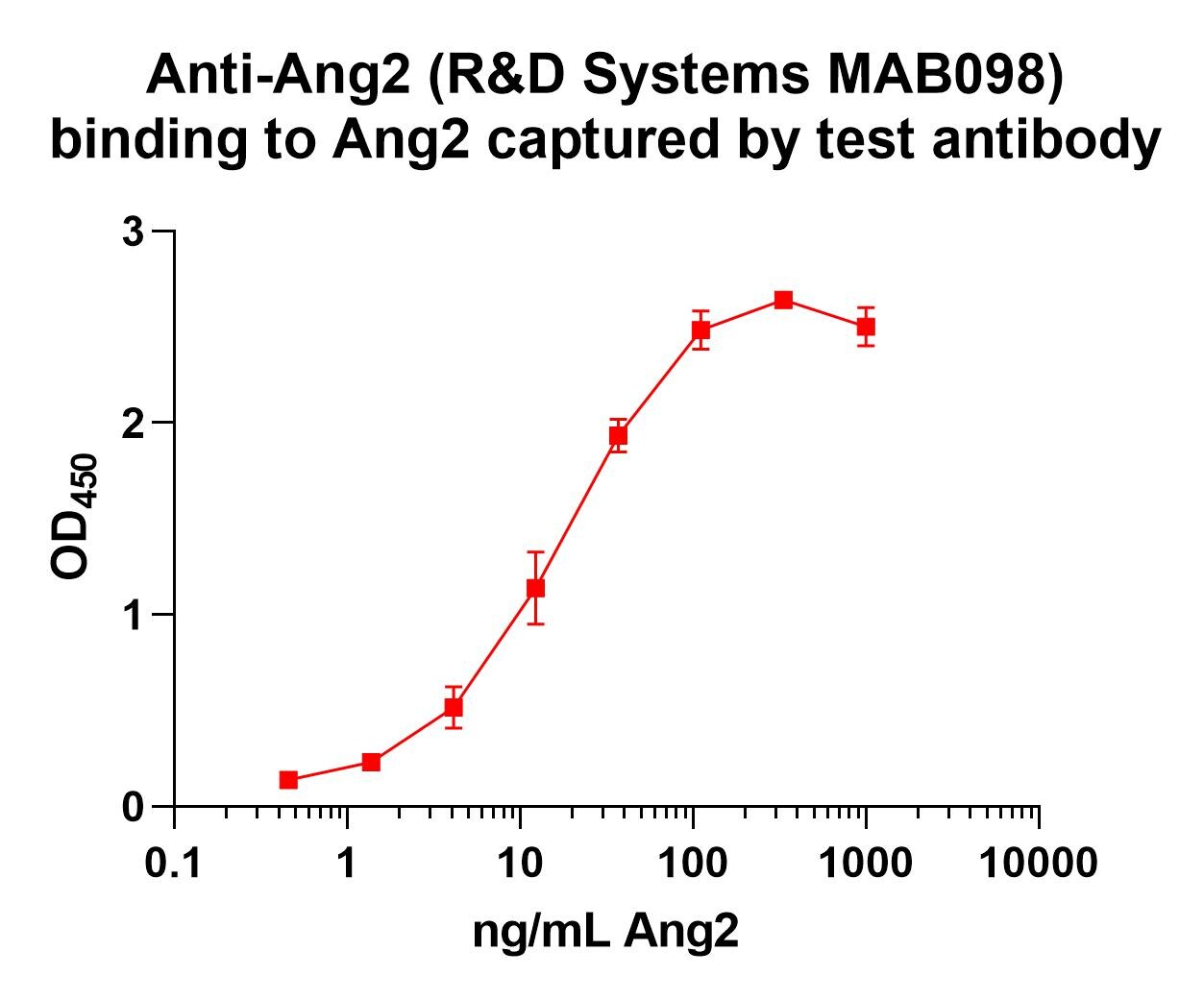
Human Angiopoietin-2 Sandwich Immunoassay
Please Note: Optimal dilutions of this antibody should be experimentally determined.
Reviewed Applications
Read 6 reviews rated 4.3 using MAB098 in the following applications:
Formulation, Preparation, and Storage
Purification
Protein A or G purified from hybridoma culture supernatant
Reconstitution
Reconstitute at 0.5 mg/mL in sterile PBS. For liquid material, refer to CoA for concentration.
Loading...
Formulation
Lyophilized from a 0.2 μm filtered solution in PBS with Trehalose. *Small pack size (SP) is supplied either lyophilized or as a 0.2 µm filtered solution in PBS.
Shipping
Lyophilized product is shipped at ambient temperature. Liquid small pack size (-SP) is shipped with polar packs. Upon receipt, store immediately at the temperature recommended below.
Stability & Storage
Use a manual defrost freezer and avoid repeated freeze-thaw cycles.
- 12 months from date of receipt, -20 to -70 °C as supplied.
- 1 month, 2 to 8 °C under sterile conditions after reconstitution.
- 6 months, -20 to -70 °C under sterile conditions after reconstitution.
Calculators
Background: Angiopoietin-2
References
- Augustin, H.G. et al. (2009) Nat. Rev. Mol. Cell Biol. 10:165.
- Murdoch, C. et al. (2007) J. Immunol. 178:7405.
- Maisonpierre, P.C. et al. (1997) Science 27:55.
- Kim, I. et al. (2000) J. Biol. Chem. 275:18550.
- Procopio, W.N. et al. (1999) J. Biol. Chem. 274:30196.
- Kim, K-T. et al. (2005) J. Biol. Chem. 280:20126.
- Gale, N.W. et al. (2002) Dev. Cell 3:411.
- Yuan, H.T. et al. (2009) Mol. Cell. Biol. 29:2011.
- Falcon, B.L. et al. (2009) Am. J. Pathol. 175:2159.
- Kim, H-Z. et al. (2009) Biochim. Biophys. Acta 1793:772.
- Kim, I. et al. (2001) Cardiovasc. Res. 49:872.
- Lobov, I.B. et al. (2002) Proc. Natl. Acad. Sci. 99:11205.
- Cao, Y. et al. (2007) Cancer Res. 67:3835.
- Nasarre, P. et al. (2009) Cancer Res. 69:1324.
- Dellinger, M. et al. (2008) Dev. Biol. 319:309.
- Fiedler, U. et al. (2006) Nat. Med. 12:235.
- Koga, K. et al. (2001) Cancer Res. 61:6248.
- Etoh, T. et al. (2001) Cancer Res. 61:2145.
- Tressel, S.L. et al. (2008) Arterioscler. Thromb. Vasc. Biol. 28:1989.
- Liu, X.S. et al. (2009) J. Biol. Chem. 284:22680.
- Hu, B. et al. (2006) Cancer Res. 66:775.
- Imanishi, Y. et al. (2007) Cancer Res. 67:4254.
Alternate Names
ANGPT2
Entrez Gene IDs
Gene Symbol
ANGPT2
UniProt
Additional Angiopoietin-2 Products
Product Documents for Angiopoietin-2 Antibody (85816)
Certificate of Analysis
To download a Certificate of Analysis, please enter a lot or batch number in the search box below.
Note: Certificate of Analysis not available for kit components.
Product Specific Notices for Angiopoietin-2 Antibody (85816)
For research use only
Citations for Angiopoietin-2 Antibody (85816)
 Powered by Bioz
Customer Reviews for Angiopoietin-2 Antibody (85816) (6)
4.3 out of 5
6 Customer Ratings
Have you used Angiopoietin-2 Antibody (85816)?
Submit a review and receive an Amazon gift card!
$25/€18/£15/$25CAN/¥2500 Yen for a review with an image
$10/€7/£6/$10CAN/¥1110 Yen for a review without an image
Submit a review
Customer Images
Showing
1
-
5 of
6 reviews
Showing All
Filter By:
-
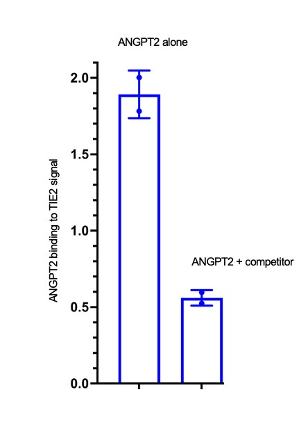
Application: ELISASample Tested: HUVEC human umbilical vein endothelial cellsSpecies: HumanVerified Customer | Posted 04/10/2024I used the antibody to do ELISA,by checking how my compound competes with 623-AN binding to TIE2. It works well, though I prefer AF623.

-
Application: ImmunohistochemistrySample Tested: Liver cancer and Liver cancer tissueSpecies: HumanVerified Customer | Posted 09/03/2021
-
Application: ELISASample Tested: Recombinant proteinSpecies: HumanVerified Customer | Posted 02/11/2021
-
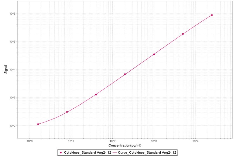
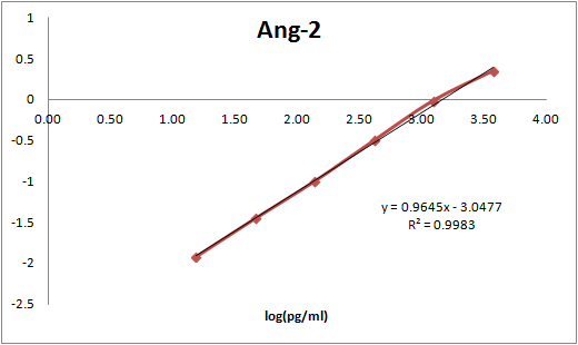
Application: MSD assaySample Tested: Vitreous humorSpecies: Cynomolgus MonkeyVerified Customer | Posted 10/30/2018After biotinylation, used as a capture reagent according to the manufacturer’s protocol (Meso Scale Diagnostics LLC). Paired with SulfoTag-modified MAB0984 as a detection antibody. Calibration curve with Recombinant Human Angiopoietin (623-AN-025/CF) is shown (dynamic range 16-250,000 pg/ml).
-
Application: ELISASample Tested: Recombinant proteinSpecies: HumanVerified Customer | Posted 12/06/2017
-
Application: ELISASample Tested: Serum and PlasmaSpecies: HumanVerified Customer | Posted 11/16/2017This antibody was paired with BAM0981 to build an ELISA to measure ANG-2 in human serum samples.
There are no reviews that match your criteria.
Protocols
Find general support by application which include: protocols, troubleshooting, illustrated assays, videos and webinars.
- Antigen Retrieval Protocol (PIER)
- Antigen Retrieval for Frozen Sections Protocol
- Appropriate Fixation of IHC/ICC Samples
- Cellular Response to Hypoxia Protocols
- Chromogenic IHC Staining of Formalin-Fixed Paraffin-Embedded (FFPE) Tissue Protocol
- Chromogenic Immunohistochemistry Staining of Frozen Tissue
- ClariTSA™ Fluorophore Kits
- Detection & Visualization of Antibody Binding
- Fluorescent IHC Staining of Frozen Tissue Protocol
- Graphic Protocol for Heat-induced Epitope Retrieval
- Graphic Protocol for the Preparation and Fluorescent IHC Staining of Frozen Tissue Sections
- Graphic Protocol for the Preparation and Fluorescent IHC Staining of Paraffin-embedded Tissue Sections
- Graphic Protocol for the Preparation of Gelatin-coated Slides for Histological Tissue Sections
- IHC Sample Preparation (Frozen sections vs Paraffin)
- Immunofluorescent IHC Staining of Formalin-Fixed Paraffin-Embedded (FFPE) Tissue Protocol
- Immunohistochemistry (IHC) and Immunocytochemistry (ICC) Protocols
- Immunohistochemistry Frozen Troubleshooting
- Immunohistochemistry Paraffin Troubleshooting
- Preparing Samples for IHC/ICC Experiments
- Preventing Non-Specific Staining (Non-Specific Binding)
- Primary Antibody Selection & Optimization
- Protocol for Heat-Induced Epitope Retrieval (HIER)
- Protocol for Making a 4% Formaldehyde Solution in PBS
- Protocol for VisUCyte™ HRP Polymer Detection Reagent
- Protocol for the Preparation & Fixation of Cells on Coverslips
- Protocol for the Preparation and Chromogenic IHC Staining of Frozen Tissue Sections
- Protocol for the Preparation and Chromogenic IHC Staining of Frozen Tissue Sections - Graphic
- Protocol for the Preparation and Chromogenic IHC Staining of Paraffin-embedded Tissue Sections
- Protocol for the Preparation and Chromogenic IHC Staining of Paraffin-embedded Tissue Sections - Graphic
- Protocol for the Preparation and Fluorescent IHC Staining of Frozen Tissue Sections
- Protocol for the Preparation and Fluorescent IHC Staining of Paraffin-embedded Tissue Sections
- Protocol for the Preparation of Gelatin-coated Slides for Histological Tissue Sections
- TUNEL and Active Caspase-3 Detection by IHC/ICC Protocol
- The Importance of IHC/ICC Controls
- Troubleshooting Guide: Immunohistochemistry
- View all Protocols, Troubleshooting, Illustrated assays and Webinars
Loading...