ATG7, also known as APG7, is a 78 kDa cytosolic ubiquitin-E1-like enzyme that plays a key role in the autophagic pathway of intracellular bulk degradation. It is required for the conjugation of ATG5 to ATG12, the lipidation of ATG8, and subsequent autophagosome formation. ATG7 is required for mitochondrial removal during erythropoiesis and for the maintenance of axonal homeostasis. Alternate splicing of human ATG7 generates an isoform that lacks 31 aa at the C-terminus, a region that is required for ATG8 lipidation. Human ATG7 shares 93% aa sequence identitiy with mouse and rat ATG7.

Key Product Details
Validated by
Biological Validation
Species Reactivity
Validated:
Human, Mouse
Cited:
Human, Mouse, Rat
Applications
Validated:
Immunohistochemistry, Western Blot, Intracellular Staining by Flow Cytometry, Immunocytochemistry, Simple Western, CyTOF-ready
Cited:
Immunohistochemistry, Western Blot, Flow Cytometry
Label
Unconjugated
Antibody Source
Monoclonal Mouse IgG1 Clone # 683906
Loading...
Product Specifications
Immunogen
E. coli-derived recombinant human ATG7
Asn387-Gln570
Accession # O95352
Asn387-Gln570
Accession # O95352
Specificity
Detects human ATG7 in direct ELISAs and Western blots.
In direct ELISAs and Western blots, no
cross-reactivity with recombinant human ATG3, 4A, 4B, 5, 10, 12, or 16L1 is
observed.
Clonality
Monoclonal
Host
Mouse
Isotype
IgG1
Scientific Data Images for ATG7 Antibody (683906)
Detection of Human ATG7 by Western Blot.
Western blot shows lysates of HeLa human cervical epithelial carcinoma cell line and HepG2 human hepatocellular carcinoma cell line. PVDF membrane was probed with 2 µg/mL of Mouse Anti-Human ATG7 Monoclonal Antibody (Catalog # MAB6608) followed by HRP-conjugated Anti-Mouse IgG Secondary Antibody (Catalog # HAF007). A specific band was detected for ATG7 at approximately 75 kDa (as indicated). This experiment was conducted under reducing conditions and using Immunoblot Buffer Group 1.
ATG7 in RAW 264.7 Mouse Cell Line.
ATG7 was detected in immersion fixed RAW 264.7 mouse monocyte/macrophage cell line using Mouse Anti-Human ATG7 Monoclonal Antibody (Catalog # MAB6608) at 25 µg/mL for 3 hours at room temperature. Cells were stained using the Northern-Lights™ 557-conjugated Anti-Mouse IgG Secondary Antibody (red; Catalog # NL007) and counterstained with DAPI (blue). Specific staining was localized to autophagosomes. View our protocol for Fluorescent ICC Staining of Non-adherent Cells.
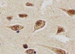
ATG7 in Human Brain.
ATG7 was detected in formalin fixed paraffin-embedded sections of human brain (cortex) using Mouse Anti-Human ATG7 Monoclonal Antibody (Catalog # MAB6608) at 15 µg/mL overnight at 4 °C. Before incubation with the primary antibody, tissue was subjected to heat-induced epitope retrieval using Antigen Retrieval Reagent-Basic (Catalog # CTS013). Tissue was stained using the Anti-Mouse HRP-DAB Cell & Tissue Staining Kit (brown; Catalog # CTS002) and counter-stained with hematoxylin (blue). Specific staining was localized to neuronal cell bodies and processes. View our protocol for Chromogenic IHC Staining of Paraffin-embedded Tissue Sections.
Detection of ATG7 in HeLa Human Cell Line by Flow Cytometry.
HeLa human cervical epithelial carcinoma cell line was stained with Mouse Anti-Human/Mouse ATG7 Monoclonal Antibody (Catalog # MAB6608, filled histogram) or isotype control antibody (Catalog # MAB002, open histogram), followed by Allophycocyanin-conjugated Anti-Mouse IgG Secondary Antibody (Catalog # F0101B). To facilitate intracellular staining, cells were fixed with Flow Cytometry Fixation Buffer (Catalog # FC004) and permeabilized with Flow Cytometry Permeabilization/Wash Buffer I (Catalog # FC005). View our protocol for Staining Intracellular Molecules.
Detection of Human ATG7 by Simple WesternTM.
Simple Western lane view shows lysates of HeLa human cervical epithelial carcinoma cell line, loaded at 0.2 mg/mL. A specific band was detected for ATG7 at approximately 72 kDa (as indicated) using 20 µg/mL of Mouse Anti-Human/Mouse ATG7 Monoclonal Antibody (Catalog # MAB6608). This experiment was conducted under reducing conditions and using the 12-230 kDa separation system.
Detection of Human ATG7 by Western Blot
ATG5 and ATG7 enhanced autophagy and inhibited ER stress in chondrocyte. a Western blotting analysis of LC3, P62, ATG5, ATG7 and ATG5-ATG12 expression after infected with Ad-ATG5, Ad-ATG7 and Ad-ATG5 + Ad-ATG7 in the C28I2 cells. beta -actin is served as an internal control. b Qualitative analysis of ATG5, ATG7, ATG5-ATG12, LC3 and P62. The values were normalized to beta -actin. c C28I2 cells were double stained with LC3 (red) and DAPI (blue) and visualized by confocal microscopy (400X) after treated with Rapamycine, Ad-ATG5, Ad-ATG7 and Ad-ATG5 + Ad-ATG7 24 h. *P < 0.05, **P < 0.01 compared with the controls. Values are means ± SD n = 3). d Qualitative analysis of LC3 fluorescence intensity of chondrocytes. The values were normalized to the NC group. e Western blotting analysis of PERK, p-PERK and Nrf2 expression after infected with Ad-ATG5, Ad-ATG7 and Ad-ATG5 + Ad-ATG7 in the C28I2 cells. beta -actin is served as an internal control. f Qualitative analysis of PERK, p-PERK and Nrf2 were normalized to beta -actin. (1:NC, 2:Ad-GFP, 3:Ad-ATG5, 4:RAPA, 5:Ad-ATG7, 6:Ad-ATG5 + RAPA, 7:Ad-ATG5 + Ad-ATG7). Rapamycin (25 μM) used as a positive control Image collected and cropped by CiteAb from the following publication (https://pubmed.ncbi.nlm.nih.gov/31060556), licensed under a CC-BY license. Not internally tested by R&D Systems.
Detection of Human ATG7 by Western Blot
Autophagy regulates the loading of LG3 on apoptotic exosome‐like vesicles (ApoExos).A Representative immunoblots and densitometric analysis of LC3, ATG7, phospho-AKT1, and total AKT1 from serum-starved (SS) HUVECs transfected with siCtrl or siATG7 or exposed to vehicle (V), wortmannin (100 nM; Wort), or bafilomycin A1 (20 nM, Baf). beta -actin (ACTB) was used as a loading control. n = 3 for each condition. B HO/PI staining of apoptosis in SS HUVECs transfected with siCtrl or siATG7 or exposed to V, Baf, or Wort. n = 3–5 for each condition. ns (nonsignificant). C Immunoblots and densitometric analysis of cleaved caspase-3 in SS HUVECs transfected with siCtrl or siATG7 or exposed to V, Baf, or Wort. beta -actin (ACTB) was used as a loading control. n = 3–5 for each condition. D Small particle flow cytometric quantifications of CMFDA+ AnnexinV+ ApoExos detected in the supernatant of HUVECs serum starved (SS) for 4 h and transfected with siCtrl or siATG7 or exposed to V, Baf, or Wort. n = 3 for each condition. E Representative immunoblots and densitometric analysis of LG3, 20S proteasome, LAMP2, and synthenin-1 in ApoExos purified from HUVECs serum starved (SS) for 4 h and transfected with siCtrl or siATG7 or exposed to V, Baf, or Wort. Ponceau red was used as a loading control. n = 3–5 for each condition. P values were obtained by the unpaired t test (*P < 0.05; **P < 0.01; ***P < 0.001). All values are expressed as the mean ± SEM. Image collected and cropped by CiteAb from the following open publication (https://pubmed.ncbi.nlm.nih.gov/35149669), licensed under a CC-BY license. Not internally tested by R&D Systems.Applications for ATG7 Antibody (683906)
Application
Recommended Usage
CyTOF-ready
Ready to be labeled using established conjugation methods. No BSA or other carrier proteins that could interfere with conjugation.
Immunocytochemistry
8-25 µg/mL
Sample: Immersion fixed RAW 264.7 mouse monocyte/macrophage cell line
Sample: Immersion fixed RAW 264.7 mouse monocyte/macrophage cell line
Immunohistochemistry
8-25 µg/mL
Sample: Immersion fixed paraffin-embedded sections of human brain (cortex)
Sample: Immersion fixed paraffin-embedded sections of human brain (cortex)
Intracellular Staining by Flow Cytometry
0.25 µg/106 cells
Sample: HeLa human cervical epithelial carcinoma cell line fixed with Flow Cytometry Fixation Buffer (Catalog # FC004) and permeabilized with Flow Cytometry Permeabilization/Wash Buffer I (Catalog # FC005)
Sample: HeLa human cervical epithelial carcinoma cell line fixed with Flow Cytometry Fixation Buffer (Catalog # FC004) and permeabilized with Flow Cytometry Permeabilization/Wash Buffer I (Catalog # FC005)
Simple Western
20 µg/mL
Sample: HeLa human cervical epithelial carcinoma cell line
Sample: HeLa human cervical epithelial carcinoma cell line
Western Blot
2 µg/mL
Sample: HeLa human cervical epithelial carcinoma cell line and HepG2 human hepatocellular carcinoma cell line
Sample: HeLa human cervical epithelial carcinoma cell line and HepG2 human hepatocellular carcinoma cell line
Reviewed Applications
Read 3 reviews rated 5 using MAB6608 in the following applications:
Flow Cytometry Panel Builder
Bio-Techne Knows Flow Cytometry
Save time and reduce costly mistakes by quickly finding compatible reagents using the Panel Builder Tool.
Advanced Features
- Spectra Viewer - Custom analysis of spectra from multiple fluorochromes
- Spillover Popups - Visualize the spectra of individual fluorochromes
- Antigen Density Selector - Match fluorochrome brightness with antigen density

Formulation, Preparation, and Storage
Purification
Protein A or G purified from hybridoma culture supernatant
Reconstitution
Sterile PBS to a final concentration of 0.5 mg/mL. For liquid material, refer to CoA for concentration.
Loading...
Formulation
Lyophilized from a 0.2 μm filtered solution in PBS with Trehalose. *Small pack size (SP) is supplied either lyophilized or as a 0.2 µm filtered solution in PBS.
Shipping
Lyophilized product is shipped at ambient temperature. Liquid small pack size (-SP) is shipped with polar packs. Upon receipt, store immediately at the temperature recommended below.
Stability & Storage
Use a manual defrost freezer and avoid repeated freeze-thaw cycles.
- 12 months from date of receipt, -20 to -70 °C as supplied.
- 1 month, 2 to 8 °C under sterile conditions after reconstitution.
- 6 months, -20 to -70 °C under sterile conditions after reconstitution.
Calculators
Background: ATG7
Long Name
ATG7 Autophagy Related 7 Homolog
Alternate Names
APG7, APG7L, GSA7
Gene Symbol
ATG7
UniProt
Additional ATG7 Products
Product Documents for ATG7 Antibody (683906)
Certificate of Analysis
To download a Certificate of Analysis, please enter a lot or batch number in the search box below.
Note: Certificate of Analysis not available for kit components.
Product Specific Notices for ATG7 Antibody (683906)
For research use only
Related Research Areas
Citations for ATG7 Antibody (683906)
 Powered by Bioz
Customer Reviews for ATG7 Antibody (683906) (3)
5 out of 5
3 Customer Ratings
Have you used ATG7 Antibody (683906)?
Submit a review and receive an Amazon gift card!
$25/€18/£15/$25CAN/¥2500 Yen for a review with an image
$10/€7/£6/$10CAN/¥1110 Yen for a review without an image
Submit a review
Customer Images
Showing
1
-
3 of
3 reviews
Showing All
Filter By:
-
Application: ImmunohistochemistrySample Tested: Brain tissueSpecies: MouseVerified Customer | Posted 03/23/2022

-
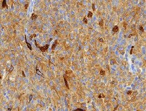
Application: ImmunohistochemistrySample Tested: Ocular melanomaSpecies: HumanVerified Customer | Posted 09/07/2021
-
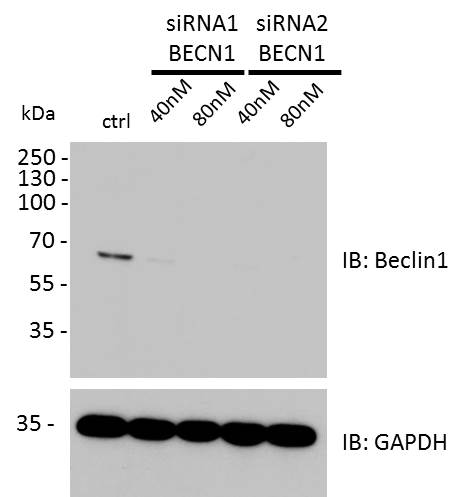
Application: Western BlotSample Tested: Human fibroblast cell lineSpecies: HumanVerified Customer | Posted 07/12/2017ctrl (scrambled-transfected) and Beclin1 siRNA-transfected cells (2 different siRNAs). IB using Beclin1 (1:1000). Beclin1 siRNA-transfected cells do not show a signal for Beclin1, whereas control cells show a signal between 55 and 70 kDa.for WB: blocking 1 h 5% skimmed milk in TBST; Ab o/n @ 4 °C 1:1000 in TBST. Beclin1-specific siRNA-transfected cells do not show a signal.
There are no reviews that match your criteria.
Protocols
Find general support by application which include: protocols, troubleshooting, illustrated assays, videos and webinars.
- 7-Amino Actinomycin D (7-AAD) Cell Viability Flow Cytometry Protocol
- Antigen Retrieval Protocol (PIER)
- Antigen Retrieval for Frozen Sections Protocol
- Appropriate Fixation of IHC/ICC Samples
- Cellular Response to Hypoxia Protocols
- Chromogenic IHC Staining of Formalin-Fixed Paraffin-Embedded (FFPE) Tissue Protocol
- Chromogenic Immunohistochemistry Staining of Frozen Tissue
- ClariTSA™ Fluorophore Kits
- Detection & Visualization of Antibody Binding
- Extracellular Membrane Flow Cytometry Protocol
- Flow Cytometry Protocol for Cell Surface Markers
- Flow Cytometry Protocol for Staining Membrane Associated Proteins
- Flow Cytometry Staining Protocols
- Flow Cytometry Troubleshooting Guide
- Fluorescent IHC Staining of Frozen Tissue Protocol
- Graphic Protocol for Heat-induced Epitope Retrieval
- Graphic Protocol for the Preparation and Fluorescent IHC Staining of Frozen Tissue Sections
- Graphic Protocol for the Preparation and Fluorescent IHC Staining of Paraffin-embedded Tissue Sections
- Graphic Protocol for the Preparation of Gelatin-coated Slides for Histological Tissue Sections
- ICC Cell Smear Protocol for Suspension Cells
- ICC Immunocytochemistry Protocol Videos
- ICC for Adherent Cells
- IHC Sample Preparation (Frozen sections vs Paraffin)
- Immunocytochemistry (ICC) Protocol
- Immunocytochemistry Troubleshooting
- Immunofluorescence of Organoids Embedded in Cultrex Basement Membrane Extract
- Immunofluorescent IHC Staining of Formalin-Fixed Paraffin-Embedded (FFPE) Tissue Protocol
- Immunohistochemistry (IHC) and Immunocytochemistry (ICC) Protocols
- Immunohistochemistry Frozen Troubleshooting
- Immunohistochemistry Paraffin Troubleshooting
- Intracellular Flow Cytometry Protocol Using Alcohol (Methanol)
- Intracellular Flow Cytometry Protocol Using Detergents
- Intracellular Nuclear Staining Flow Cytometry Protocol Using Detergents
- Intracellular Staining Flow Cytometry Protocol Using Alcohol Permeabilization
- Intracellular Staining Flow Cytometry Protocol Using Detergents to Permeabilize Cells
- Preparing Samples for IHC/ICC Experiments
- Preventing Non-Specific Staining (Non-Specific Binding)
- Primary Antibody Selection & Optimization
- Propidium Iodide Cell Viability Flow Cytometry Protocol
- Protocol for Heat-Induced Epitope Retrieval (HIER)
- Protocol for Liperfluo
- Protocol for Making a 4% Formaldehyde Solution in PBS
- Protocol for VisUCyte™ HRP Polymer Detection Reagent
- Protocol for the Characterization of Human Th22 Cells
- Protocol for the Characterization of Human Th9 Cells
- Protocol for the Fluorescent ICC Staining of Cell Smears - Graphic
- Protocol for the Fluorescent ICC Staining of Cultured Cells on Coverslips - Graphic
- Protocol for the Preparation & Fixation of Cells on Coverslips
- Protocol for the Preparation and Chromogenic IHC Staining of Frozen Tissue Sections
- Protocol for the Preparation and Chromogenic IHC Staining of Frozen Tissue Sections - Graphic
- Protocol for the Preparation and Chromogenic IHC Staining of Paraffin-embedded Tissue Sections
- Protocol for the Preparation and Chromogenic IHC Staining of Paraffin-embedded Tissue Sections - Graphic
- Protocol for the Preparation and Fluorescent ICC Staining of Cells on Coverslips
- Protocol for the Preparation and Fluorescent ICC Staining of Non-adherent Cells
- Protocol for the Preparation and Fluorescent ICC Staining of Stem Cells on Coverslips
- Protocol for the Preparation and Fluorescent IHC Staining of Frozen Tissue Sections
- Protocol for the Preparation and Fluorescent IHC Staining of Paraffin-embedded Tissue Sections
- Protocol for the Preparation of Gelatin-coated Slides for Histological Tissue Sections
- Protocol for the Preparation of a Cell Smear for Non-adherent Cell ICC - Graphic
- Protocol: Annexin V and PI Staining by Flow Cytometry
- Protocol: Annexin V and PI Staining for Apoptosis by Flow Cytometry
- R&D Systems Quality Control Western Blot Protocol
- TUNEL and Active Caspase-3 Detection by IHC/ICC Protocol
- The Importance of IHC/ICC Controls
- Troubleshooting Guide: Fluorokine Flow Cytometry Kits
- Troubleshooting Guide: Immunohistochemistry
- Troubleshooting Guide: Western Blot Figures
- Western Blot Conditions
- Western Blot Protocol
- Western Blot Protocol for Cell Lysates
- Western Blot Troubleshooting
- Western Blot Troubleshooting Guide
- View all Protocols, Troubleshooting, Illustrated assays and Webinars
Loading...