Cleaved Caspase-3 (Asp175) Antibody (269518)
R&D Systems | Catalog # MAB835
 by Western Blot.")
Key Product Details
Validated by
Biological Validation
Species Reactivity
Validated:
Human, Mouse
Cited:
Human, Mouse, Rat, Porcine, Transgenic Mouse, Xenograft
Applications
Validated:
Immunohistochemistry, Western Blot, Intracellular Staining by Flow Cytometry, Immunocytochemistry
Cited:
Immunohistochemistry, Immunohistochemistry-Paraffin, Immunohistochemistry-Frozen, Western Blot, Flow Cytometry, Immunofluorescence, Immunocytochemistry, Immunocytochemistry/ Immunofluorescence, Dot Blot, IHC Paraffin-embedded, Westen Blot
Label
Unconjugated
Antibody Source
Monoclonal Rabbit IgG Clone # 269518
Loading...
Product Specifications
Immunogen
KLH-conjugated human Caspase-3 synthetic peptide
CRGTELDCGIETD
Accession # U26943
CRGTELDCGIETD
Accession # U26943
Specificity
Detects human and mouse Caspase-3 cleaved at Asp175. No cross-reactivity was detected with the full-length procaspase-3 or other caspases.
Clonality
Monoclonal
Host
Rabbit
Isotype
IgG
Scientific Data Images for Cleaved Caspase-3 (Asp175) Antibody (269518)
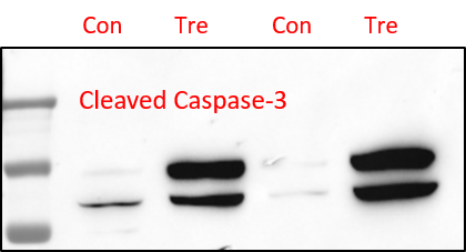
Detection of Human and Mouse Cleaved Caspase‑3 (Asp175) by Western Blot.
Western blot shows lysates of Jurkat human acute T cell leukemia cell line and DA3 mouse myeloma cell line untreated (-) or treated (+) with 1 µM staurosporine (STS) for the indicated times. PVDF membrane was probed with 0.5 µg/mL of Human/Mouse Cleaved Caspase-3 (Asp175) Monoclonal Antibody (MAB835), followed by HRP-conjugated Anti-Rabbit IgG Secondary Antibody (Catalog # HAF008). A specific band was detected for Cleaved Caspase-3 (Asp175) at approximately 18 kDa (as indicated). This experiment was conducted under reducing conditions and using Immunoblot Buffer Group 3.
Caspase‑3 in Jurkat Human Cell Line.
Caspase-3 was detected in immersion fixed Jurkat human acute T cell leukemia cell line treated with staurosporin using Human/Mouse Cleaved Caspase-3 (Asp175) Monoclonal Antibody (Catalog # MAB835) at 10 µg/mL for 3 hours at room temperature. Cells were stained using the NorthernLights™ 557-conjugated Anti-Rabbit IgG Secondary Antibody (red; Catalog # NL004) and counterstained with DAPI (blue). View our protocol for Fluorescent ICC Staining of Non-adherent Cells.
Caspase‑3 in Human Colon Cancer Tissue.
Caspase-3 was detected in immersion fixed paraffin-embedded sections of human colon cancer tissue using Rabbit Anti-Human/Mouse Cleaved Caspase-3 (Asp175) Monoclonal Antibody (Catalog # MAB835) at 0.3 µg/mL for 1 hour at room temperature followed by incubation with the Anti-Rabbit IgG VisUCyte™ HRP Polymer Antibody (Catalog # VC003). Before incubation with the primary antibody, tissue was subjected to heat-induced epitope retrieval using Antigen Retrieval Reagent-Basic (Catalog # CTS013). Tissue was stained using DAB (brown) and counterstained with hematoxylin (blue). Specific staining was localized to cytoplasm. View our protocol for IHC Staining with VisUCyte HRP Polymer Detection Reagents.
Detection of Cleaved Caspase‑3 in Jurkat Human Cell Line by Flow Cytometry.
Jurkat human acute T cell leukemia cell line untreated (open histogram) or treated with 3 µM Staurosporine for 3 hours (filled histogram) was stained with Rabbit Anti-Human/Mouse Caspase-3 Monoclonal Antibody (Catalog # MAB835, filled histogram) followed by anti-Rabbit IgG FITC-conjugated secondary antibody (Catalog # F0112). To facilitate intracellular staining, cells were fixed with Flow Cytometry Fixation Buffer (Catalog # FC004) and permeabilized with 90% methanol. View our protocol for Staining Intracellular Molecules.
Detection of Human Caspase-3 by Immunocytochemistry/Immunofluorescence
Caspase-3 activation and actin cytoskeletal organization in melanoma A375 cells cultured in the presence of PFII.Cells were grown on glass coverslips in the presence of 65, 130, 260 µM or 1 µM staurosporine (STS). (A–E) Active caspase-3 was visualized with anti-active caspase-3 antibody followed by a FITC-conjugated secondary antibody (green). (F–J) Actin was visualized using laser scanning confocal microscope (LSCM) after staining with Alexa Fluor 568 - conjugated phalloidin (red). Scale bar - 50 µm. Image collected and cropped by CiteAb from the following publication (https://dx.plos.org/10.1371/journal.pone.0057991), licensed under a CC-BY license. Not internally tested by R&D Systems.
Detection of Mouse Caspase-3 by Western Blot
Epidermis-specific deletion of Rac1 increases UV-light-induced keratinocyte apoptosis in vivo. (a) H/E staining of UV-irradiated skin of Rac1 fl/fl and Rac1-EKO mice at 12 h after UV-irradiation. Black arrows indicate sunburn cells. (b) Graph shows the percentage number of sunburn cells at 12 h with (red bars) or without (blue bars) UV-irradiation in Rac1 fl/fl (n=4) and Rac1-EKO (n=5) mice. The percentage of sunburn cells within the epidermis after UV-irradiation in Rac1 fl/fl mice was 3.4%, whereas in Rac1-EKO mice it was 8.6% of total epidermal keratinocytes. Non-irradiated samples showed <1% sunburn cells in both the genotypes. (c) Immunostainings against cleaved caspase-3 (green) of UV-irradiated skin of Rac1 fl/fl and Rac1-EKO mice at 12 h after UV-irradiation. Nuclei are stained in blue. Scale bar=100 μm. (d) Western blot analysis of cleaved caspase-3 from epidermal lysates of untreated (no UV) and UV-light treated (UV) Rac1 fl/fl and Rac1-EKO mice. Non-irradiated controls showed no bands for cleaved capsase-3, whereas samples of irradiated epidermis showed cleaved caspase-3-specific bands at ~25 kDa and 23 kDa. Numbers on the left denote molecular weights in kDa. (e) Graph shows densitometry analysis of western blot in (d). Error bars show S.D. Asterisks show P-value<0.001 Image collected and cropped by CiteAb from the following publication (https://pubmed.ncbi.nlm.nih.gov/28277539), licensed under a CC-BY license. Not internally tested by R&D Systems.
Detection of Human Caspase-3 by Immunocytochemistry/Immunofluorescence
Apoptotic markers in PFII treated NHDFs.(A–C) [Ca2+]i fluorescence visualizations in cells as revealed by using an LSCM with fluorescent probe Fluo3/AM, scale bar - 100 µm. (D–F) Externalized phosphatidylserine by annexin V-fluorescein binding after PFII treatment, scale bar - 100 µm. Third (G–I) and fourth (J–L) panels show actin cytoskeletal organization and caspase-3 activation. Scale bar - 50 µm. Image collected and cropped by CiteAb from the following publication (https://dx.plos.org/10.1371/journal.pone.0057991), licensed under a CC-BY license. Not internally tested by R&D Systems.
Detection of Mouse Caspase-3 by Western Blot
Rac1 deficiency increases sensitivity towards UV-light-induced keratinocyte apoptosis in vitro. (a and b) Western blot analysis of cleaved caspase-3 from Rac1 fl/fl and Rac1-EKO cultured keratinocytes at 6 h (a) and at 12 h (b) with (UV) or without (no UV) UV-irradiation. (c) Densitometry analysis of cleaved caspase-3 at 6 h after UV-irradiation from Rac1 fl/fl and Rac1-EKO keratinocytes. (d) Quantification of CPDs by CPD ELISA carried out from genomic DNA isolated from Rac1 fl/fl and Rac1-EKO mouse primary keratinocytes immediately after UV-irradiation. Error bars show S.D. Asterisks show P-value<0.01 Image collected and cropped by CiteAb from the following publication (https://pubmed.ncbi.nlm.nih.gov/28277539), licensed under a CC-BY license. Not internally tested by R&D Systems.
Detection of Mouse Caspase-3 by Western Blot
Increase in UV-light-induced apoptosis in Rac1-deficient keratinocytes requires activation of caspase-8. (a) Western blot analysis of cleaved caspase-8 from Rac1 fl/fl and Rac1-EKO cultured keratinocytes at 6 h with (UV) or without (no UV) UV-irradiation. (b) Densitometry analysis of fold change of cleaved caspase-8 in (a) normalized to GAPDH in Rac1 fl/fl and Rac1-EKO samples after UV-irradiation. (c) Western blot analysis of cleaved caspase-8 from CPDPL/Rac1-EKO epidermal lysates from mice kept in the dark or under the photoreactivtion lamp (PR). (d) Densitometric analysis of cleaved caspase-8 western blots in c. Error bars show S.D. (e) Western blot analysis of cleaved caspase-3 from wild-type (WT) and TNF receptor-1-deficient (TNFR-1 KO) cultured keratinocytes at 6 h with (UV) or without (no UV) UV-irradiation. (f) Western blot analysis of cleaved caspase-3 from TNFR-1 KO cultured keratinocytes incubated with DMSO or Rac1 inhibitor (EHT 1864) at 6 h with (UV) or without (no UV) UV-irradiation. GAPDH is used as a loading control. Numbers on the left denote molecular weights in kDa. * and *** represent P-value<0.05 and <0.001, respectively Image collected and cropped by CiteAb from the following publication (https://pubmed.ncbi.nlm.nih.gov/28277539), licensed under a CC-BY license. Not internally tested by R&D Systems.
Detection of Rat Caspase-3 by Western Blot
Western blot analysis of high-mobility group box-1 (HMGB1), brain-derived neurotrophic factor (BDNF), synaptophysin, and cleaved caspase-3 in rat retinas. There is a significant increase in the expression of HMGB1 and cleaved caspase-3 and a significant decrease in the expression of BDNF and synaptophysin in the retinas of diabetic rats (D) compared with the nondiabetic control rats (N). Each experiment was repeated 2 to 3 times with fresh samples (n = 6). Image collected and cropped by CiteAb from the following publication (https://pubmed.ncbi.nlm.nih.gov/23766563), licensed under a CC-BY license. Not internally tested by R&D Systems.
Detection of Human Caspase-3 by Immunocytochemistry/Immunofluorescence
BFSE decreases macrophage viability. THP-1 macrophage, or MDM cells, were exposure to air control or 1%, 2.5%, 5%, 10% BFSE or 10% CSE for 24 h. LDH from THP-1 macrophage or MDM cells was measured in supernatants after 24 h (n = 4) (A). THP-1 macrophage expression of Bcl-2 and PARP was assessed by western blot (B), band densitometry analysis of Bcl-2 (C) and PARP (D) was performed, data is a representation of three independent experiments, and was baselined to the air treated control sample, and normalized to beta -actin expression. Error bars represent 95% confidence intervals. Representative confocal images of active caspase-3 (E) and PAR (F), in THP-1 macrophage with quantitative MFI measurement of active caspase-3 (G) and PAR (H). (n = 4) *p < 0.05, **p < 0.01. Image collected and cropped by CiteAb from the following publication (https://pubmed.ncbi.nlm.nih.gov/30194323), licensed under a CC-BY license. Not internally tested by R&D Systems.
Detection of Human Caspase-3 by Western Blot
Rac1 deficiency increases sensitivity towards UV-light-induced keratinocyte apoptosis in vitro. (a and b) Western blot analysis of cleaved caspase-3 from Rac1 fl/fl and Rac1-EKO cultured keratinocytes at 6 h (a) and at 12 h (b) with (UV) or without (no UV) UV-irradiation. (c) Densitometry analysis of cleaved caspase-3 at 6 h after UV-irradiation from Rac1 fl/fl and Rac1-EKO keratinocytes. (d) Quantification of CPDs by CPD ELISA carried out from genomic DNA isolated from Rac1 fl/fl and Rac1-EKO mouse primary keratinocytes immediately after UV-irradiation. Error bars show S.D. Asterisks show P-value<0.01 Image collected and cropped by CiteAb from the following publication (https://pubmed.ncbi.nlm.nih.gov/28277539), licensed under a CC-BY license. Not internally tested by R&D Systems.
Detection of Human Caspase-3 by Immunocytochemistry/Immunofluorescence
Caspase-3 activation and actin cytoskeletal organization in melanoma A375 cells cultured in the presence of PFII.Cells were grown on glass coverslips in the presence of 65, 130, 260 µM or 1 µM staurosporine (STS). (A–E) Active caspase-3 was visualized with anti-active caspase-3 antibody followed by a FITC-conjugated secondary antibody (green). (F–J) Actin was visualized using laser scanning confocal microscope (LSCM) after staining with Alexa Fluor 568 - conjugated phalloidin (red). Scale bar - 50 µm. Image collected and cropped by CiteAb from the following publication (https://dx.plos.org/10.1371/journal.pone.0057991), licensed under a CC-BY license. Not internally tested by R&D Systems.
Detection of Rat Caspase-3 by Western Blot
Western blot analysis of rat retinas. Intravitreal administration of high-mobility group box-1 (HMGB1) induced a significant upregulation of the expression of HMGB1 and cleaved caspase-3 and a significant downregulation of the expression of brain-derived neurotrophic factor (BDNF) and synaptophysin compared with intravitreal administration of phosphate buffer saline (PBS). Each experiment was repeated 2 to 3 times with fresh samples (n = 6). Image collected and cropped by CiteAb from the following publication (https://pubmed.ncbi.nlm.nih.gov/23766563), licensed under a CC-BY license. Not internally tested by R&D Systems.
Detection of Human Caspase-3 by Immunocytochemistry/Immunofluorescence
Apoptotic markers in PFII treated NHDFs.(A–C) [Ca2+]i fluorescence visualizations in cells as revealed by using an LSCM with fluorescent probe Fluo3/AM, scale bar - 100 µm. (D–F) Externalized phosphatidylserine by annexin V-fluorescein binding after PFII treatment, scale bar - 100 µm. Third (G–I) and fourth (J–L) panels show actin cytoskeletal organization and caspase-3 activation. Scale bar - 50 µm. Image collected and cropped by CiteAb from the following publication (https://dx.plos.org/10.1371/journal.pone.0057991), licensed under a CC-BY license. Not internally tested by R&D Systems.
Detection of Human Caspase-3 by Immunocytochemistry/Immunofluorescence
Caspase-3 activation and actin cytoskeletal organization in melanoma A375 cells cultured in the presence of PFII.Cells were grown on glass coverslips in the presence of 65, 130, 260 µM or 1 µM staurosporine (STS). (A–E) Active caspase-3 was visualized with anti-active caspase-3 antibody followed by a FITC-conjugated secondary antibody (green). (F–J) Actin was visualized using laser scanning confocal microscope (LSCM) after staining with Alexa Fluor 568 - conjugated phalloidin (red). Scale bar - 50 µm. Image collected and cropped by CiteAb from the following publication (https://dx.plos.org/10.1371/journal.pone.0057991), licensed under a CC-BY license. Not internally tested by R&D Systems.
Detection of Mouse Caspase-3 by Western Blot
Increase in UV-light-induced apoptosis in Rac1-deficient keratinocytes requires activation of caspase-8. (a) Western blot analysis of cleaved caspase-8 from Rac1 fl/fl and Rac1-EKO cultured keratinocytes at 6 h with (UV) or without (no UV) UV-irradiation. (b) Densitometry analysis of fold change of cleaved caspase-8 in (a) normalized to GAPDH in Rac1 fl/fl and Rac1-EKO samples after UV-irradiation. (c) Western blot analysis of cleaved caspase-8 from CPDPL/Rac1-EKO epidermal lysates from mice kept in the dark or under the photoreactivtion lamp (PR). (d) Densitometric analysis of cleaved caspase-8 western blots in c. Error bars show S.D. (e) Western blot analysis of cleaved caspase-3 from wild-type (WT) and TNF receptor-1-deficient (TNFR-1 KO) cultured keratinocytes at 6 h with (UV) or without (no UV) UV-irradiation. (f) Western blot analysis of cleaved caspase-3 from TNFR-1 KO cultured keratinocytes incubated with DMSO or Rac1 inhibitor (EHT 1864) at 6 h with (UV) or without (no UV) UV-irradiation. GAPDH is used as a loading control. Numbers on the left denote molecular weights in kDa. * and *** represent P-value<0.05 and <0.001, respectively Image collected and cropped by CiteAb from the following publication (https://pubmed.ncbi.nlm.nih.gov/28277539), licensed under a CC-BY license. Not internally tested by R&D Systems. Antibody by Western Blot")
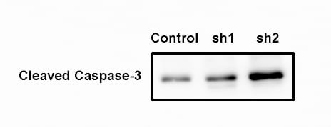
Detection of Mouse Human/Mouse Cleaved Caspase-3 (Asp175) Antibody by Western Blot
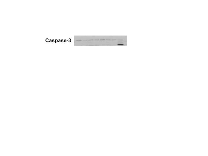
Gamma herpesvirus 68 ( gamma HV68) replicates in alveolar epithelial cells (AECs). (A) Wild-type mice were infected with 5 × 10⁴ PFU gamma HV68 on day 0. On day 7 after infection, frozen sections were prepared, and stained with a rabbit polyclonal antisera against gamma HV68, or with non-immune rabbit sera as control. The goat anti-rabbit secondary was linked to alkaline phosphatase. Vivid replication of gamma HV68 is visible in alveolar lining cells (original magnification × 100). Sections shown are representative of four mice examined. (B) AECs were isolated from lungs of Balb/c or TLR-9-/- mice treated with bleomycin plus gamma HV68 on day 21, and were cultured on fibronectin-coated slides (TiterTek). Sections were stained with antibodies (M30 Cytodeath), and the number of positive cells per high power field (HPF; ×400) were calculated (n = 30 HPF per genotype). (C) AECs were isolated from Balb/c or TLR-9-/- mice and cells were infected in vitro with 0.01 or 0.001 PFU gamma HV68 for 48 hours. Cell lysates were then analyzed for cleaved caspase 3 by western blotting. Data are from one experiment, representative of two. Image collected and cropped by CiteAb from the following publication (https://pubmed.ncbi.nlm.nih.gov/21810214), licensed under a CC-BY license. Not internally tested by R&D Systems. Antibody by Immunohistochemistry")
Detection of Human Human/Mouse Cleaved Caspase-3 (Asp175) Antibody by Immunohistochemistry
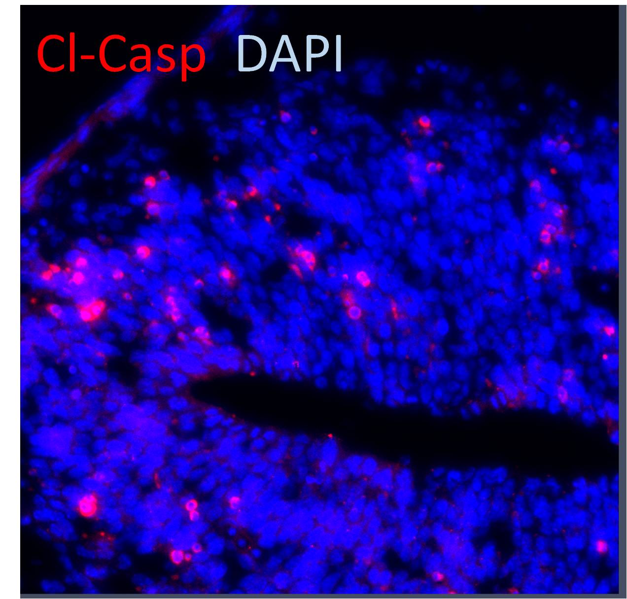
Assessment of the skin structure and apoptosis upon treatment with wound gels. Representative images and zoom-ins of the boxed areas of haematoxylin and eosin (H&E)-stained tape-stripped (TS) sections from biopsies (age 38 years) either left untreated or treated with indicated wound gels using a light microscope. The presence of caspase-3 expressing cells was evaluated using a caspase-3 (CASP3) antibody and visualisation with a secondary Alexa Fluor™ 546 AB (red). Nuclei were counterstained with DAPI (blue). Scale bar = 50 μm. Image collected and cropped by CiteAb from the following publication (https://pubmed.ncbi.nlm.nih.gov/36261541), licensed under a CC-BY license. Not internally tested by R&D Systems.Applications for Cleaved Caspase-3 (Asp175) Antibody (269518)
Application
Recommended Usage
Immunocytochemistry
8-25 µg/mL
Sample: Immersion fixed Jurkat human acute T cell leukemia cell line treated with staurosporin
Sample: Immersion fixed Jurkat human acute T cell leukemia cell line treated with staurosporin
Immunohistochemistry
0.3-25 µg/mL
Sample: Immersion fixed paraffin-embedded sections of human colon cancer tissue
Sample: Immersion fixed paraffin-embedded sections of human colon cancer tissue
Intracellular Staining by Flow Cytometry
0.25 µg/106 cells
Sample: Jurkat human acute T cell leukemia cell line treated with Staurosporine was fixed with Flow Cytometry Fixation Buffer (Catalog # FC004) and permeabilized with 90% methanol
Sample: Jurkat human acute T cell leukemia cell line treated with Staurosporine was fixed with Flow Cytometry Fixation Buffer (Catalog # FC004) and permeabilized with 90% methanol
Western Blot
0.5 µg/mL
Sample: Staurosporine-treated Jurkat human acute T cell leukemia cell line and DA3 mouse myeloma cell line
Sample: Staurosporine-treated Jurkat human acute T cell leukemia cell line and DA3 mouse myeloma cell line
Reviewed Applications
Read 13 reviews rated 4.5 using MAB835 in the following applications:
Flow Cytometry Panel Builder
Bio-Techne Knows Flow Cytometry
Save time and reduce costly mistakes by quickly finding compatible reagents using the Panel Builder Tool.
Advanced Features
- Spectra Viewer - Custom analysis of spectra from multiple fluorochromes
- Spillover Popups - Visualize the spectra of individual fluorochromes
- Antigen Density Selector - Match fluorochrome brightness with antigen density

Formulation, Preparation, and Storage
Purification
Protein A or G purified from hybridoma culture supernatant
Reconstitution
Reconstitute at 0.5 mg/mL in sterile PBS. For liquid material, refer to CoA for concentration.
Loading...
Formulation
Lyophilized from a 0.2 μm filtered solution in PBS with Trehalose. *Small pack size (SP) is supplied either lyophilized or as a 0.2 µm filtered solution in PBS.
Shipping
Lyophilized product is shipped at ambient temperature. Liquid small pack size (-SP) is shipped with polar packs. Upon receipt, store immediately at the temperature recommended below.
Stability & Storage
Use a manual defrost freezer and avoid repeated freeze-thaw cycles.
- 12 months from date of receipt, -20 to -70 °C as supplied.
- 1 month, 2 to 8 °C under sterile conditions after reconstitution.
- 6 months, -20 to -70 °C under sterile conditions after reconstitution.
Calculators
Background: Caspase-3
Alternate Names
Apopain, CASP3, Caspase3, CPP32, LICE-1, YAMA
Gene Symbol
CASP3
UniProt
Additional Caspase-3 Products
Product Documents for Cleaved Caspase-3 (Asp175) Antibody (269518)
Certificate of Analysis
To download a Certificate of Analysis, please enter a lot or batch number in the search box below.
Note: Certificate of Analysis not available for kit components.
Product Specific Notices for Cleaved Caspase-3 (Asp175) Antibody (269518)
For research use only
Related Research Areas
Citations for Cleaved Caspase-3 (Asp175) Antibody (269518)
 Powered by Bioz
Customer Reviews for Cleaved Caspase-3 (Asp175) Antibody (269518) (13)
4.5 out of 5
13 Customer Ratings
Have you used Cleaved Caspase-3 (Asp175) Antibody (269518)?
Submit a review and receive an Amazon gift card!
$25/€18/£15/$25CAN/¥2500 Yen for a review with an image
$10/€7/£6/$10CAN/¥1110 Yen for a review without an image
Submit a review
Customer Images

Showing
1
-
5 of
13 reviews
Showing All
Filter By:
-
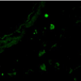
Application: Immunocytochemistry/ImmunofluorescenceSample Tested: Adult lungSpecies: MouseVerified Customer | Posted 03/23/2025

-
Application: Western BlotSample Tested: Breast cancer cellsSpecies: HumanVerified Customer | Posted 09/23/2023In the immunoblot, our goal is to see the expressing differences within different treatments, and we clearly saw the bands. Although there were some non-specific bands, but they did not affect the discrimination between groups.
-
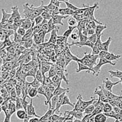
Application: Block/NeutralizeSample Tested: HEK293 human embryonic kidney cell lineSpecies: HumanVerified Customer | Posted 09/22/2022I used it to block this protein.
-
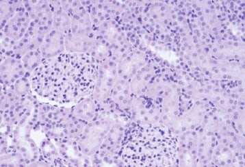
Application: ImmunohistochemistrySample Tested: Kidney tissueSpecies: MouseVerified Customer | Posted 08/04/2021
-
Application: Western BlotSample Tested: MDA-MB-231 human breast cancer cell lineSpecies: HumanVerified Customer | Posted 02/16/2020
-
Application: Western BlotSample Tested: MDA-MB-231 human breast cancer cell lineSpecies: HumanVerified Customer | Posted 01/17/2018
-
Application: Flow CytometrySample Tested: Liver tissueSpecies: MouseVerified Customer | Posted 09/27/2017
-
Application: ImmunohistochemistrySample Tested: Embryonic brainSpecies: MouseVerified Customer | Posted 09/07/2017
-
Application: Immunocytochemistry/ImmunofluorescenceSample Tested: IPS2 induced pluripotent stem cells derived cellsSpecies: HumanVerified Customer | Posted 02/08/2017
-
Application: Western BlotSample Tested: See PMID 22160308Species: HumanVerified Customer | Posted 02/25/2015
-
Application: ImmunofluorescenceSample Tested: See PMID 23483962Species: HumanVerified Customer | Posted 02/25/2015
-
Application: Western BlotSample Tested: See PMID 23536519Species: HumanVerified Customer | Posted 02/25/2015
-
Application: Immunohistochemistry-ParaffinSample Tested: See PMID 23637762Species: MouseVerified Customer | Posted 02/25/2015
There are no reviews that match your criteria.
Protocols
Find general support by application which include: protocols, troubleshooting, illustrated assays, videos and webinars.
- 7-Amino Actinomycin D (7-AAD) Cell Viability Flow Cytometry Protocol
- Antigen Retrieval Protocol (PIER)
- Antigen Retrieval for Frozen Sections Protocol
- Appropriate Fixation of IHC/ICC Samples
- Cellular Response to Hypoxia Protocols
- Chromogenic IHC Staining of Formalin-Fixed Paraffin-Embedded (FFPE) Tissue Protocol
- Chromogenic Immunohistochemistry Staining of Frozen Tissue
- ClariTSA™ Fluorophore Kits
- Detection & Visualization of Antibody Binding
- Extracellular Membrane Flow Cytometry Protocol
- Flow Cytometry Protocol for Cell Surface Markers
- Flow Cytometry Protocol for Staining Membrane Associated Proteins
- Flow Cytometry Staining Protocols
- Flow Cytometry Troubleshooting Guide
- Fluorescent IHC Staining of Frozen Tissue Protocol
- Graphic Protocol for Heat-induced Epitope Retrieval
- Graphic Protocol for the Preparation and Fluorescent IHC Staining of Frozen Tissue Sections
- Graphic Protocol for the Preparation and Fluorescent IHC Staining of Paraffin-embedded Tissue Sections
- Graphic Protocol for the Preparation of Gelatin-coated Slides for Histological Tissue Sections
- ICC Cell Smear Protocol for Suspension Cells
- ICC Immunocytochemistry Protocol Videos
- ICC for Adherent Cells
- IHC Sample Preparation (Frozen sections vs Paraffin)
- Immunocytochemistry (ICC) Protocol
- Immunocytochemistry Troubleshooting
- Immunofluorescence of Organoids Embedded in Cultrex Basement Membrane Extract
- Immunofluorescent IHC Staining of Formalin-Fixed Paraffin-Embedded (FFPE) Tissue Protocol
- Immunohistochemistry (IHC) and Immunocytochemistry (ICC) Protocols
- Immunohistochemistry Frozen Troubleshooting
- Immunohistochemistry Paraffin Troubleshooting
- Intracellular Flow Cytometry Protocol Using Alcohol (Methanol)
- Intracellular Flow Cytometry Protocol Using Detergents
- Intracellular Nuclear Staining Flow Cytometry Protocol Using Detergents
- Intracellular Staining Flow Cytometry Protocol Using Alcohol Permeabilization
- Intracellular Staining Flow Cytometry Protocol Using Detergents to Permeabilize Cells
- Preparing Samples for IHC/ICC Experiments
- Preventing Non-Specific Staining (Non-Specific Binding)
- Primary Antibody Selection & Optimization
- Propidium Iodide Cell Viability Flow Cytometry Protocol
- Protocol for Heat-Induced Epitope Retrieval (HIER)
- Protocol for Liperfluo
- Protocol for Making a 4% Formaldehyde Solution in PBS
- Protocol for VisUCyte™ HRP Polymer Detection Reagent
- Protocol for the Characterization of Human Th22 Cells
- Protocol for the Characterization of Human Th9 Cells
- Protocol for the Fluorescent ICC Staining of Cell Smears - Graphic
- Protocol for the Fluorescent ICC Staining of Cultured Cells on Coverslips - Graphic
- Protocol for the Preparation & Fixation of Cells on Coverslips
- Protocol for the Preparation and Chromogenic IHC Staining of Frozen Tissue Sections
- Protocol for the Preparation and Chromogenic IHC Staining of Frozen Tissue Sections - Graphic
- Protocol for the Preparation and Chromogenic IHC Staining of Paraffin-embedded Tissue Sections
- Protocol for the Preparation and Chromogenic IHC Staining of Paraffin-embedded Tissue Sections - Graphic
- Protocol for the Preparation and Fluorescent ICC Staining of Cells on Coverslips
- Protocol for the Preparation and Fluorescent ICC Staining of Non-adherent Cells
- Protocol for the Preparation and Fluorescent ICC Staining of Stem Cells on Coverslips
- Protocol for the Preparation and Fluorescent IHC Staining of Frozen Tissue Sections
- Protocol for the Preparation and Fluorescent IHC Staining of Paraffin-embedded Tissue Sections
- Protocol for the Preparation of Gelatin-coated Slides for Histological Tissue Sections
- Protocol for the Preparation of a Cell Smear for Non-adherent Cell ICC - Graphic
- Protocol: Annexin V and PI Staining by Flow Cytometry
- Protocol: Annexin V and PI Staining for Apoptosis by Flow Cytometry
- R&D Systems Quality Control Western Blot Protocol
- TUNEL and Active Caspase-3 Detection by IHC/ICC Protocol
- The Importance of IHC/ICC Controls
- Troubleshooting Guide: Fluorokine Flow Cytometry Kits
- Troubleshooting Guide: Immunohistochemistry
- Troubleshooting Guide: Western Blot Figures
- Western Blot Conditions
- Western Blot Protocol
- Western Blot Protocol for Cell Lysates
- Western Blot Troubleshooting
- Western Blot Troubleshooting Guide
- View all Protocols, Troubleshooting, Illustrated assays and Webinars
Loading...
Associated Pathways

