EZH2 (Enhancer of zeste homolog 2; also ENX-1 and Lys N-methyltransferase 6) is an 80 kDa member of the EZ family of chromatin-dependent gene regulators. It is a nuclear protein that represses gene transcription through histone methylation. Human EZH2 is 746 amino acids (aa) in length. It contains an NLS (aa 490‑495), a Cys-rich region, and a methyltransferase SET (Suppressor/Enhancer/Trithorax) domain (aa 606‑729). There are four potential splice variants. One shows a premature truncation after Cys286, a second shows a 6 aa substitution for aa 329‑746, a third shows a deletion of aa 83‑121, and the fourth exhibits a 5 aa insertion after His297. Over aa 512‑645, human, mouse and canine EZH2 are identical in amino acid sequence.

Key Product Details
Validated by
Knockout/Knockdown
Species Reactivity
Validated:
Human, Mouse
Cited:
Human
Applications
Validated:
Western Blot, Immunocytochemistry
Cited:
Immunohistochemistry-Frozen, Western Blot, Immunoprecipitation
Label
Unconjugated
Antibody Source
Polyclonal Goat IgG
Loading...
Product Specifications
Immunogen
E. coli-derived recombinant human EZH2
Gly512-Ile645
Accession # Q15910
Gly512-Ile645
Accession # Q15910
Specificity
Detects human and mouse EZH2 in direct ELISAs and Western blots.
Clonality
Polyclonal
Host
Goat
Isotype
IgG
Scientific Data Images for EZH2 Antibody
Detection of Human and Mouse EZH2 by Western Blot.
Western blot shows lysates of HeLa human cervical epithelial carcinoma cell line, Jurkat human acute T cell leukemia cell line, and mouse spleen tissue. PVDF membrane was probed with 1 µg/mL of Goat Anti-Human/Mouse EZH2 Antigen Affinity-purified Polyclonal Antibody (Catalog # AF4767) followed by HRP-conjugated Anti-Goat IgG Secondary Antibody (Catalog # HAF019). A specific band was detected for EZH2 at approximately 80 kDa (as indicated). This experiment was conducted under reducing conditions and using Immunoblot Buffer Group 8.
Detection of Human EZH2/KMT6 by Western Blot
Measurement of EZH2 protein under conditions of SIRT1 knockdown. SIRT1 knockdown in Caco-2 cells was achieved using two different siRNAs and protein was analysed by Western blotting using anti-EZH2 antibody (a) or anti-SIRT1 antibody (to confirm efficacy of knockdown) (b). Blots were probed with anti-alpha -tubulin antibody to confirm equal protein loading and transfer. Approximately 10 μg of protein was loaded in each lane. Three biological replicates are presented for each condition. Approximate molecular weights are indicated Image collected and cropped by CiteAb from the following publication (https://humgenomics.biomedcentral.com/articles/10.1186/s40246-015-0036-0), licensed under a CC-BY license. Not internally tested by R&D Systems.
Detection of Human EZH2/KMT6 by Western Blot
EZH2 elevates at S phase in hDPCs and interacts with PCNA. a Schematic chart illustrating the serum deprivation approach used for G0/G1 phase synchronization of hDPCs. b Western blot shows the expression of EZH2 in hDPCs at the indicated time points after release from serum deprivation. Immunofluorescence shows the subcellular localization of EZH2 in hDPCs in G1 or S phase. c Co-immunoprecipitation (Co-IP) of EZH2 and PCNA in hDPCs (synchronized at S phase) and proximity ligation assay (PLA) after co-incubating anti-EZH2 and anti-PCNA antibodies in hDPCs in S phase. IgGs were used as control antibodies for the IP. Antibodies used for IP and western blot are labelled as IP and IB, respectively. Total lysate (10 μg) was used as an input control. Scale bars represent 20 μm Image collected and cropped by CiteAb from the following publication (https://pubmed.ncbi.nlm.nih.gov/30071900), licensed under a CC-BY license. Not internally tested by R&D Systems.
Detection of Human EZH2/KMT6 by Proximity Ligation Assay
EZH2 elevates at S phase in hDPCs and interacts with PCNA. a Schematic chart illustrating the serum deprivation approach used for G0/G1 phase synchronization of hDPCs. b Western blot shows the expression of EZH2 in hDPCs at the indicated time points after release from serum deprivation. Immunofluorescence shows the subcellular localization of EZH2 in hDPCs in G1 or S phase. c Co-immunoprecipitation (Co-IP) of EZH2 and PCNA in hDPCs (synchronized at S phase) and proximity ligation assay (PLA) after co-incubating anti-EZH2 and anti-PCNA antibodies in hDPCs in S phase. IgGs were used as control antibodies for the IP. Antibodies used for IP and western blot are labelled as IP and IB, respectively. Total lysate (10 μg) was used as an input control. Scale bars represent 20 μm Image collected and cropped by CiteAb from the following publication (https://pubmed.ncbi.nlm.nih.gov/30071900), licensed under a CC-BY license. Not internally tested by R&D Systems.
Detection of Human EZH2/KMT6 by Western Blot
Measurement of EZH2 protein under conditions of SIRT1 knockdown. SIRT1 knockdown in Caco-2 cells was achieved using two different siRNAs and protein was analysed by Western blotting using anti-EZH2 antibody (a) or anti-SIRT1 antibody (to confirm efficacy of knockdown) (b). Blots were probed with anti-alpha -tubulin antibody to confirm equal protein loading and transfer. Approximately 10 μg of protein was loaded in each lane. Three biological replicates are presented for each condition. Approximate molecular weights are indicated Image collected and cropped by CiteAb from the following open publication (https://pubmed.ncbi.nlm.nih.gov/26104761), licensed under a CC-BY license. Not internally tested by R&D Systems.Applications for EZH2 Antibody
Application
Recommended Usage
Immunocytochemistry
5-15 µg/mL
Sample: Immersion fixed HL-60 human acute promyelocytic leukemia cell line
Sample: Immersion fixed HL-60 human acute promyelocytic leukemia cell line
Western Blot
1 µg/mL
Sample: HeLa human cervical epithelial carcinoma cell line, Jurkat human acute T cell leukemia cell line, and mouse spleen tissue
Sample: HeLa human cervical epithelial carcinoma cell line, Jurkat human acute T cell leukemia cell line, and mouse spleen tissue
Reviewed Applications
Read 1 review rated 5 using AF4767 in the following applications:
Formulation, Preparation, and Storage
Purification
Antigen Affinity-purified
Reconstitution
Reconstitute at 0.2 mg/mL in sterile PBS. For liquid material, refer to CoA for concentration.
Loading...
Formulation
Lyophilized from a 0.2 μm filtered solution in PBS with Trehalose. *Small pack size (SP) is supplied either lyophilized or as a 0.2 µm filtered solution in PBS.
Shipping
Lyophilized product is shipped at ambient temperature. Liquid small pack size (-SP) is shipped with polar packs. Upon receipt, store immediately at the temperature recommended below.
Stability & Storage
Use a manual defrost freezer and avoid repeated freeze-thaw cycles.
- 12 months from date of receipt, -20 to -70 °C as supplied.
- 1 month, 2 to 8 °C under sterile conditions after reconstitution.
- 6 months, -20 to -70 °C under sterile conditions after reconstitution.
Calculators
Background: EZH2
Long Name
Enhancer of Zeste Homolog 2 [Drosophila]
Alternate Names
ENX1, KMT6
Gene Symbol
EZH2
UniProt
Additional EZH2 Products
Product Documents for EZH2 Antibody
Certificate of Analysis
To download a Certificate of Analysis, please enter a lot or batch number in the search box below.
Note: Certificate of Analysis not available for kit components.
Product Specific Notices for EZH2 Antibody
For research use only
Related Research Areas
Citations for EZH2 Antibody
 Powered by Bioz
Customer Reviews for EZH2 Antibody (1)
5 out of 5
1 Customer Rating
Have you used EZH2 Antibody?
Submit a review and receive an Amazon gift card!
$25/€18/£15/$25CAN/¥2500 Yen for a review with an image
$10/€7/£6/$10CAN/¥1110 Yen for a review without an image
Submit a review
Customer Images
Showing
1
-
1 of
1 review
Showing All
Filter By:
-
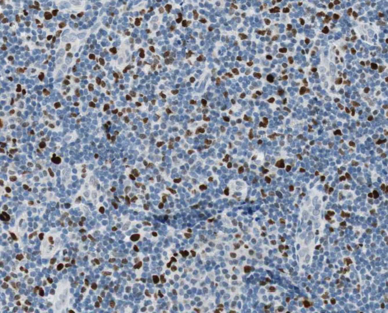
Application: ImmunohistochemistrySample Tested: Spleen tissueSpecies: HumanVerified Customer | Posted 10/26/2021

There are no reviews that match your criteria.
Protocols
Find general support by application which include: protocols, troubleshooting, illustrated assays, videos and webinars.
- Appropriate Fixation of IHC/ICC Samples
- Cellular Response to Hypoxia Protocols
- ClariTSA™ Fluorophore Kits
- Detection & Visualization of Antibody Binding
- ICC Cell Smear Protocol for Suspension Cells
- ICC Immunocytochemistry Protocol Videos
- ICC for Adherent Cells
- Immunocytochemistry (ICC) Protocol
- Immunocytochemistry Troubleshooting
- Immunofluorescence of Organoids Embedded in Cultrex Basement Membrane Extract
- Immunohistochemistry (IHC) and Immunocytochemistry (ICC) Protocols
- Preparing Samples for IHC/ICC Experiments
- Preventing Non-Specific Staining (Non-Specific Binding)
- Primary Antibody Selection & Optimization
- Protocol for VisUCyte™ HRP Polymer Detection Reagent
- Protocol for the Fluorescent ICC Staining of Cell Smears - Graphic
- Protocol for the Fluorescent ICC Staining of Cultured Cells on Coverslips - Graphic
- Protocol for the Preparation and Fluorescent ICC Staining of Cells on Coverslips
- Protocol for the Preparation and Fluorescent ICC Staining of Non-adherent Cells
- Protocol for the Preparation and Fluorescent ICC Staining of Stem Cells on Coverslips
- Protocol for the Preparation of a Cell Smear for Non-adherent Cell ICC - Graphic
- R&D Systems Quality Control Western Blot Protocol
- TUNEL and Active Caspase-3 Detection by IHC/ICC Protocol
- The Importance of IHC/ICC Controls
- Troubleshooting Guide: Western Blot Figures
- Western Blot Conditions
- Western Blot Protocol
- Western Blot Protocol for Cell Lysates
- Western Blot Troubleshooting
- Western Blot Troubleshooting Guide
- View all Protocols, Troubleshooting, Illustrated assays and Webinars
Loading...
Associated Pathways
