Loading...
Key Product Details
Species Reactivity
Validated:
Human, Mouse
Cited:
Human, Mouse, Rat, Transgenic Mouse
Applications
Validated:
Immunohistochemistry, Western Blot, Direct ELISA, Immunoprecipitation
Cited:
Immunohistochemistry, Western Blot, Immunoprecipitation
Label
Unconjugated
Antibody Source
Monoclonal Mouse IgG2A Clone # B0422
Loading...
Product Specifications
Immunogen
Recombinant mouse GLP/EHMT1
aa 134-234
aa 134-234
Specificity
This antibody specifically recognizes mouse GLP and cross-reacts with human GLP. Not yet tested in other species.
Clonality
Monoclonal
Host
Mouse
Isotype
IgG2A
Applications for GLP/EHMT1 Antibody (B0422)
Application
Recommended Usage
Direct ELISA
This antibody can be used at 0.1 μg/mL with the appropriate secondary reagents to detect mouse GLP.
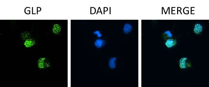
Immunohistochemistry
This antibody can be used at 5 μg/mL with the appropriate secondary reagents to detect mouse GLP.
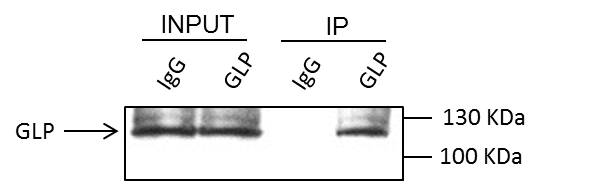
Immunoprecipitation
Optimal dilutions should be determined by each laboratory.
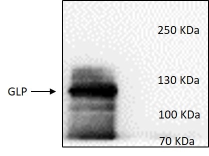
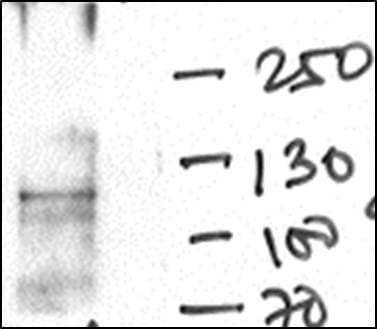
Western Blot
This antibody can be used at 0.5 μg/mL with the appropriate secondary reagents to detect mouse GLP.
Reviewed Applications
Read 8 reviews rated 4.3 using PP-B0422-00 in the following applications:
Formulation, Preparation, and Storage
Purification
Protein A or G purified from hybridoma culture supernatant
Formulation
A liquid formulation in physiologic saline with 0.1% NaN3.
Shipping
The product is shipped with dry ice or equivalent. Upon receipt, store it immediately at the temperature recommended below.
Stability & Storage
This antibody is stable for greater than six months when held at -20 °C in a manual defrost freezer or at -70 °C. Upon thawing, the antibody can be stored at 2-8 °C for at least 1 month without detectable loss of activity. Avoid repeated freeze-thaw cycles.
Background: GLP/EHMT1
Long Name
G9a-like Protein 1/Euchromatic Histone-Lysine N-Methyltransferase 1
Alternate Names
DEL9q34, EHMT1, Eu-HMTase1, KMT1D, Lysine N-methyltransferase 1D
Gene Symbol
EHMT1
Additional GLP/EHMT1 Products
Product Documents for GLP/EHMT1 Antibody (B0422)
Certificate of Analysis
To download a Certificate of Analysis, please enter a lot or batch number in the search box below.
Note: Certificate of Analysis not available for kit components.
Product Specific Notices for GLP/EHMT1 Antibody (B0422)
For research use only
Related Research Areas
Citations for GLP/EHMT1 Antibody (B0422)
 Powered by Bioz
Customer Reviews for GLP/EHMT1 Antibody (B0422) (8)
4.3 out of 5
8 Customer Ratings
Have you used GLP/EHMT1 Antibody (B0422)?
Submit a review and receive an Amazon gift card!
$25/€18/£15/$25CAN/¥2500 Yen for a review with an image
$10/€7/£6/$10CAN/¥1110 Yen for a review without an image
Submit a review
Customer Images
Showing
1
-
5 of
8 reviews
Showing All
Filter By:
-
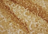
Application: ImmunohistochemistrySample Tested: Brain tissueSpecies: HumanVerified Customer | Posted 09/07/2021

-
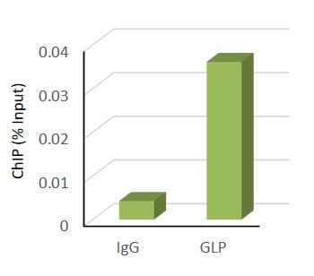
Application: Chromatin ImmunoprecipitationSample Tested: B cellsSpecies: HumanVerified Customer | Posted 06/26/2018ChIP was performed using PP-B0422-00 or isotype IgG (1 ug antibody/ 100 ug Chromatin). Realtime-PCR was performed using primers specific for promoter of gene of interest and the results are expressed as % input.
-
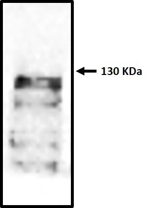
Application: Western BlotSample Tested: 293TSpecies: HumanVerified Customer | Posted 06/22/2018293T whole cell lysate was immunoblotted with anti GLP anitibody.
-
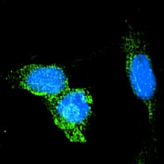
Application: Immunocytochemistry/ImmunofluorescenceSample Tested: CCD-1070Sk human foreskin fibroblast cell lineSpecies: HumanVerified Customer | Posted 06/19/2018
-
Application: Western BlotSample Tested: RL human non-Hodgkin's lymphoma B cell lineSpecies: HumanVerified Customer | Posted 01/04/2018
-
Application: ImmunocytochemistrySample Tested: Human Dermal Microvascular Endothelial cells - Paraformaldehyde fixed cellsSpecies: HumanVerified Customer | Posted 07/11/2017Immunofluorescence staining of paraformaldehyde fixed Human Dermal Microvascular Endothelial cells. PP-B0422-00 used at 1:100 dilution.
-
Application: ImmunoprecipitationSample Tested: Human Dermal Microvascular Endothelial whole cell lysateSpecies: HumanVerified Customer | Posted 04/21/2017
-
Application: Western BlotSample Tested: Human Dermal Microvascular Endothelial whole cell lysateSpecies: HumanVerified Customer | Posted 04/21/2017
There are no reviews that match your criteria.
Protocols
Find general support by application which include: protocols, troubleshooting, illustrated assays, videos and webinars.
- Antigen Retrieval Protocol (PIER)
- Antigen Retrieval for Frozen Sections Protocol
- Appropriate Fixation of IHC/ICC Samples
- Cellular Response to Hypoxia Protocols
- Chromogenic IHC Staining of Formalin-Fixed Paraffin-Embedded (FFPE) Tissue Protocol
- Chromogenic Immunohistochemistry Staining of Frozen Tissue
- ClariTSA™ Fluorophore Kits
- Detection & Visualization of Antibody Binding
- ELISA Sample Preparation & Collection Guide
- ELISA Troubleshooting Guide
- Fluorescent IHC Staining of Frozen Tissue Protocol
- Graphic Protocol for Heat-induced Epitope Retrieval
- Graphic Protocol for the Preparation and Fluorescent IHC Staining of Frozen Tissue Sections
- Graphic Protocol for the Preparation and Fluorescent IHC Staining of Paraffin-embedded Tissue Sections
- Graphic Protocol for the Preparation of Gelatin-coated Slides for Histological Tissue Sections
- How to Run an R&D Systems DuoSet ELISA
- How to Run an R&D Systems Quantikine ELISA
- How to Run an R&D Systems Quantikine™ QuicKit™ ELISA
- IHC Sample Preparation (Frozen sections vs Paraffin)
- Immunofluorescent IHC Staining of Formalin-Fixed Paraffin-Embedded (FFPE) Tissue Protocol
- Immunohistochemistry (IHC) and Immunocytochemistry (ICC) Protocols
- Immunohistochemistry Frozen Troubleshooting
- Immunohistochemistry Paraffin Troubleshooting
- Immunoprecipitation Protocol
- Preparing Samples for IHC/ICC Experiments
- Preventing Non-Specific Staining (Non-Specific Binding)
- Primary Antibody Selection & Optimization
- Protocol for Heat-Induced Epitope Retrieval (HIER)
- Protocol for Making a 4% Formaldehyde Solution in PBS
- Protocol for VisUCyte™ HRP Polymer Detection Reagent
- Protocol for the Preparation & Fixation of Cells on Coverslips
- Protocol for the Preparation and Chromogenic IHC Staining of Frozen Tissue Sections
- Protocol for the Preparation and Chromogenic IHC Staining of Frozen Tissue Sections - Graphic
- Protocol for the Preparation and Chromogenic IHC Staining of Paraffin-embedded Tissue Sections
- Protocol for the Preparation and Chromogenic IHC Staining of Paraffin-embedded Tissue Sections - Graphic
- Protocol for the Preparation and Fluorescent IHC Staining of Frozen Tissue Sections
- Protocol for the Preparation and Fluorescent IHC Staining of Paraffin-embedded Tissue Sections
- Protocol for the Preparation of Gelatin-coated Slides for Histological Tissue Sections
- Quantikine HS ELISA Kit Assay Principle, Alkaline Phosphatase
- Quantikine HS ELISA Kit Principle, Streptavidin-HRP Polymer
- R&D Systems Quality Control Western Blot Protocol
- Sandwich ELISA (Colorimetric) – Biotin/Streptavidin Detection Protocol
- Sandwich ELISA (Colorimetric) – Direct Detection Protocol
- TUNEL and Active Caspase-3 Detection by IHC/ICC Protocol
- The Importance of IHC/ICC Controls
- Troubleshooting Guide: ELISA
- Troubleshooting Guide: Immunohistochemistry
- Troubleshooting Guide: Western Blot Figures
- Western Blot Conditions
- Western Blot Protocol
- Western Blot Protocol for Cell Lysates
- Western Blot Troubleshooting
- Western Blot Troubleshooting Guide
- View all Protocols, Troubleshooting, Illustrated assays and Webinars
Loading...
Associated Pathways
