Annexin A2 (ANXA2), also known as Annexin II and Lipocortin II (LPC2), is a 38.6 kDa member of the Annexin family of proteins. The Annexins are a family of Calcium-dependent phospholipid-binding proteins that are preferentially located on the cytosolic face of the plasma membrane. The Annexin’s have a molecular weight of approximately 35‑40 kDa and consist of a unique amino terminal domain followed by a homologous C-terminal core domain containing the calcium-dependent phospholipid-binding sites. The C-terminal domain is comprised of four 60‑70 amino acid (aa) repeats, known as annexin repeats or an endonexin fold (Annexin A6 contains 8 annexin repeats). The four annexin repeats form a highly alpha -helical, tightly packed disc known as the annexin domain, which binds to phospholipids in the membrane in a calcium-dependent manner. Members of the annexin family play a role in cytoskeletal interactions, phospholipase inhibition, regulation of cellular growth, and intracellular signal transduction pathways. Annexin A2 was identified as an inhibitor of phospholipase A2 activity. Annexin A2 also functions as an autocrine factor to enhance osteoclast formation and bone resorption and is a major cellular substrate of the tyrosine kinase encoded by the SRC oncogene. Human Annexin A2 shares 97% identity with mouse and rat Annexin A2.

Key Product Details
Validated by
Knockout/Knockdown
Species Reactivity
Validated:
Human, Mouse, Rat
Cited:
Human, Mouse, Transgenic Mouse
Applications
Validated:
Knockout Validated, Immunohistochemistry, Western Blot, Simple Western
Cited:
Immunohistochemistry, Western Blot, Immunoelectron Microscopy
Label
Unconjugated
Antibody Source
Polyclonal Goat IgG
Loading...
Product Specifications
Immunogen
E. coli-derived recombinant human Annexin A2
Met1-Asp339
Accession # P07355
Met1-Asp339
Accession # P07355
Specificity
Detects human, mouse, and rat Annexin A2 in Western blots. In Western blots, no cross-reactivity with recombinant human Annexin A1, A3, A4, A5, A6, A7, A8, A9, A10, A11, or A13 is observed.
Clonality
Polyclonal
Host
Goat
Isotype
IgG
Scientific Data Images for Annexin A2 Antibody
Detection of Human/Mouse/Rat Annexin A2 by Western Blot.
Western blot shows lysates of A431 human epithelial carcinoma cell line, NIH-3T3 mouse embryonic fibroblast cell line, and L6 rat myoblast cell line. PVDF membrane was probed with 1 µg/mL of Goat Anti-Human/Mouse/Rat Annexin A2 Antigen Affinity-purified Polyclonal Antibody (Catalog # AF3928) followed by HRP-conjugated Anti-Goat IgG Secondary Antibody (Catalog # HAF017). A specific band was detected for Annexin A2 at approximately 39 kDa (as indicated). This experiment was conducted using Immunoblot Buffer Group 2.
Detection of Human and Mouse Annexin A2 by Simple WesternTM.
Simple Western shows lysates of Exosome Standards (HT‑29) (NBP3-11685) and COLO 205 human colorectal adenocarcinoma cell line, loaded at 0.5 mg/ml. A specific band was detected for Annexin A2 at approximately 44 kDa (as indicated) using 100 µg/mL of Goat Anti-Human/Mouse/Rat Annexin A2 Antigen Affinity-purified Polyclonal Antibody (Catalog # AF3928). This experiment was conducted under reducing conditions and using the 12-230kDa separation system.
Annexin A2 in Human Liver.
Annexin A2 was detected in immersion fixed paraffin-embedded sections of normal human liver using Goat Anti-Human/Mouse/Rat Annexin A2 Antigen Affinity-purified Polyclonal Antibody (Catalog # AF3928) at 3 µg/mL overnight at 4 °C. Before incubation with the primary antibody, tissue was subjected to heat-induced epitope retrieval using Antigen Retrieval Reagent-Basic (Catalog # CTS013). Tissue was stained using the Anti-Goat HRP-DAB Cell & Tissue Staining Kit (brown; Catalog # CTS008) and counterstained with hematoxylin (blue). Specific staining was localized to plasma membranes of hepatocytes. View our protocol for Chromogenic IHC Staining of Paraffin-embedded Tissue Sections.
Western Blot Shows Human Annexin A2 Specificity by Using Knockout Cell Line.
Western blot shows lysates of HeLa human cervical epithelial carcinoma parental cell line and Annexin A2 knockout HeLa cell line (KO). PVDF membrane was probed with 1 µg/mL of Goat Anti-Human/Mouse/Rat Annexin A2 Antigen Affinity-purified Polyclonal Antibody (Catalog # AF3928) followed by HRP-conjugated Anti-Goat IgG Secondary Antibody (Catalog # HAF017). A specific band was detected for Annexin A2 at approximately 37 kDa (as indicated) in the parental HeLa cell line, but is not detectable in knockout HeLa cell line. GAPDH (Catalog # AF5718) is shown as a loading control. This experiment was conducted under reducing conditions and using Immunoblot Buffer Group 1.
Detection of Mouse Annexin A2 by Western Blot
MIPs induce Src-dependent tyrosine phosphorylation and translocation of A2 to the cell surface. A ARPE-19 cells were incubated for 18 h with basic medium (Basic), activated THP-1 cell conditioned medium (CM), or THP-1 CM with RPE CM with or without addition of anti-human MIP-1 alpha / beta or non-immune IgG. Cell surface A2 (sA2) was labeled using membrane impermeable biotinylation and captured with NeutrAvidin agarose beads. Labeled cell surface proteins were resolved by SDS-PAGE and cell surface (sA2) and total cell lysate A2 (tA2) probed by immunoblot with mouse (BD #610069) or rabbit (Cell Signaling #8235) anti-A2 IgG. b Pixel density of immunoblotted bands from 3-5 separate experiments were quantified and normalized to the unstimulated sample (Basic). c ARPE-19 cells were incubated with basic media with or without recombinant human MIP-1 alpha / beta (MIPs, 200 ng/ml each), and with or without the Src kinase inhibitor PP2 or its control PP3 (10 µM, 3 h). Cell lysates were probed for pY23-A2, tA2, pY416 activated Src, total Src (tSrc), and GAPDH, as a loading control. d Pixel density of immunoblotted bands from 3-6 separate experiments were quantified and normalized to the unstimulated sample (Basic). e ARPE-19 cells were incubated with or without recombinant human MIP-1 alpha / beta (MIPs, 200 ng/ml each) in the presence or absence of PP2 or PP3, as in (c). f Pixel density of immunoblotted bands from 3–7 separate experiments were quantified and normalized to the unstimulated sample (Basic). Shown for (b, d, f) are mean values ± SE, with p values determined by unpaired one-way ANOVA and the post hoc Tukey test. Source data for (b, d, and f) are provided as a Source File. Image collected and cropped by CiteAb from the following open publication (https://pubmed.ncbi.nlm.nih.gov/39384746), licensed under a CC-BY license. Not internally tested by R&D Systems.
Detection of Mouse Annexin A2 by Flow Cytometry
Effect of anti-annexin A2 IgG pretreatment on dispase-induced PVR.a, b Wild type C57Bl/6 mice were pretreated with an anterior chamber injection of anti-A2 IgG (1A7 or 2E6), control IgG (1D4), or PBS, two days prior to intravitreal dispase injection. At 2 (a) and 4 (b) weeks, the eyes were harvested, fixed, embedded, sectioned, and stained with H and E. Scale bars for (a and b) are 200 um. PVR histology (c) and RPE migration (d) scores were recorded by trained, masked observers. Shown for (c and d) are mean ± SE, n = 7 animals/group. p values were determined by unpaired one-way ANOVA and the post hoc Tukey test. Source data for (c and d), are provided as a Source File. Image collected and cropped by CiteAb from the following open publication (https://pubmed.ncbi.nlm.nih.gov/39384746), licensed under a CC-BY license. Not internally tested by R&D Systems.Applications for Annexin A2 Antibody
Application
Recommended Usage
Immunohistochemistry
5-15 µg/mL
Sample: Immersion fixed paraffin-embedded sections of normal human liver
Sample: Immersion fixed paraffin-embedded sections of normal human liver
Knockout Validated
Annexin A2
is specifically detected in HeLa human cervical epithelial carcinoma parental cell line but is not detectable in
Annexin A2 knockout HeLa cell line.
Simple Western
100 µg/mL
Sample: Exosome Standard (HT-29) (Catalog # NBP3-11685) and COLO 205 human colorectal adenocarcinoma cell line
Sample: Exosome Standard (HT-29) (Catalog # NBP3-11685) and COLO 205 human colorectal adenocarcinoma cell line
Western Blot
1 µg/mL
Sample: A431 human epithelial carcinoma cell line, NIH-3T3 mouse embryonic fibroblast cell line, and L6 rat myoblast cell line
Sample: A431 human epithelial carcinoma cell line, NIH-3T3 mouse embryonic fibroblast cell line, and L6 rat myoblast cell line
Reviewed Applications
Read 1 review rated 5 using AF3928 in the following applications:
Formulation, Preparation, and Storage
Purification
Antigen Affinity-purified
Reconstitution
Reconstitute at 0.2 mg/mL in sterile PBS. For liquid material, refer to CoA for concentration.
Loading...
Formulation
Lyophilized from a 0.2 μm filtered solution in PBS with Trehalose. See Certificate of Analysis for details.
*Small pack size (-SP) is supplied either lyophilized or as a 0.2 µm filtered solution in PBS.
*Small pack size (-SP) is supplied either lyophilized or as a 0.2 µm filtered solution in PBS.
Shipping
Lyophilized product is shipped at ambient temperature. Liquid small pack size (-SP) is shipped with polar packs. Upon receipt, store immediately at the temperature recommended below.
Stability & Storage
Use a manual defrost freezer and avoid repeated freeze-thaw cycles.
- 12 months from date of receipt, -20 to -70 °C as supplied.
- 1 month, 2 to 8 °C under sterile conditions after reconstitution.
- 6 months, -20 to -70 °C under sterile conditions after reconstitution.
Calculators
Background: Annexin A2
Alternate Names
ANX2L4, ANXA2, CAL1H, LIP2, Lipocortin-2, LPC2D, PAP-IV
Gene Symbol
ANXA2
UniProt
Additional Annexin A2 Products
Product Documents for Annexin A2 Antibody
Certificate of Analysis
To download a Certificate of Analysis, please enter a lot or batch number in the search box below.
Note: Certificate of Analysis not available for kit components.
Product Specific Notices for Annexin A2 Antibody
For research use only
Citations for Annexin A2 Antibody
 Powered by Bioz
Customer Reviews for Annexin A2 Antibody (1)
5 out of 5
1 Customer Rating
Have you used Annexin A2 Antibody?
Submit a review and receive an Amazon gift card!
$25/€18/£15/$25CAN/¥2500 Yen for a review with an image
$10/€7/£6/$10CAN/¥1110 Yen for a review without an image
Submit a review
Customer Images
Showing
1
-
1 of
1 review
Showing All
Filter By:
-
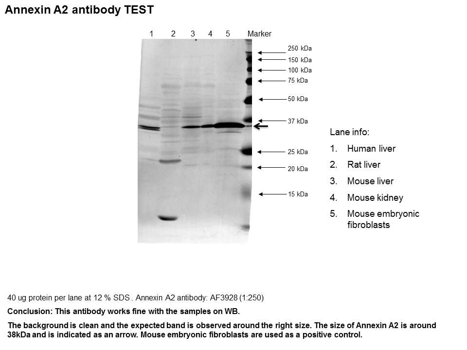
Application: Western BlotSample Tested: Liver tissue, Kidney tissue and Mouse embryonic fibroblastsSpecies: Human, Mouse and RatVerified Customer | Posted 11/01/2016

There are no reviews that match your criteria.
Protocols
Find general support by application which include: protocols, troubleshooting, illustrated assays, videos and webinars.
- Antigen Retrieval Protocol (PIER)
- Antigen Retrieval for Frozen Sections Protocol
- Appropriate Fixation of IHC/ICC Samples
- Cellular Response to Hypoxia Protocols
- Chromogenic IHC Staining of Formalin-Fixed Paraffin-Embedded (FFPE) Tissue Protocol
- Chromogenic Immunohistochemistry Staining of Frozen Tissue
- ClariTSA™ Fluorophore Kits
- Detection & Visualization of Antibody Binding
- Fluorescent IHC Staining of Frozen Tissue Protocol
- Graphic Protocol for Heat-induced Epitope Retrieval
- Graphic Protocol for the Preparation and Fluorescent IHC Staining of Frozen Tissue Sections
- Graphic Protocol for the Preparation and Fluorescent IHC Staining of Paraffin-embedded Tissue Sections
- Graphic Protocol for the Preparation of Gelatin-coated Slides for Histological Tissue Sections
- IHC Sample Preparation (Frozen sections vs Paraffin)
- Immunofluorescent IHC Staining of Formalin-Fixed Paraffin-Embedded (FFPE) Tissue Protocol
- Immunohistochemistry (IHC) and Immunocytochemistry (ICC) Protocols
- Immunohistochemistry Frozen Troubleshooting
- Immunohistochemistry Paraffin Troubleshooting
- Preparing Samples for IHC/ICC Experiments
- Preventing Non-Specific Staining (Non-Specific Binding)
- Primary Antibody Selection & Optimization
- Protocol for Heat-Induced Epitope Retrieval (HIER)
- Protocol for Making a 4% Formaldehyde Solution in PBS
- Protocol for VisUCyte™ HRP Polymer Detection Reagent
- Protocol for the Preparation & Fixation of Cells on Coverslips
- Protocol for the Preparation and Chromogenic IHC Staining of Frozen Tissue Sections
- Protocol for the Preparation and Chromogenic IHC Staining of Frozen Tissue Sections - Graphic
- Protocol for the Preparation and Chromogenic IHC Staining of Paraffin-embedded Tissue Sections
- Protocol for the Preparation and Chromogenic IHC Staining of Paraffin-embedded Tissue Sections - Graphic
- Protocol for the Preparation and Fluorescent IHC Staining of Frozen Tissue Sections
- Protocol for the Preparation and Fluorescent IHC Staining of Paraffin-embedded Tissue Sections
- Protocol for the Preparation of Gelatin-coated Slides for Histological Tissue Sections
- R&D Systems Quality Control Western Blot Protocol
- TUNEL and Active Caspase-3 Detection by IHC/ICC Protocol
- The Importance of IHC/ICC Controls
- Troubleshooting Guide: Immunohistochemistry
- Troubleshooting Guide: Western Blot Figures
- Western Blot Conditions
- Western Blot Protocol
- Western Blot Protocol for Cell Lysates
- Western Blot Troubleshooting
- Western Blot Troubleshooting Guide
- View all Protocols, Troubleshooting, Illustrated assays and Webinars
Loading...