Cells have evolved complex mechanisms to maintain redox balance and defend against oxidative stress. Catalase is a tetrameric enzyme comprised of four 60 kDa subunits. Catalase is typically localized in the peroxisome where it functions as an antioxidant, protecting cells from damage due to oxidative stress. Catalase converts reactive oxygen species, such as H2O2, into water and O2. Human Catalase shares 89% homology to mouse and rat Catalase. The cells redox environment can serve as an important signaling switch or trigger to initiate a number of cellular processes, including gene expression, differentiation, proliferation and apoptosis.

Key Product Details
Validated by
Knockout/Knockdown
Species Reactivity
Validated:
Human, Mouse, Rat
Cited:
Human, Mouse, Rat
Applications
Validated:
Knockout Validated, Western Blot, Immunocytochemistry, Simple Western
Cited:
Western Blot, Immunocytochemistry
Label
Unconjugated
Antibody Source
Polyclonal Goat IgG
Loading...
Product Specifications
Immunogen
E. coli-derived recombinant human Catalase
Met1-Leu527
Accession # P04040
Met1-Leu527
Accession # P04040
Specificity
Detects human, mouse and rat Catalase in Western blots.
Clonality
Polyclonal
Host
Goat
Isotype
IgG
Scientific Data Images for Catalase Antibody
Detection of Human/Mouse/Rat Catalase by Western Blot.
Western blot shows lysates of Jurkat human acute T cell leukemia cell line, Raji human Burkitt's lymphoma cell line, HeLa human cervical epithelial carcinoma cell line, NIH-3T3 mouse embryonic fibroblast cell line, A20 mouse B cell lymphoma cell line, and Rat-2 rat embryonic fibroblast cell line. PVDF membrane was probed with 0.5 µg/mL of Goat Anti-Human/Mouse/Rat Catalase Antigen Affinity-purified Polyclonal Antibody (Catalog # AF3398) followed by HRP-conjugated Anti-Goat IgG Secondary Antibody (Catalog # HAF109). A specific band was detected for Catalase at approximately 64 kDa (as indicated). This experiment was conducted using Immunoblot Buffer Group 2.
Detection of Human Catalase by Simple WesternTM.
Simple Western lane view shows lysates of Exosome Standards (HEK293) (NBP3-11684) and Jurkat human acute T cell leukemia cell line, loaded at 0.5 mg/ml. A specific band was detected for Catalase at approximately 61 kDa (as indicated) using 10 µg/ml of Goat Anti-Human/Mouse/Rat Catalase Antigen Affinity-purified Polyclonal Antibody (Catalog # AF3398) followed by HRP-conjugated Donkey Anti-Goat Secondary Antibody (Catalog # 042-206). This experiment was conducted under reducing conditions and using the 12-230kDa separation system.
Detection of Human Catalase by Simple WesternTM.
Simple Western lane view shows lysates of Jurkat human acute T cell leukemia cell line and Raji human Burkitt's lymphoma cell line, loaded at 0.2 mg/mL. A specific band was detected for Catalase at approximately 62 kDa (as indicated) using 5 µg/mL of Goat Anti-Human/Mouse/Rat Catalase Antigen Affinity-purified Polyclonal Antibody (Catalog # AF3398) followed by 1:50 dilution of HRP-conjugated Anti-Goat IgG Secondary Antibody (Catalog # HAF109). This experiment was conducted under reducing conditions and using the 12-230 kDa separation system.
Catalase in HL‑60 Human Cell Line.
Catalase was detected in immersion fixed HL-60 human acute promyelocytic leukemia cell line using Goat Anti-Human/Mouse/Rat Catalase Antigen Affinity-purified Polyclonal Antibody (Catalog # AF3398) at 1.7 µg/mL for 3 hours at room temperature. Cells were stained using the NorthernLights™ 557-conjugated Anti-Goat IgG Secondary Antibody (red; Catalog # NL001) and counterstained with DAPI (blue). Specific staining was localized to peroxisomes. View our protocol for Fluorescent ICC Staining of Non-adherent Cells.
Western Blot Shows Human Catalase Specificity by Using Knockout Cell Line.
Western blot shows lysates of HeLa human cervical epithelial carcinoma parental cell line and Catalase knockout HeLa cell line (KO). PVDF membrane was probed with 0.5 µg/mL of Goat Anti-Human/Mouse/Rat Catalase Antigen Affinity-purified Polyclonal Antibody (Catalog # AF3398) followed by HRP-conjugated Anti-Goat IgG Secondary Antibody (Catalog # HAF017). A specific band was detected for Catalase at approximately 64 kDa (as indicated) in the parental HeLa cell line, but is not detectable in knockout HeLa cell line. GAPDH (Catalog # AF5718) is shown as a loading control. This experiment was conducted under reducing conditions and using Immunoblot Buffer Group 1.
Detection of Catalase by Western Blot
Representative western blots showing the effects of grape pomace extract (GPE) on the expression of (A) gamma-glutamylcysteine synthetase (GCS), (B) catalase (CAT), (C) superoxide dismutase (SOD) and (D) heme oxygenase 1 (HO-1) in EA.hy926 endothelial cells. The results of densitometric quantification for all enzymes are also shown. The cells were incubated with GPE at 0.068 and 0.250 µg/ml for 3, 6, 12, 18 and 24 h. The expression of GAPDH was used as a loading control for normalization. *P<0.05, statistically significant difference compared to the control (untreated cells). The results are presented as the means ± SEM. Image collected and cropped by CiteAb from the following open publication (https://pubmed.ncbi.nlm.nih.gov/26082074), licensed under a CC-BY license. Not internally tested by R&D Systems.
Detection of Catalase by Western Blot
Representative western blots showing the effects of grape pomace extract (GPE) on the expression of (A) gamma-glutamylcysteine synthetase (GCS), (B) catalase (CAT), (C) superoxide dismutase (SOD) and (D) heme oxygenase 1 (HO-1) in C2C12 muscle cells. The results of densitometric quantification for all enzymes are also shown. The cells were incubated with GPE at 2.5 and 10 µg/ml for 3, 6, 12, 18 and 24 h. The expression of GAPDH was used as a loading control for normalization. *P<0.05, statistically significant difference compared to the control (untreated cells). The results are presented as the mean ± SEM. Image collected and cropped by CiteAb from the following open publication (https://pubmed.ncbi.nlm.nih.gov/26082074), licensed under a CC-BY license. Not internally tested by R&D Systems.
Detection of Catalase by Western Blot
Effect of argan oil, olive oil or colza oil treatment on brain expressions of peroxisomal proteins, CAT (A), ACOX1 (B), ABCD1 (C), ABCD2 (D) and the heatmap for all protein expression (E). Male C57BL/6 mice received for 28 days a standard diet (control (CTRL)), a diet enriched with 6% (w/w) AO, OO, or CO, and intravenous injection of LPS (100 µg) four hours antemortem. Brain homogenates were separated in PAGE-SDS electrophoresis and subjected to immunoblotting as described in Section 4. Band intensities were analyzed by densitometry and standardized to beta -actine expression level. Tables represent the standardized densitometric analysis obtained after signal intensity quantification of different proteins. Image collected and cropped by CiteAb from the following open publication (https://pubmed.ncbi.nlm.nih.gov/35455460), licensed under a CC-BY license. Not internally tested by R&D Systems.
Detection of Catalase by Western Blot
Effect of argan oil, olive oil or colza oil treatment on liver expressions of peroxisomal proteins, CAT (A), ACOX1 (B), ABCD1 (C), ABCD2 (D) and the heatmap for all protein expression (E). Male C57BL/6 mice received for 28 days a standard diet (control (CTRL)), a diet enriched with 6% (w/w) AO, OO, or CO, and intravenous injection of LPS (100 µg) four hours antemortem. Liver homogenates were prepared as described in Section 4. Band intensities were analyzed by densitometry and standardized to beta -actin expression level. Tables represent the standardized densitometric analysis obtained after signal intensity quantification of different proteins. Image collected and cropped by CiteAb from the following open publication (https://pubmed.ncbi.nlm.nih.gov/35455460), licensed under a CC-BY license. Not internally tested by R&D Systems.Applications for Catalase Antibody
Application
Recommended Usage
Immunocytochemistry
1-15 µg/mL
Sample: Immersion fixed HL-60 human acute promyelocytic leukemia cell line
Sample: Immersion fixed HL-60 human acute promyelocytic leukemia cell line
Knockout Validated
Catalase
is specifically detected in HeLa human cervical epithelial carcinoma parental cell line but is not detectable in
Catalase knockout HeLa cell line.
Simple Western
5-10 µg/mL
Sample: Exosome Standards (HEK293) (Catalog # NBP3-11684), Jurkat human acute T cell leukemia cell line and Raji human Burkitt's lymphoma cell line
Sample: Exosome Standards (HEK293) (Catalog # NBP3-11684), Jurkat human acute T cell leukemia cell line and Raji human Burkitt's lymphoma cell line
Western Blot
0.5 µg/mL
Sample: Jurkat human acute T cell leukemia cell line, Raji human Burkitt's lymphoma cell line, HeLa human cervical epithelial carcinoma cell line, NIH-3T3 mouse embryonic fibroblast cell line, A20 mouse B cell lymphoma cell line, and Rat-2 rat embryonic fibroblast cell line
Sample: Jurkat human acute T cell leukemia cell line, Raji human Burkitt's lymphoma cell line, HeLa human cervical epithelial carcinoma cell line, NIH-3T3 mouse embryonic fibroblast cell line, A20 mouse B cell lymphoma cell line, and Rat-2 rat embryonic fibroblast cell line
Reviewed Applications
Read 7 reviews rated 4.6 using AF3398 in the following applications:
Formulation, Preparation, and Storage
Purification
Antigen Affinity-purified
Reconstitution
Reconstitute at 0.2 mg/mL in sterile PBS. For liquid material, refer to CoA for concentration.
Loading...
Formulation
Lyophilized from a 0.2 μm filtered solution in PBS with Trehalose. See Certificate of Analysis for details.
*Small pack size (-SP) is supplied either lyophilized or as a 0.2 µm filtered solution in PBS.
*Small pack size (-SP) is supplied either lyophilized or as a 0.2 µm filtered solution in PBS.
Shipping
Lyophilized product is shipped at ambient temperature. Liquid small pack size (-SP) is shipped with polar packs. Upon receipt, store immediately at the temperature recommended below.
Stability & Storage
Use a manual defrost freezer and avoid repeated freeze-thaw cycles.
- 12 months from date of receipt, -20 to -70 °C as supplied.
- 1 month, 2 to 8 °C under sterile conditions after reconstitution.
- 6 months, -20 to -70 °C under sterile conditions after reconstitution.
Calculators
Background: Catalase
Alternate Names
Cas1, CAT, Cs-1
Gene Symbol
CAT
UniProt
Additional Catalase Products
Product Documents for Catalase Antibody
Certificate of Analysis
To download a Certificate of Analysis, please enter a lot or batch number in the search box below.
Note: Certificate of Analysis not available for kit components.
Product Specific Notices for Catalase Antibody
For research use only
Related Research Areas
Citations for Catalase Antibody
 Powered by Bioz
Customer Reviews for Catalase Antibody (7)
4.6 out of 5
7 Customer Ratings
Have you used Catalase Antibody?
Submit a review and receive an Amazon gift card!
$25/€18/£15/$25CAN/¥2500 Yen for a review with an image
$10/€7/£6/$10CAN/¥1110 Yen for a review without an image
Submit a review
Customer Images


Showing
1
-
5 of
7 reviews
Showing All
Filter By:
-
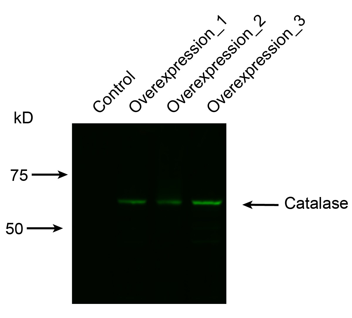
Application: Western BlotSample Tested: HEK293 human embryonic kidney cell lineSpecies: HumanVerified Customer | Posted 09/23/2021we overexpressed catalase contained plasmid in HEK293 cells and used 1:1000 dilution to detect this antibody.

-
Application: Western BlotSample Tested: Kidney tissue and HK-2 human proximal tubule epithelial cell lineSpecies: Mouse and HumanVerified Customer | Posted 01/05/2021
-
Application: Western BlotSample Tested: Hek 293T cell lysate and RAW264.7 whole cell lysateSpecies: Human and MouseVerified Customer | Posted 10/01/2019Restained with goat-anti-Catalase (1:50) - 2 nights 4C 2nd antibody Donkey-aa=nti-Goat-800 (1:5000) 30 min RT
-
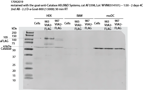
Application: Western BlotSample Tested: HEK 293 cell line, HEK 293 cell lysate, Monocyte-derived dendritic cells, RAW264.7 whole cell lysate, Hek 293T cell lysate and Monocyte-derived immature dendritic cellsSpecies: HumanVerified Customer | Posted 10/01/2019The blot was restained with the goat-anti-Catalase AB (R&D Systems, cat AF3398, Lot WVM0314101) - 1:50 - 2 days 4CThe blot was restained with the goat-anti-Catalase AB (R&D Systems, cat AF3398, Lot WVM0314101) - 1:50 - 2 days 4C (anti-FLAG-bands are from previous staining) 2nd AB - Donkey-anti-Goat-800 (1:5000) 30 min at RT The blot shows clear expression of Catalase by HEKs and human moDC (65kDa band)
-
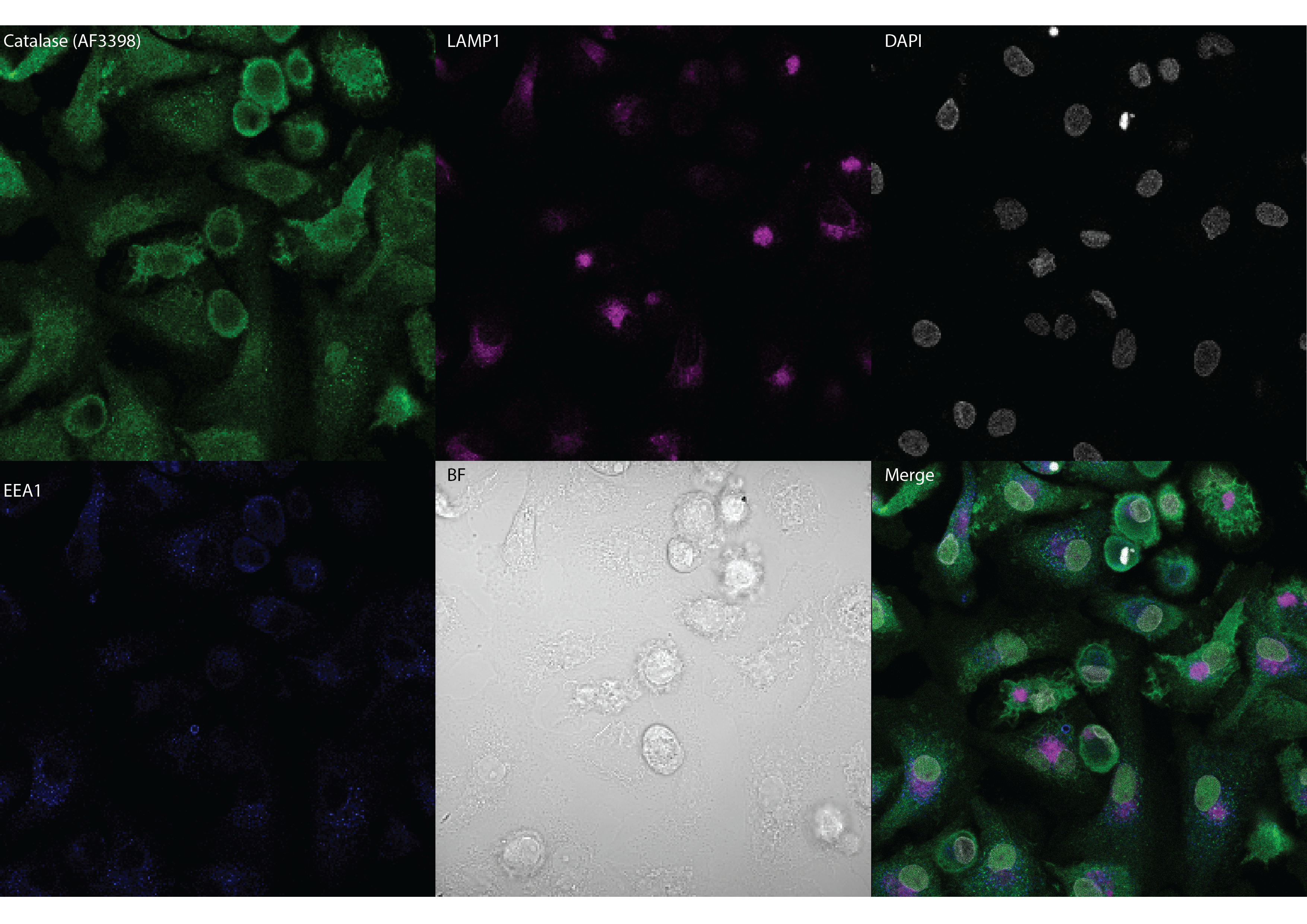
Application: ImmunocytochemistrySample Tested: Monocyte-derived immature dendritic cellsSpecies: HumanVerified Customer | Posted 10/01/2019Human moDCs stained with goat anti-Catalase antibody (AF3398) at 1:20 (10 ug/mL), rabbit anti-LAMP1 and mouse anti-EEA1.
-
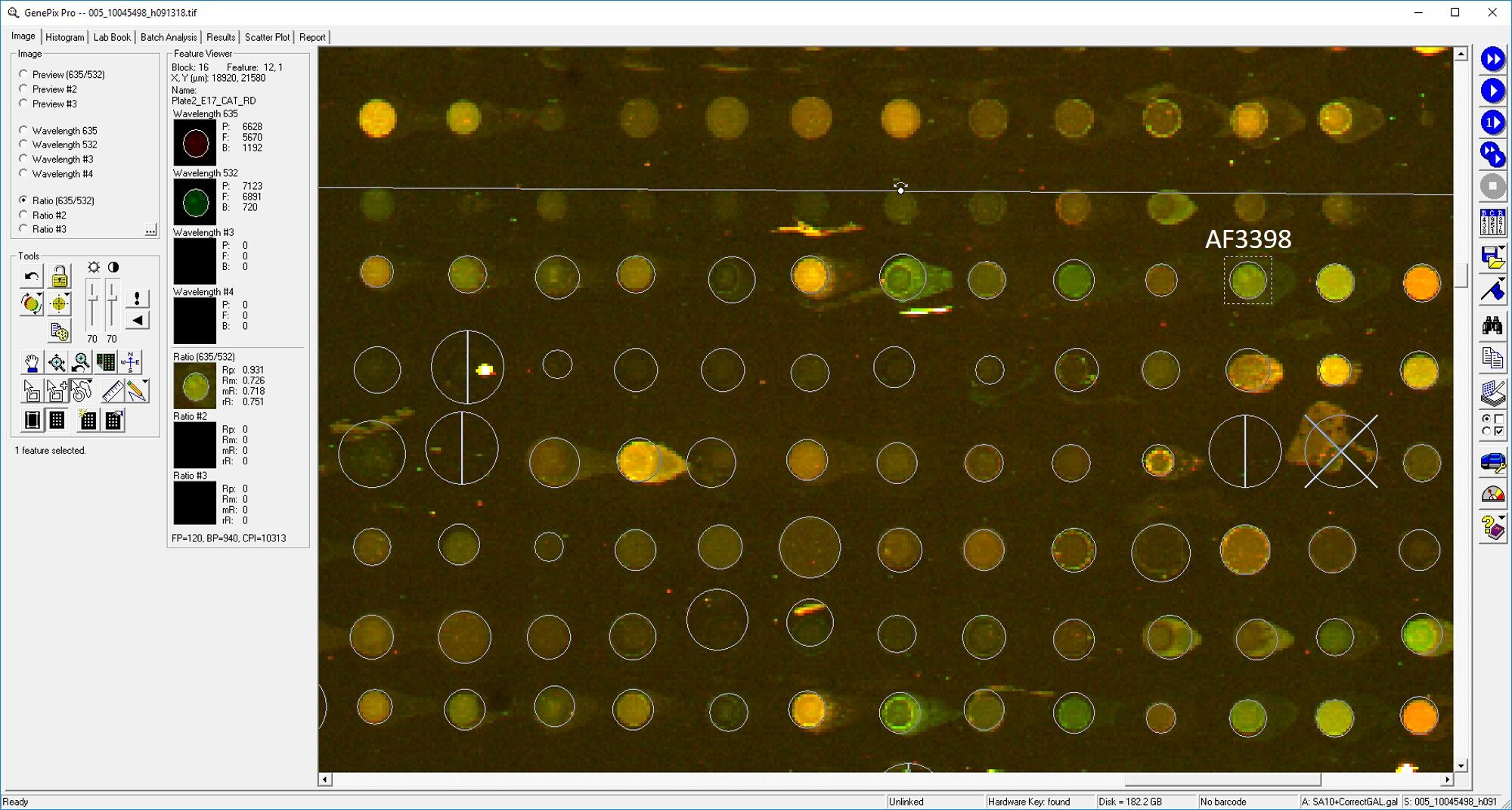
Application: MicroarraysSample Tested: EDTA PlasmaSpecies: HumanVerified Customer | Posted 03/11/2019
-
Application: MicroarraySample Tested: EDTA PlasmaSpecies: HumanVerified Customer | Posted 11/02/2018
There are no reviews that match your criteria.
Protocols
Find general support by application which include: protocols, troubleshooting, illustrated assays, videos and webinars.
- Appropriate Fixation of IHC/ICC Samples
- Cellular Response to Hypoxia Protocols
- ClariTSA™ Fluorophore Kits
- Detection & Visualization of Antibody Binding
- ICC Cell Smear Protocol for Suspension Cells
- ICC Immunocytochemistry Protocol Videos
- ICC for Adherent Cells
- Immunocytochemistry (ICC) Protocol
- Immunocytochemistry Troubleshooting
- Immunofluorescence of Organoids Embedded in Cultrex Basement Membrane Extract
- Immunohistochemistry (IHC) and Immunocytochemistry (ICC) Protocols
- Preparing Samples for IHC/ICC Experiments
- Preventing Non-Specific Staining (Non-Specific Binding)
- Primary Antibody Selection & Optimization
- Protocol for VisUCyte™ HRP Polymer Detection Reagent
- Protocol for the Fluorescent ICC Staining of Cell Smears - Graphic
- Protocol for the Fluorescent ICC Staining of Cultured Cells on Coverslips - Graphic
- Protocol for the Preparation and Fluorescent ICC Staining of Cells on Coverslips
- Protocol for the Preparation and Fluorescent ICC Staining of Non-adherent Cells
- Protocol for the Preparation and Fluorescent ICC Staining of Stem Cells on Coverslips
- Protocol for the Preparation of a Cell Smear for Non-adherent Cell ICC - Graphic
- R&D Systems Quality Control Western Blot Protocol
- TUNEL and Active Caspase-3 Detection by IHC/ICC Protocol
- The Importance of IHC/ICC Controls
- Troubleshooting Guide: Western Blot Figures
- Western Blot Conditions
- Western Blot Protocol
- Western Blot Protocol for Cell Lysates
- Western Blot Troubleshooting
- Western Blot Troubleshooting Guide
- View all Protocols, Troubleshooting, Illustrated assays and Webinars
Loading...