Cell Division Cycle 25A (CDC25A) phosphatase removes inorganic phosphate groups covalently attached to tyrosine, serine and threonine residues in proteins (1). Overexpression of CDC25A in small mammary carcinomas has been associated with a poor patient survival prognosis (2). Levels of CDC25A activity are highest in S phase of the cell cycle, where it is suspected to be involved in “checkpoint” control of cell cycle progression (3). Induction of DNA damage with ultraviolet or ionizing radiation causes an increase in CDC25A ubiquinylation and proteosomal degradation, leading to cell cycle block between G1 and S phases (4). One of its major substrates, the kinase CDC2, is activated by dephosphorylation (5). Recombinant CDC25A is truncated to remove the N-terminal regulatory domains and is fully active.

Key Product Details
Species Reactivity
Validated:
Human, Mouse, Rat
Cited:
Human, Mouse
Applications
Validated:
Immunohistochemistry, Western Blot
Cited:
Western Blot
Label
Unconjugated
Antibody Source
Monoclonal Mouse IgG2B Clone # 336445
Loading...
Product Specifications
Immunogen
E. coli-derived recombinant human CDC25A
Glu2-Leu523
Accession # AAA58415
Glu2-Leu523
Accession # AAA58415
Specificity
Detects human, mouse, and rat CDC25A in direct ELISAs and Western blots. In direct ELISA and Western blots, this antibody does not cross-react with recombinant human (rh) CDC25B or rhCDC25C.
Clonality
Monoclonal
Host
Mouse
Isotype
IgG2B
Scientific Data Images for CDC25A Antibody (336445)
Detection of Human/Mouse/Rat CDC25A by Western Blot.
Western blot shows lysates of MCF-7 human breast cancer cell line, CTLL-2 mouse cytotoxic T cell line, and Nb2-11 rat lymphoma cell line. PVDF membrane was probed with 1 µg/mL of Human/Mouse/Rat CDC25A Monoclonal Antibody (Catalog # MAB1648) followed by HRP-conjugated Anti-Mouse IgG Secondary Antibody (Catalog # HAF007). Specific bands were detected for CDC25A at approximately 55-60 kDa (as indicated). This experiment was conducted under reducing conditions and using Immunoblot Buffer Group 1.Applications for CDC25A Antibody (336445)
Application
Recommended Usage
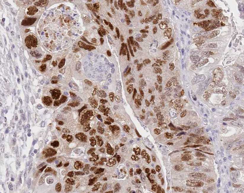
Immunohistochemistry
8-25 µg/mL
Sample: Immersion fixed paraffin-embedded sections of human breast cancer tissue
Sample: Immersion fixed paraffin-embedded sections of human breast cancer tissue
Western Blot
1 µg/mL
Sample: MCF-7 human breast cancer cell line, CTLL-2 mouse cytotoxic T cell line, and Nb2-11 rat lymphoma cell line
Sample: MCF-7 human breast cancer cell line, CTLL-2 mouse cytotoxic T cell line, and Nb2-11 rat lymphoma cell line
Reviewed Applications
Read 1 review rated 5 using MAB1648 in the following applications:
Formulation, Preparation, and Storage
Purification
Protein A or G purified from hybridoma culture supernatant
Reconstitution
Reconstitute at 0.5 mg/mL in sterile PBS. For liquid material, refer to CoA for concentration.
Loading...
Formulation
Lyophilized from a 0.2 μm filtered solution in PBS with Trehalose. *Small pack size (SP) is supplied either lyophilized or as a 0.2 µm filtered solution in PBS.
Shipping
Lyophilized product is shipped at ambient temperature. Liquid small pack size (-SP) is shipped with polar packs. Upon receipt, store immediately at the temperature recommended below.
Stability & Storage
Use a manual defrost freezer and avoid repeated freeze-thaw cycles.
- 12 months from date of receipt, -20 to -70 °C as supplied.
- 1 month, 2 to 8 °C under sterile conditions after reconstitution.
- 6 months, -20 to -70 °C under sterile conditions after reconstitution.
Calculators
Background: CDC25A
References
- Draetta, G. and J. Eckstein (1997) Biochim. Biophys. Acta. 1332:M53.
- Cangi, M. et al. (2000) J. Clin. Invest. 106:753.
- Hoffmann, I. et al. (1995) EMBO J. 13:4302.
- Sagata, N. (2002) Science 298:1905.
- Gautier, J. et al. (1991) Cell 67:197.
Long Name
Cell Division Cycle 25A
Alternate Names
CDC25A2, CDC25A2-CAG isoform, cell division cycle 25 homolog A (S. cerevisiae), cell division cycle 25 homolog A (S. pombe), cell division cycle 25A, Dual specificity phosphatase Cdc25A, EC 3.1.3.48, M-phase inducer phosphatase 1
Entrez Gene IDs
993 (Human)
Gene Symbol
CDC25A
UniProt
Additional CDC25A Products
Product Documents for CDC25A Antibody (336445)
Certificate of Analysis
To download a Certificate of Analysis, please enter a lot or batch number in the search box below.
Note: Certificate of Analysis not available for kit components.
Product Specific Notices for CDC25A Antibody (336445)
For research use only
Related Research Areas
Citations for CDC25A Antibody (336445)
 Powered by Bioz
Customer Reviews for CDC25A Antibody (336445) (1)
5 out of 5
1 Customer Rating
Have you used CDC25A Antibody (336445)?
Submit a review and receive an Amazon gift card!
$25/€18/£15/$25CAN/¥2500 Yen for a review with an image
$10/€7/£6/$10CAN/¥1110 Yen for a review without an image
Submit a review
Customer Images
Showing
1
-
1 of
1 review
Showing All
Filter By:
-
Application: ImmunohistochemistrySample Tested: Colon cancer cell lineSpecies: HumanVerified Customer | Posted 09/22/2021

There are no reviews that match your criteria.
Protocols
Find general support by application which include: protocols, troubleshooting, illustrated assays, videos and webinars.
- Antigen Retrieval Protocol (PIER)
- Antigen Retrieval for Frozen Sections Protocol
- Appropriate Fixation of IHC/ICC Samples
- Cellular Response to Hypoxia Protocols
- Chromogenic IHC Staining of Formalin-Fixed Paraffin-Embedded (FFPE) Tissue Protocol
- Chromogenic Immunohistochemistry Staining of Frozen Tissue
- ClariTSA™ Fluorophore Kits
- Detection & Visualization of Antibody Binding
- Fluorescent IHC Staining of Frozen Tissue Protocol
- Graphic Protocol for Heat-induced Epitope Retrieval
- Graphic Protocol for the Preparation and Fluorescent IHC Staining of Frozen Tissue Sections
- Graphic Protocol for the Preparation and Fluorescent IHC Staining of Paraffin-embedded Tissue Sections
- Graphic Protocol for the Preparation of Gelatin-coated Slides for Histological Tissue Sections
- IHC Sample Preparation (Frozen sections vs Paraffin)
- Immunofluorescent IHC Staining of Formalin-Fixed Paraffin-Embedded (FFPE) Tissue Protocol
- Immunohistochemistry (IHC) and Immunocytochemistry (ICC) Protocols
- Immunohistochemistry Frozen Troubleshooting
- Immunohistochemistry Paraffin Troubleshooting
- Preparing Samples for IHC/ICC Experiments
- Preventing Non-Specific Staining (Non-Specific Binding)
- Primary Antibody Selection & Optimization
- Protocol for Heat-Induced Epitope Retrieval (HIER)
- Protocol for Making a 4% Formaldehyde Solution in PBS
- Protocol for VisUCyte™ HRP Polymer Detection Reagent
- Protocol for the Preparation & Fixation of Cells on Coverslips
- Protocol for the Preparation and Chromogenic IHC Staining of Frozen Tissue Sections
- Protocol for the Preparation and Chromogenic IHC Staining of Frozen Tissue Sections - Graphic
- Protocol for the Preparation and Chromogenic IHC Staining of Paraffin-embedded Tissue Sections
- Protocol for the Preparation and Chromogenic IHC Staining of Paraffin-embedded Tissue Sections - Graphic
- Protocol for the Preparation and Fluorescent IHC Staining of Frozen Tissue Sections
- Protocol for the Preparation and Fluorescent IHC Staining of Paraffin-embedded Tissue Sections
- Protocol for the Preparation of Gelatin-coated Slides for Histological Tissue Sections
- R&D Systems Quality Control Western Blot Protocol
- TUNEL and Active Caspase-3 Detection by IHC/ICC Protocol
- The Importance of IHC/ICC Controls
- Troubleshooting Guide: Immunohistochemistry
- Troubleshooting Guide: Western Blot Figures
- Western Blot Conditions
- Western Blot Protocol
- Western Blot Protocol for Cell Lysates
- Western Blot Troubleshooting
- Western Blot Troubleshooting Guide
- View all Protocols, Troubleshooting, Illustrated assays and Webinars
Loading...