
Key Product Details
Species Reactivity
Validated:
Human, Mouse, Rat
Cited:
Human, Mouse, Rat, Rabbit
Applications
Validated:
Immunohistochemistry, Western Blot, Neutralization
Cited:
Immunohistochemistry, Immunohistochemistry-Frozen, Western Blot, Neutralization, Immunocytochemistry, In vivo assay
Label
Unconjugated
Antibody Source
Polyclonal Goat IgG
Loading...
Product Specifications
Immunogen
E. coli-derived recombinant mouse GDF-8/Myostatin
Asp268-Ser376
Accession # O08689
Asp268-Ser376
Accession # O08689
Specificity
Detects human, mouse, and rat GDF-8/Myostatin in direct ELISAs and Western blots.
Clonality
Polyclonal
Host
Goat
Isotype
IgG
Endotoxin Level
<0.10 EU per 1 μg of the antibody by the LAL method.
Scientific Data Images for GDF-8/Myostatin Antibody
Detection of Recombinant Human, Mouse, and Rat GDF-8/Myostatin by Western Blot.
Western blot shows 25 ng of Recombinant Human/Mouse/Rat GDF-8/Myostatin (788-G8), Recombinant Human/Mouse/Rat GDF-11/BMP-11 (1958-GD), Recombinant Human BMP-6 (507-BP), and Recombinant Mouse BMP-6 (6325-BM). PVDF Membrane was probed with 0.1 µg/mL of Goat Anti-Human/Mouse/Rat GDF-8/Myostatin Antigen Affinity-purified Polyclonal Antibody (Catalog # AF788) followed by HRP-conjugated Anti-Goat IgG Secondary Antibody (HAF109). A specific band was detected for GDF-8/Myostatin at approximately 14 kDa (as indicated). This experiment was conducted under reducing conditions and using Immunoblot Buffer Group 3.
GDF‑8/Myostatin in Mouse Embryo.
GDF-8/Myostatin was detected in immersion fixed frozen sections of mouse embryo (10 d.p.c., section through neural tube) using Goat Anti-Human/Mouse/Rat GDF-8/Myostatin Antigen Affinity-purified Polyclonal Antibody (Catalog # AF788) at 15 µg/mL overnight at 4 °C. Tissue was stained using the Anti-Goat HRP-DAB Cell & Tissue Staining Kit (brown; CTS008) and counterstained with hematoxylin (blue). View our protocol for Chromogenic IHC Staining of Frozen Tissue Sections.
Hemoglobin Expression Induced by GDF‑8/Myostatin and Neutralization by Mouse GDF‑8/Myostatin Antibody.
Recombinant Mouse GDF-8/Myostatin (788-G8) increases hemoglobin expression in the K562 human chronic myelogenous leukemia cell line in a dose-dependent manner (orange line), as measured by the psuedoperoxidase assay. Hemoglobin expression elicited by Recombinant Mouse GDF-8/Myostatin (30 ng/mL) is neutralized (green line) by increasing concentrations of Goat Anti-Human/Mouse/Rat GDF-8/Myostatin Antigen Affinity-purified Polyclonal Antibody (Catalog # AF788). The ND50 is typically 0.6-3 µg/mL.Applications for GDF-8/Myostatin Antibody
Application
Recommended Usage
Immunohistochemistry
5-15 µg/mL
Sample: Perfusion fixed frozen sections of mouse embryo (10 d.p.c., section through neural tube)
Sample: Perfusion fixed frozen sections of mouse embryo (10 d.p.c., section through neural tube)
Western Blot
0.1 µg/mL
Sample: Recombinant Human/Mouse/Rat GDF‑8/Myostatin (Catalog # 788-G8)
Sample: Recombinant Human/Mouse/Rat GDF‑8/Myostatin (Catalog # 788-G8)
Neutralization
Measured by its ability to neutralize GDF‑8/Myostatin-induced hemoglobin expression in the K562 human chronic myelogenous leukemia cell line. The Neutralization Dose (ND50) is typically 0.6-3 µg/mL in the presence of 30 ng/mL Recombinant Mouse GDF‑8/Myostatin.
Reviewed Applications
Read 4 reviews rated 3.5 using AF788 in the following applications:
Formulation, Preparation, and Storage
Purification
Antigen Affinity-purified
Reconstitution
Reconstitute at 0.2 mg/mL in sterile PBS. For liquid material, refer to CoA for concentration.
Loading...
Formulation
Lyophilized from a 0.2 μm filtered solution in PBS with Trehalose. See Certificate of Analysis for details.
*Small pack size (-SP) is supplied either lyophilized or as a 0.2 µm filtered solution in PBS.
*Small pack size (-SP) is supplied either lyophilized or as a 0.2 µm filtered solution in PBS.
Shipping
Lyophilized product is shipped at ambient temperature. Liquid small pack size (-SP) is shipped with polar packs. Upon receipt, store immediately at the temperature recommended below.
Stability & Storage
Use a manual defrost freezer and avoid repeated freeze-thaw cycles.
- 12 months from date of receipt, -20 to -70 °C as supplied.
- 1 month, 2 to 8 °C under sterile conditions after reconstitution.
- 6 months, -20 to -70 °C under sterile conditions after reconstitution.
Calculators
Background: GDF-8/Myostatin
References
- Storm, E.E. et al. (1994) Nature 368:639.
- Sharma, M. et al. (1999) J. Cell Physiol. 180:1.
- McPherron, A.C. et al. (1997) Nature 387:83.
- Lee, S.J. et al. (2001) Proc. Natl. Acad. Sci. USA 98:9306.
- Kim, H.S. et al. (2001) Biochem. Biophys. Res. Commun. 281:902.
Long Name
Growth Differentiation Factor 8
Alternate Names
GDF8, MSLHP, MSTN, Myostatin
Gene Symbol
MSTN
UniProt
Additional GDF-8/Myostatin Products
Product Documents for GDF-8/Myostatin Antibody
Certificate of Analysis
To download a Certificate of Analysis, please enter a lot or batch number in the search box below.
Note: Certificate of Analysis not available for kit components.
Product Specific Notices for GDF-8/Myostatin Antibody
For research use only
Related Research Areas
Citations for GDF-8/Myostatin Antibody
 Powered by Bioz
Customer Reviews for GDF-8/Myostatin Antibody (4)
3.5 out of 5
4 Customer Ratings
Have you used GDF-8/Myostatin Antibody?
Submit a review and receive an Amazon gift card!
$25/€18/£15/$25CAN/¥2500 Yen for a review with an image
$10/€7/£6/$10CAN/¥1110 Yen for a review without an image
Submit a review
Customer Images
Showing
1
-
4 of
4 reviews
Showing All
Filter By:
-
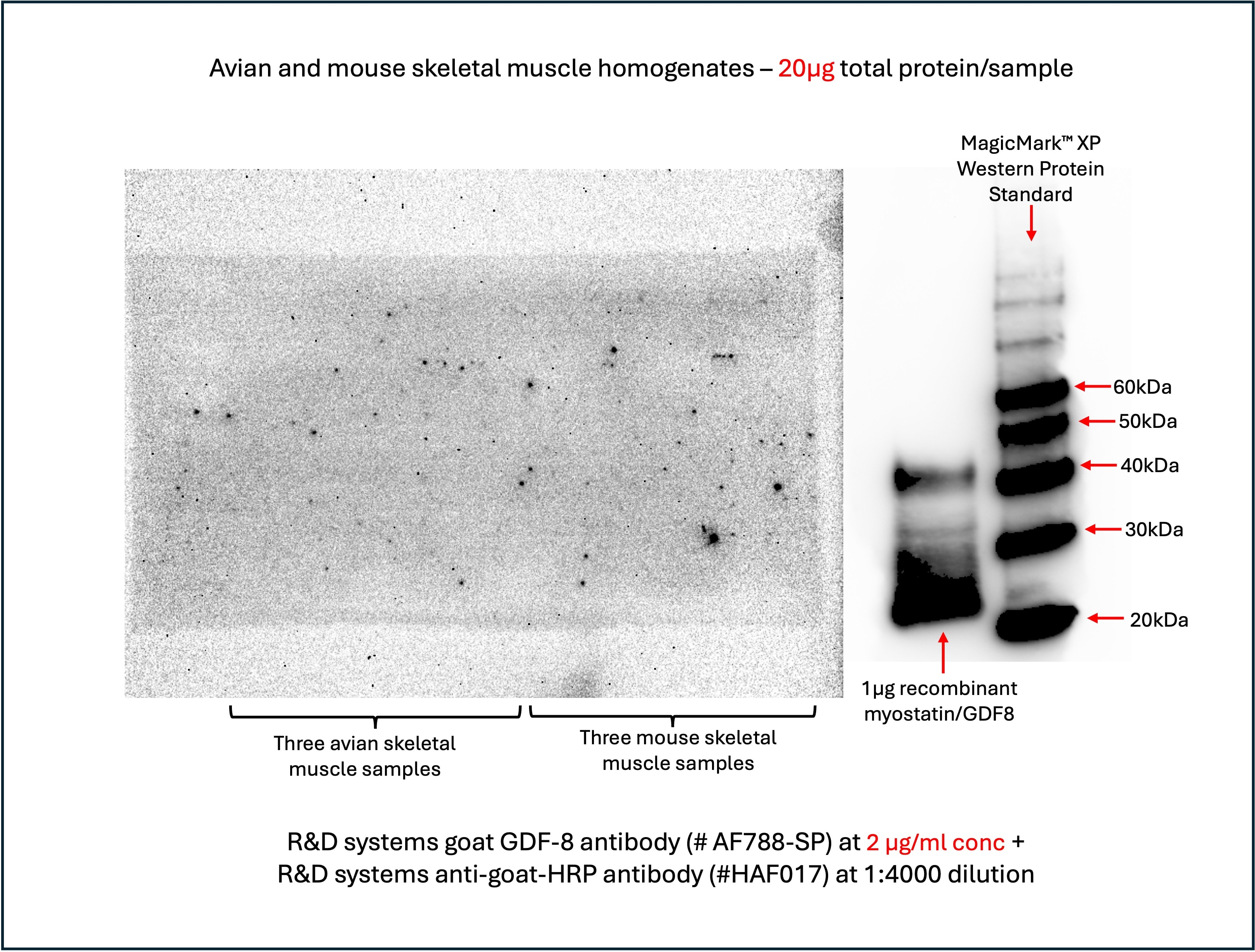
Application: Western BlotSample Tested: Skeletal muscle tissueSpecies: Avian and MouseVerified Customer | Posted 04/15/2024The test samples consisted of skeletal muscle tissue homogenates derived from mouse and avian muscle tissues. The homogenisation buffer contained protease inhibitor cocktail. The positive control used was 1μg of R&D systems recombinant myostatin/GDF8 (#788-G8). I had loaded 20μg of total protein for each of the avian and mouse samples, along with1μg of R&D systems recombinant myostatin/GDF8 (#788-G8) into a 4-15% gel. The electrophoresis was performed with MES running buffer at 90V constant current, and the proteins were subsequently blotted onto a 0.2μm nitrocellulose membrane. To check for transfer efficiency and protein integrity, i had stained the blot with Ponceau S and found that the transfer was efficient and the proteins were not degraded. Subsequently, i destained the blot with TBS-T and deionised water, and blocked the blots with 5% milk in TBS-T. Prior to incubating the membrane in primary antibody, i had cut the membrane such that one section contained all the animal tissue samples and the other section contained the recombinant GDF-8/myostatin and the western standard. The cut sections were incubated separately with identical concentrations of primary and secondary antibodies - R&D systems goat polyclonal GDF8 antibody (AF788-SP) at 2 µg/mL concentration, and R&D system anti-goat secondary antibody (HAF017) at 1:4000 dilution. The recombinant GDF-8/Myostatin positive control and the western standards produced clear and robust signals. However, the sections containing the samples derived from animal tissues (avian and mouse) failed to produce any discernible signal. I had repeated the same experiment above three times with increasing concentrations of primary antibody - 0.1, 0.25 and 2 µg/mL. However, all three experiments yielded the same result - strong signal for the recombinant GDF-8/Myostatin, but no visible signal detected with the avian and mouse tissue samples.
 Bio-Techne ResponseThank you for reviewing our product. We are sorry to hear that this product did not perform as expected. We have been in touch with the customer to resolve this issue according to our Product Guarantee and to the customer’s satisfaction.
 -
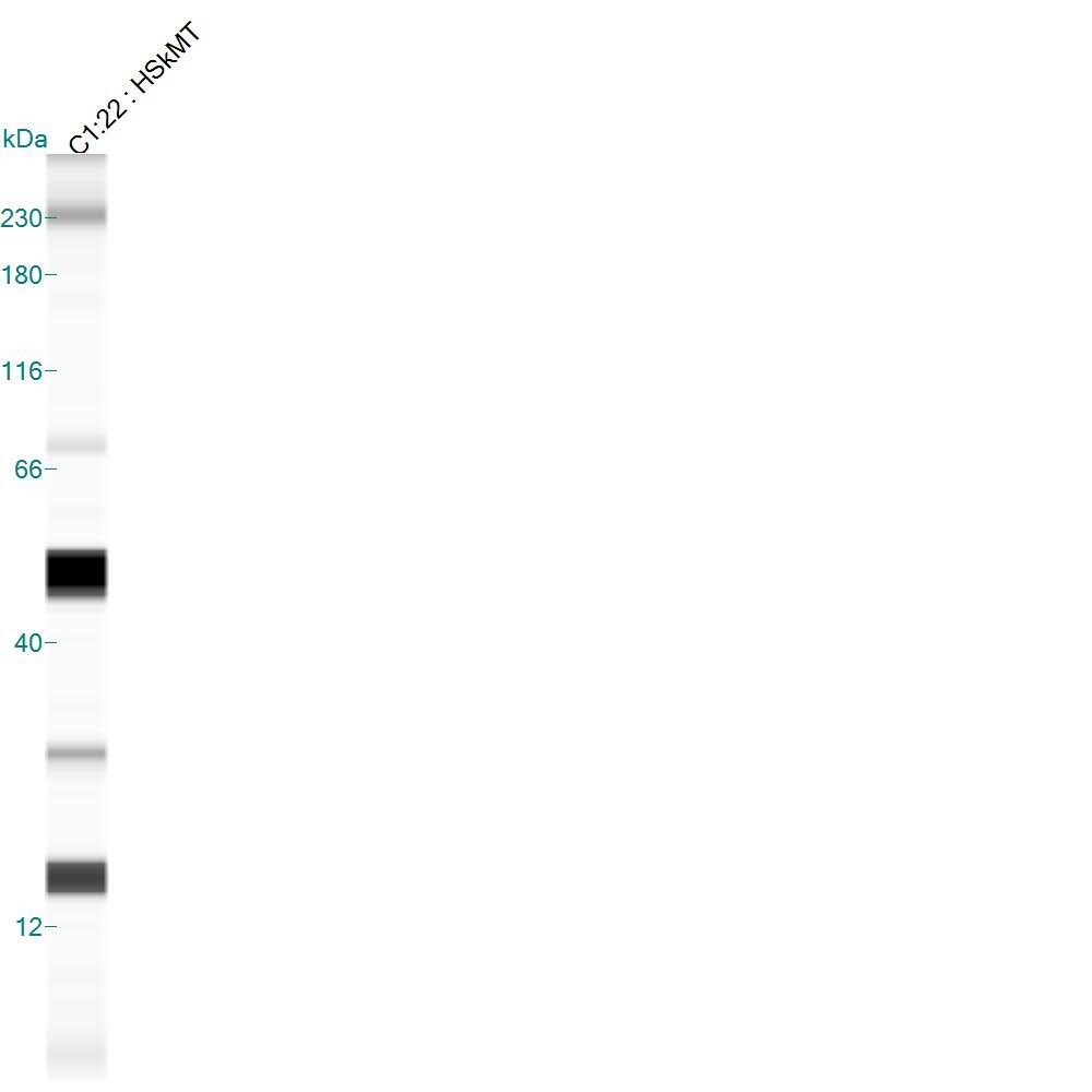
Application: Simple WesternSample Tested: Human Skeletal Muscle LysateSpecies: HumanVerified Customer | Posted 06/23/2015Simple Western. 0.2 ug/uL human skeletal muscle tissue lysate. Precursor observed ~51 kDa; mature form observed at ~29 kDa.
-
Application: Western BlotSample Tested: See PMID 23829672Species: HumanVerified Customer | Posted 01/09/2015
-
Application: Western BlotSample Tested: See PMID 23297411Species: MouseVerified Customer | Posted 01/09/2015
There are no reviews that match your criteria.
Protocols
Find general support by application which include: protocols, troubleshooting, illustrated assays, videos and webinars.
- Antigen Retrieval Protocol (PIER)
- Antigen Retrieval for Frozen Sections Protocol
- Appropriate Fixation of IHC/ICC Samples
- Cellular Response to Hypoxia Protocols
- Chromogenic IHC Staining of Formalin-Fixed Paraffin-Embedded (FFPE) Tissue Protocol
- Chromogenic Immunohistochemistry Staining of Frozen Tissue
- ClariTSA™ Fluorophore Kits
- Detection & Visualization of Antibody Binding
- Fluorescent IHC Staining of Frozen Tissue Protocol
- Graphic Protocol for Heat-induced Epitope Retrieval
- Graphic Protocol for the Preparation and Fluorescent IHC Staining of Frozen Tissue Sections
- Graphic Protocol for the Preparation and Fluorescent IHC Staining of Paraffin-embedded Tissue Sections
- Graphic Protocol for the Preparation of Gelatin-coated Slides for Histological Tissue Sections
- IHC Sample Preparation (Frozen sections vs Paraffin)
- Immunofluorescent IHC Staining of Formalin-Fixed Paraffin-Embedded (FFPE) Tissue Protocol
- Immunohistochemistry (IHC) and Immunocytochemistry (ICC) Protocols
- Immunohistochemistry Frozen Troubleshooting
- Immunohistochemistry Paraffin Troubleshooting
- Preparing Samples for IHC/ICC Experiments
- Preventing Non-Specific Staining (Non-Specific Binding)
- Primary Antibody Selection & Optimization
- Protocol for Heat-Induced Epitope Retrieval (HIER)
- Protocol for Making a 4% Formaldehyde Solution in PBS
- Protocol for VisUCyte™ HRP Polymer Detection Reagent
- Protocol for the Preparation & Fixation of Cells on Coverslips
- Protocol for the Preparation and Chromogenic IHC Staining of Frozen Tissue Sections
- Protocol for the Preparation and Chromogenic IHC Staining of Frozen Tissue Sections - Graphic
- Protocol for the Preparation and Chromogenic IHC Staining of Paraffin-embedded Tissue Sections
- Protocol for the Preparation and Chromogenic IHC Staining of Paraffin-embedded Tissue Sections - Graphic
- Protocol for the Preparation and Fluorescent IHC Staining of Frozen Tissue Sections
- Protocol for the Preparation and Fluorescent IHC Staining of Paraffin-embedded Tissue Sections
- Protocol for the Preparation of Gelatin-coated Slides for Histological Tissue Sections
- R&D Systems Quality Control Western Blot Protocol
- TUNEL and Active Caspase-3 Detection by IHC/ICC Protocol
- The Importance of IHC/ICC Controls
- Troubleshooting Guide: Immunohistochemistry
- Troubleshooting Guide: Western Blot Figures
- Western Blot Conditions
- Western Blot Protocol
- Western Blot Protocol for Cell Lysates
- Western Blot Troubleshooting
- Western Blot Troubleshooting Guide
- View all Protocols, Troubleshooting, Illustrated assays and Webinars
Loading...