
Key Product Details
Species Reactivity
Human, Mouse, Rat
Applications
Western Blot, Immunocytochemistry, Simple Western
Label
Unconjugated
Antibody Source
Polyclonal Goat IgG
Loading...
Product Specifications
Immunogen
E. coli-derived recombinant human IKK gamma
Met1-Glu419
Accession # Q9Y6K9
Met1-Glu419
Accession # Q9Y6K9
Specificity
Detects human, mouse, and rat IKK gamma in Western blots.
Clonality
Polyclonal
Host
Goat
Isotype
IgG
Scientific Data Images for IKK gamma Antibody
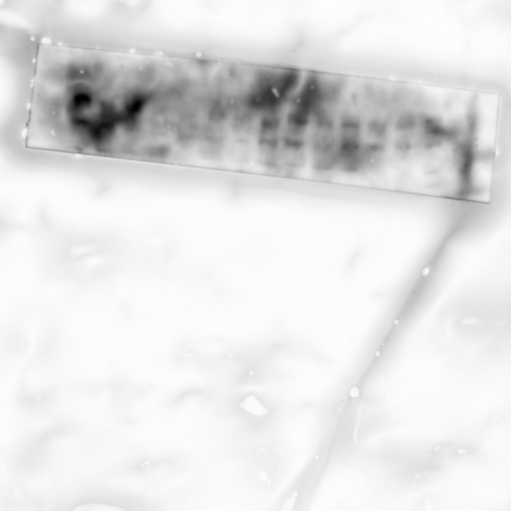
Detection of Human/Mouse/Rat IKK gamma by Western Blot.
Western blot shows lysates of MDA-MB-468 human breast cancer cell line and HeLa human cervical epithelial carcinoma cell line. PVDF membrane was probed with 0.5 µg/mL Goat Anti-Human/Mouse/Rat IKK gamma Antigen Affinity-purified Polyclonal Antibody (Catalog # AF2684) followed by HRP-conjugated Anti-Goat IgG Secondary Antibody (Catalog # HAF109). For additional reference, recombinant human IKK gamma (1 ng) was included. Specific bands for IKK gamma were detected at approximately 50 and 55 kDa (as indicated). This experiment was conducted under reducing conditions and using Immunoblot Buffer Group 1.
Detection of Human IKK gamma by Simple WesternTM.
Simple Western lane view shows lysates of MDA-MB-468 human breast cancer cell line and HeLa human cervical epithelial carcinoma cell line, loaded at 0.2 mg/mL. Specific bands were detected for IKK gamma at approximately 47-56 kDa (as indicated) using 5 µg/mL of Goat Anti-Human/Mouse/Rat IKK gamma Antigen Affinity-purified Polyclonal Antibody (Catalog # AF2684) followed by 1:50 dilution of HRP-conjugated Anti-Goat IgG Secondary Antibody (Catalog # HAF109). This experiment was conducted under reducing conditions and using the 12-230 kDa separation system.
IKK gamma in MCF‑7 Human Cell Line.
IKK gamma was detected in immersion fixed MCF-7 human breast cancer cell line using Goat Anti-Human/Mouse/Rat IKK gamma Antigen Affinity-purified Polyclonal Antibody (Catalog # AF2684) at 15 µg/mL for 3 hours at room temperature. Cells were stained using the NorthernLights™ 557-conjugated Anti-Goat IgG Secondary Antibody (red; Catalog # NL001) and counterstained with DAPI (blue). Specific staining was localized to cytoplasm. View our protocol for Fluorescent ICC Staining of Cells on Coverslips.Applications for IKK gamma Antibody
Application
Recommended Usage
Immunocytochemistry
5-15 µg/mL
Sample: Immersion fixed MCF-7 human breast cancer cell line and HeLa human cervical epithelial carcinoma cell line
Sample: Immersion fixed MCF-7 human breast cancer cell line and HeLa human cervical epithelial carcinoma cell line
Simple Western
5 µg/mL
Sample: MDA‑MB‑468 human breast cancer cell line and HeLa human cervical epithelial carcinoma cell line
Sample: MDA‑MB‑468 human breast cancer cell line and HeLa human cervical epithelial carcinoma cell line
Western Blot
0.5 µg/mL
Sample: MDA-MB-468 human breast cancer cell line and HeLa human cervical epithelial carcinoma cell line
Sample: MDA-MB-468 human breast cancer cell line and HeLa human cervical epithelial carcinoma cell line
Reviewed Applications
Read 1 review rated 2 using AF2684 in the following applications:
Formulation, Preparation, and Storage
Purification
Antigen Affinity-purified
Reconstitution
Reconstitute at 0.2 mg/mL in sterile PBS. For liquid material, refer to CoA for concentration.
Loading...
Formulation
Lyophilized from a 0.2 μm filtered solution in PBS with Trehalose. *Small pack size (SP) is supplied either lyophilized or as a 0.2 µm filtered solution in PBS.
Shipping
Lyophilized product is shipped at ambient temperature. Liquid small pack size (-SP) is shipped with polar packs. Upon receipt, store immediately at the temperature recommended below.
Stability & Storage
Use a manual defrost freezer and avoid repeated freeze-thaw cycles.
- 12 months from date of receipt, -20 to -70 °C as supplied.
- 1 month, 2 to 8 °C under sterile conditions after reconstitution.
- 6 months, -20 to -70 °C under sterile conditions after reconstitution.
Calculators
Background: IKK gamma
Long Name
IkB Kinase gamma
Alternate Names
FIP3P, IKBKG, IKKAP1, NEMO
Gene Symbol
IKBKG
UniProt
Additional IKK gamma Products
Product Documents for IKK gamma Antibody
Certificate of Analysis
To download a Certificate of Analysis, please enter a lot or batch number in the search box below.
Note: Certificate of Analysis not available for kit components.
Product Specific Notices for IKK gamma Antibody
For research use only
Citations for IKK gamma Antibody
 Powered by Bioz
Customer Reviews for IKK gamma Antibody (1)
2 out of 5
1 Customer Rating
Have you used IKK gamma Antibody?
Submit a review and receive an Amazon gift card!
$25/€18/£15/$25CAN/¥2500 Yen for a review with an image
$10/€7/£6/$10CAN/¥1110 Yen for a review without an image
Submit a review
Customer Images
Showing
1
-
1 of
1 review
Showing All
Filter By:
-
Application: Western BlotSample Tested: Lung single-cell suspensionSpecies: MouseVerified Customer | Posted 06/06/2019
 Bio-Techne ResponseThank you for reviewing our product. We are sorry to hear that this product did not perform as expected. We have been in touch with the customer to resolve this issue according to our Product Guarantee and to the customer’s satisfaction.
There are no reviews that match your criteria.
Protocols
Find general support by application which include: protocols, troubleshooting, illustrated assays, videos and webinars.
- Appropriate Fixation of IHC/ICC Samples
- Cellular Response to Hypoxia Protocols
- ClariTSA™ Fluorophore Kits
- Detection & Visualization of Antibody Binding
- ICC Cell Smear Protocol for Suspension Cells
- ICC Immunocytochemistry Protocol Videos
- ICC for Adherent Cells
- Immunocytochemistry (ICC) Protocol
- Immunocytochemistry Troubleshooting
- Immunofluorescence of Organoids Embedded in Cultrex Basement Membrane Extract
- Immunohistochemistry (IHC) and Immunocytochemistry (ICC) Protocols
- Preparing Samples for IHC/ICC Experiments
- Preventing Non-Specific Staining (Non-Specific Binding)
- Primary Antibody Selection & Optimization
- Protocol for VisUCyte™ HRP Polymer Detection Reagent
- Protocol for the Fluorescent ICC Staining of Cell Smears - Graphic
- Protocol for the Fluorescent ICC Staining of Cultured Cells on Coverslips - Graphic
- Protocol for the Preparation and Fluorescent ICC Staining of Cells on Coverslips
- Protocol for the Preparation and Fluorescent ICC Staining of Non-adherent Cells
- Protocol for the Preparation and Fluorescent ICC Staining of Stem Cells on Coverslips
- Protocol for the Preparation of a Cell Smear for Non-adherent Cell ICC - Graphic
- R&D Systems Quality Control Western Blot Protocol
- TUNEL and Active Caspase-3 Detection by IHC/ICC Protocol
- The Importance of IHC/ICC Controls
- Troubleshooting Guide: Western Blot Figures
- Western Blot Conditions
- Western Blot Protocol
- Western Blot Protocol for Cell Lysates
- Western Blot Troubleshooting
- Western Blot Troubleshooting Guide
- View all Protocols, Troubleshooting, Illustrated assays and Webinars


