The c-Jun N-terminal kinases (JNKs) are encoded by three genes: JNK1, JNK2, and JNK3. Members of the MAPK superfamily, JNKs are activated by environmental stresses and inflammatory cytokines. JNK1, also known as SAPK1 gamma and MAPK8, is expressed as four isoforms generated by alternative splicing. JNK1 is activated by dual phosphorylation at T183 and Y185 by the MAPK kinases MKK4 and/or MKK7.

Key Product Details
Species Reactivity
Validated:
Human, Mouse, Rat
Cited:
Human, Mouse
Applications
Validated:
Western Blot, Immunocytochemistry
Cited:
Western Blot, ELISA Development
Label
Unconjugated
Antibody Source
Monoclonal Mouse IgG1 Clone # 228601
Loading...
Product Specifications
Immunogen
E. coli-derived recombinant human JNK1 isoform 2
Ser2-Ile384
Accession # P45983-1
Ser2-Ile384
Accession # P45983-1
Specificity
Detects human JNK1 in direct ELISAs and human, mouse, and rat JNK1 expressed predominantly as p46 JNK (isoforms 2 and/or 3, both 384 aa), with lesser amounts of p54 JNK (isoforms 1 and/or 4, both 427 aa), in Western blots. In Western blots, no cross-reactivity with recombinant human (rh) JNK2 or rhJNK3 is observed.
Clonality
Monoclonal
Host
Mouse
Isotype
IgG1
Scientific Data Images for JNK1 Antibody (228601)
Detection of Human JNK1 by Western Blot.
Western blot shows recombinant human JNK1, JNK2, and JNK3 (1 ng/lane). PVDF membrane was probed with 1 µg/mL Mouse Anti-Human/Mouse/Rat JNK1 Monoclonal Antibody (Catalog # MAB17761) followed by HRP-conjugated Anti-Mouse IgG Secondary Antibody (Catalog # HAF007). A specific band for JNK1 was detected at approximately 46 kDa (as indicated). This experiment was conducted under reducing conditions and using Immunoblot Buffer Group 4.
Detection of Human, Mouse, and Rat JNK1 by Western Blot.
Western blot shows lysates of CHP-100 human neuroblastoma cell line, C6 rat glioma cell line, and C2C12 mouse myoblast cell line. PVDF membrane was probed with 1 µg/mL Mouse Anti-Human/Mouse/Rat JNK1 Monoclonal Antibody (Catalog # MAB17761) followed by HRP-conjugated Anti-Mouse IgG Secondary Antibody (Catalog # HAF007). A specific band for JNK1 was detected at approximately 46 kDa and 54 kDa (as indicated). This experiment was conducted under reducing conditions and using Immunoblot Buffer Group 4.
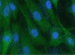
JNK1 in MCF‑7 Human Cell Line.
JNK1 was detected in immersion fixed MCF-7 human breast cancer cell line using Mouse Anti-Human/Mouse/Rat JNK1 Monoclonal Antibody (Catalog # MAB17761) at 10 µg/mL for 3 hours at room temperature. Cells were stained using the NorthernLights™ 557-conjugated Anti-Mouse IgG Secondary Antibody (red; Catalog # NL007) and counterstained with DAPI (blue). Specific staining was localized to cell surfaces. View our protocol for Fluorescent ICC Staining of Cells on Coverslips.Applications for JNK1 Antibody (228601)
Application
Recommended Usage
Immunocytochemistry
8-25 µg/mL
Sample: Immersion fixed MCF-7 human breast cancer cell line
Sample: Immersion fixed MCF-7 human breast cancer cell line
Western Blot
1 µg/mL
Sample: CHP-100 human neuroblastoma cell line, C6 rat glioma cell line, and C2C12 mouse myoblast cell line
Sample: CHP-100 human neuroblastoma cell line, C6 rat glioma cell line, and C2C12 mouse myoblast cell line
Reviewed Applications
Read 3 reviews rated 4.3 using MAB17761 in the following applications:
Formulation, Preparation, and Storage
Purification
Protein A or G purified from hybridoma culture supernatant
Reconstitution
Reconstitute at 0.5 mg/mL in sterile PBS containing 0.02% NaN3. For liquid material, refer to CoA for concentration.
Formulation
Lyophilized from a 0.2 μm filtered solution in PBS with Trehalose. *Small pack size (SP) is supplied either lyophilized or as a 0.2 µm filtered solution in PBS.
Shipping
Lyophilized product is shipped at ambient temperature. Liquid small pack size (-SP) is shipped with polar packs. Upon receipt, store immediately at the temperature recommended below.
Stability & Storage
Use a manual defrost freezer and avoid repeated freeze-thaw cycles.
- 12 months from date of receipt, -20 to -70 °C as supplied.
- 1 month, 2 to 8 °C under sterile conditions after reconstitution.
- 6 months, -20 to -70 °C under sterile conditions after reconstitution.
Calculators
Background: JNK1
Long Name
C-Jun N-terminal Kinase 1
Alternate Names
MAPK8, PRKM8, SAPK1
Gene Symbol
MAPK8
UniProt
Additional JNK1 Products
Product Documents for JNK1 Antibody (228601)
Certificate of Analysis
To download a Certificate of Analysis, please enter a lot or batch number in the search box below.
Note: Certificate of Analysis not available for kit components.
Product Specific Notices for JNK1 Antibody (228601)
For research use only
Citations for JNK1 Antibody (228601)
 Powered by Bioz
Customer Reviews for JNK1 Antibody (228601) (3)
4.3 out of 5
3 Customer Ratings
Have you used JNK1 Antibody (228601)?
Submit a review and receive an Amazon gift card!
$25/€18/£15/$25CAN/¥2500 Yen for a review with an image
$10/€7/£6/$10CAN/¥1110 Yen for a review without an image
Submit a review
Customer Images
Showing
1
-
3 of
3 reviews
Showing All
Filter By:
-
Application: Immunocytochemistry/ImmunofluorescenceSample Tested: Epithelial cellsSpecies: MouseVerified Customer | Posted 01/26/2022

-
Application: Western BlotSample Tested: Prostate cancerSpecies: HumanVerified Customer | Posted 01/25/2018
-
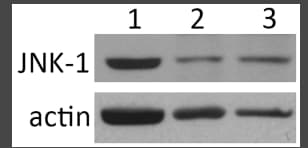
Application: Western BlotSample Tested: RetinaSpecies: Rat and MouseVerified Customer | Posted 12/29/2016Rat (lane 1), wild-type mouse (lane 2), and JNK2 ko mouse (lane 3) retinal tissue lysates were probed for JNK1 (upper panel) followed by reprobing for actin (lower panel) to compare sample load.JNK1 antibody used at 1:2000 over night, followed by donkey anti mouse secondary antibody conjugated to peroxidase and chemiluminescence detection
There are no reviews that match your criteria.
Protocols
Find general support by application which include: protocols, troubleshooting, illustrated assays, videos and webinars.
- Appropriate Fixation of IHC/ICC Samples
- Cellular Response to Hypoxia Protocols
- ClariTSA™ Fluorophore Kits
- Detection & Visualization of Antibody Binding
- ICC Cell Smear Protocol for Suspension Cells
- ICC Immunocytochemistry Protocol Videos
- ICC for Adherent Cells
- Immunocytochemistry (ICC) Protocol
- Immunocytochemistry Troubleshooting
- Immunofluorescence of Organoids Embedded in Cultrex Basement Membrane Extract
- Immunohistochemistry (IHC) and Immunocytochemistry (ICC) Protocols
- Preparing Samples for IHC/ICC Experiments
- Preventing Non-Specific Staining (Non-Specific Binding)
- Primary Antibody Selection & Optimization
- Protocol for VisUCyte™ HRP Polymer Detection Reagent
- Protocol for the Fluorescent ICC Staining of Cell Smears - Graphic
- Protocol for the Fluorescent ICC Staining of Cultured Cells on Coverslips - Graphic
- Protocol for the Preparation and Fluorescent ICC Staining of Cells on Coverslips
- Protocol for the Preparation and Fluorescent ICC Staining of Non-adherent Cells
- Protocol for the Preparation and Fluorescent ICC Staining of Stem Cells on Coverslips
- Protocol for the Preparation of a Cell Smear for Non-adherent Cell ICC - Graphic
- R&D Systems Quality Control Western Blot Protocol
- TUNEL and Active Caspase-3 Detection by IHC/ICC Protocol
- The Importance of IHC/ICC Controls
- Troubleshooting Guide: Western Blot Figures
- Western Blot Conditions
- Western Blot Protocol
- Western Blot Protocol for Cell Lysates
- Western Blot Troubleshooting
- Western Blot Troubleshooting Guide
- View all Protocols, Troubleshooting, Illustrated assays and Webinars
Loading...
Associated Pathways


TGF-beta Signaling Pathways
 Toll-Like Receptor Signaling Pathways
 Wnt Signaling Pathways: beta-Catenin-dependent Wnt Signaling
