
Kelch-like ECH-associated protein 1 (Keap1) is a 624 amino acid, 69 kDa protein which interacts with the transcription factor NF-E2-related factor 2 (Nrf2). Keap1 represses Nrf2 function by sequestering Nrf2 in the cytoplasm. Keap1 contains an N-terminal BTB domain and six C-terminal KELCH domains (aa 327‑611) that interact with Nrf2. Dissociation of the two proteins in response to redox-sensitive cell stress is followed by the translocation of Nrf2 to the nucleus and transcription of detoxifying/oxidative stress enzyme genes.


Scientific images may be resized, cropped, or adjusted for brightness or contrast for presentation purposes. These updates are limited to presentation and do not affect the underlying scientific interpretation of the data. Where available, additional source data may be provided upon request.
Key Product Details
Validated by
Knockout/Knockdown
Species Reactivity
Validated:
Human, Mouse, Rat
Cited:
Mouse, Rat
Applications
Validated:
Knockout Validated, Western Blot, Simple Western
Cited:
Western Blot
Label
Unconjugated
Antibody Source
Monoclonal Mouse IgG2B Clone # 333116
Loading...
Product Specifications
Immunogen
E. coli-derived recombinant human Keap1
Ala90-Ile250
Accession # Q14145
Ala90-Ile250
Accession # Q14145
Specificity
Detects human, mouse and rat Keap1 in Western blots.
Clonality
Monoclonal
Host
Mouse
Isotype
IgG2B
Scientific Data Images for Keap1 Antibody (333116)
Detection of Human/Mouse/Rat Keap1 by Western Blot.
Western blot shows lysates of Jurkat human acute T cell leukemia cell line, Raji human Burkitt's lymphoma cell line, Neuro-2A mouse neuroblastoma cell line, and L6 rat myoblast cell line. PVDF membrane was probed with 1 µg/mL of Human/Mouse/Rat Keap1 Monoclonal Antibody (Catalog # MAB3024) followed by HRP-conjugated Anti-Mouse IgG Secondary Antibody (Catalog # HAF007). A specific band was detected for Keap1 at approximately 66 kDa (as indicated). This experiment was conducted under reducing conditions and using Immunoblot Buffer Group 4.
Detection of Human Keap1 by Simple WesternTM.
Simple Western lane view shows lysates of Jurkat human acute T cell leukemia cell line, loaded at 0.2 mg/mL. A specific band was detected for Keap1 at approximately 75 kDa (as indicated) using 20 µg/mL of Mouse Anti-Human/Mouse/Rat Keap1 Monoclonal Antibody (Catalog # MAB3024). This experiment was conducted under reducing conditions and using the 12-230 kDa separation system. Non-specific interaction with the 230 kDa Simple Western standard may be seen with this antibody.
Western Blot Shows Human Keap1 Specificity by Using Knockout Cell Line.
Western blot shows lysates of Jurkat human acute T cell leukemia parental cell line and Keap1 Jurkat cell line (KO). PVDF membrane was probed with 1 µg/mL of Mouse Anti-Human/Mouse/Rat Keap1 Monoclonal Antibody (Catalog # MAB3024) followed by HRP-conjugated Anti-Mouse IgG Secondary Antibody (Catalog # HAF018). A specific band was detected for Keap1 at approximately 66 kDa (as indicated) in the parental Jurkat cell line, but is not detectable in knockout Jurkat cell line. GAPDH (Catalog # MAB5718) is shown as a loading control. This experiment was conducted under reducing conditions and using Immunoblot Buffer Group 1.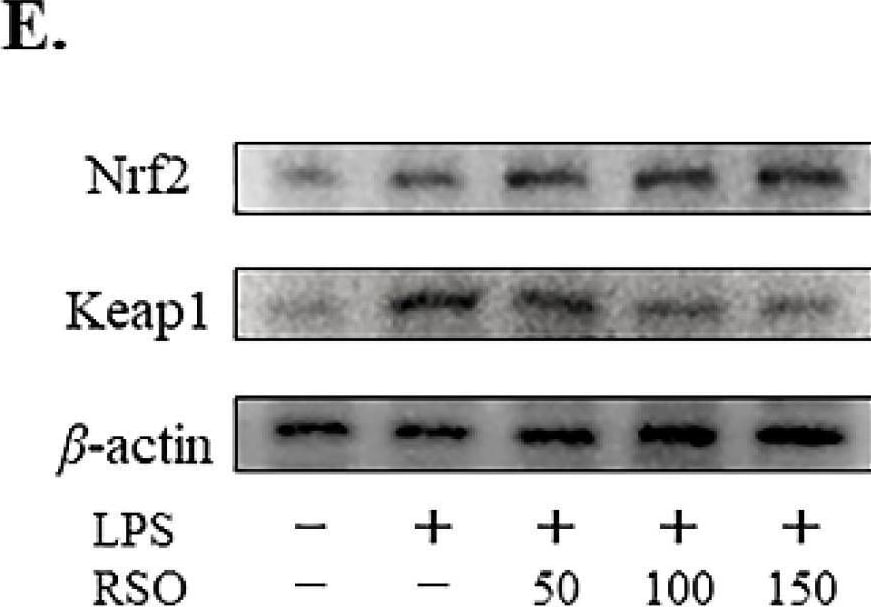
Detection of Keap1 by Western Blot
Effect of RSO on Nrf2-Keap1 pathway of LPS-induced RAW 264.7 cells. Relative mRNA expression of Nrf2 (A), HO-1 (B), NQO1 (C), and Keap1 (D) genes normalized to beta -actin and expressed as fold change of the control group, n = 3. (E) The protein levels of Nrf2 and Keap1 were analyzed by immunoblotting with beta -actin as the internal reference protein for normalization, n = 3. Relative protein expression of Nrf2 (F) and Keap1 (G) were expressed as fold change of the LPS-induced group. ** p < 0.01 vs control group; # p < 0.05, ## p < 0.01 vs LPS-induced group. Image collected and cropped by CiteAb from the following open publication (https://www.mdpi.com/2072-6643/14/7/1349), licensed under a CC-BY license. Not internally tested by R&D Systems.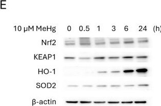
Detection of Keap1 by Western Blot
MeHg induced the expression of antioxidant enzymes in C8-D1A astrocytic cells. Astrocytic C8-D1A cells were treated with MeHg (0, 0.1, 0.5, 1, 5, 10, or 20 µM). Western blot analysis was used to measure changes in the antioxidant proteins Nrf2 (A), KEAP1 (B), HO-1 (C), and SOD2 (D) in C8-D1A cells exposed to 10 µM MeHg for 30 min, 1, 3, 6, or 24 h. (E) shows representative densitometry images. Hmox1 (F), Nqo1 (G), Sod2 (H), and Slc7a11 (I) gene expression were measured using qPCR in cells exposed to 10 µM MeHg for 1, 3, or 6 h. Data are presented as mean ± SD. Statistical significance was determined using one-way ANOVA followed by Bonferroni’s post-hoc analysis, or with the Kruskal-Wallis test followed by Dunn’s post hoc test, adjusted by Bonferroni correction when normality was not achieved. p < 0.05 was considered statistically significant, * denotes a significant difference Image collected and cropped by CiteAb from the following open publication (https://link.springer.com/10.1007/s11064-025-04507-7), licensed under a CC-BY license. Not internally tested by R&D Systems.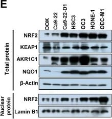
Detection of Keap1 by Western Blot
NRF2 expression is positively associated with malignancy in HNSCC. (A, C, D) Representative IHC staining of NRF2 in the specimens obtained from HNSCC patients. Brown staining indicates NRF2 positivity. The cell nuclei and background were counterstained with hematoxylin (blue coloration). (A) NRF2 protein expression in HNSCC specimens with different clinicopathological stages. Magnification, 100× (upper panel) and 400× (lower panel). (B) The correlation between the level of nuclear and cytosolic expression of NRF2 and tumor stage was estimated using the immunoreactivity scoring system (IRS). The IRS was calculated by multiplying the intensity of NRF2 staining (scale, 0-3) by the percentage of positive cells (4, > 80%; 3, 51-80%; 2, 10-50%; 1, < 10%; 0, 0%), which resulted in values ranging from 0 to 12. Groups with different letters are significant different (p < 0.05) from each other. (C) NRF2 protein expression was positively correlated with the degree of tumor cell differentiation in human head and neck squamous cell carcinoma. Magnification, 50× (upper panel) and 400× (lower panel). (D) NRF2 expression was highest in the invasive carcinoma nests. Magnification 50×. (E) Levels of NRF2 and NRF2 downstream targets in dysplasia oral keratinocyte (DOK) and HNSCC cell lines. (F) Motility of DOK and various HNSCC cell lines via trans-well migration assay. * p < 0.05; ** p < 0.01; *** p < 0.001. Image collected and cropped by CiteAb from the following open publication (https://pubmed.ncbi.nlm.nih.gov/33859744), licensed under a CC-BY license. Not internally tested by R&D Systems.Applications for Keap1 Antibody (333116)
Application
Recommended Usage
Knockout Validated
Keap1
is specifically detected in Jurkat human acute T cell leukemia parental cell line but is not detectable in
Keap1 knockout Jurkat cell line.
Simple Western
20 µg/mL
Sample: Jurkat human acute T cell leukemia cell line
Sample: Jurkat human acute T cell leukemia cell line
Western Blot
1 µg/mL
Sample: Jurkat human acute T cell leukemia cell line, Raji human Burkitt's lymphoma cell line, Neuro-2A mouse neuroblastoma cell line, and L6 rat myoblast cell line
Sample: Jurkat human acute T cell leukemia cell line, Raji human Burkitt's lymphoma cell line, Neuro-2A mouse neuroblastoma cell line, and L6 rat myoblast cell line
Reviewed Applications
Read 1 review rated 4 using MAB3024 in the following applications:
Formulation, Preparation, and Storage
Purification
Protein A or G purified from hybridoma culture supernatant
Reconstitution
Reconstitute at 0.5 mg/mL in sterile PBS. For liquid material, refer to CoA for concentration.
Loading...
Formulation
Lyophilized from a 0.2 μm filtered solution in PBS with Trehalose. *Small pack size (SP) is supplied either lyophilized or as a 0.2 µm filtered solution in PBS.
Shipping
Lyophilized product is shipped at ambient temperature. Liquid small pack size (-SP) is shipped with polar packs. Upon receipt, store immediately at the temperature recommended below.
Stability & Storage
Use a manual defrost freezer and avoid repeated freeze-thaw cycles.
- 12 months from date of receipt, -20 to -70 °C as supplied.
- 1 month, 2 to 8 °C under sterile conditions after reconstitution.
- 6 months, -20 to -70 °C under sterile conditions after reconstitution.
Calculators
Background: Keap1
Long Name
Kelch-like ECH-associated Protein 1
Alternate Names
INrf2
Entrez Gene IDs
9817 (Human)
Gene Symbol
KEAP1
UniProt
Additional Keap1 Products
Product Documents for Keap1 Antibody (333116)
Certificate of Analysis
To download a Certificate of Analysis, please enter a lot or batch number in the search box below.
Note: Certificate of Analysis not available for kit components.
Product Specific Notices for Keap1 Antibody (333116)
For research use only
Related Research Areas
Citations for Keap1 Antibody (333116)
 Powered by Bioz
Customer Reviews for Keap1 Antibody (333116) (1)
4 out of 5
1 Customer Rating
Have you used Keap1 Antibody (333116)?
Submit a review and receive an Amazon gift card!
$25/€18/£15/$25CAN/¥2500 for a review with an image
$10/€7/£6/$10CAN/¥1110 for a review without an image
Submit a review
Showing
1
-
1 of
1 review
Showing All
Filter By:
-
Application: Western BlotSample Tested: See PMID 22836768Species: HumanVerified Customer | Posted 02/19/2015
There are no reviews that match your criteria.
Protocols
Find general support by application which include: protocols, troubleshooting, illustrated assays, videos and webinars.
- Cellular Response to Hypoxia Protocols
- R&D Systems Quality Control Western Blot Protocol
- Troubleshooting Guide: Western Blot Figures
- Western Blot Conditions
- Western Blot Protocol
- Western Blot Protocol for Cell Lysates
- Western Blot Troubleshooting
- Western Blot Troubleshooting Guide
- View all Protocols, Troubleshooting, Illustrated assays and Webinars
Loading...