Myelin Basic Protein (MBP) is the most abundant protein component of the myelin membrane in the central nervous system. MBP has a role in both the formation and stabilization of this compact multilayer arrangement of bilayers. In vitro, MBP is suitable as a substrate for numerous protein kinases, including the ERK and p38 MAP kinases that phosphorylate MBP at T98.

Key Product Details
Species Reactivity
Validated:
Human, Mouse, Rat
Cited:
Human, Mouse, Rat, Transgenic Mouse
Applications
Validated:
Immunohistochemistry, Western Blot, Immunocytochemistry
Cited:
Immunohistochemistry, Western Blot, Immunocytochemistry
Label
Unconjugated
Antibody Source
Monoclonal Mouse IgG1 Clone # 932908
Loading...
Product Specifications
Immunogen
Purified
MBP from bovine brain
Accession # P02686
Accession # P02686
Specificity
Detects human, mouse, and rat MBP
Clonality
Monoclonal
Host
Mouse
Isotype
IgG1
Scientific Data Images for MBP Antibody (932908)
Detection of Human MBP by Western Blot.
Western blot shows lysates of mouse brain (cerebellum) tissue and rat brain (cerebellum). PVDF membrane was probed with 0.1 µg/mL of Mouse Anti-Human/Mouse/Rat MBP Monoclonal Antibody (Catalog # MAB42282) followed by HRP-conjugated Anti-Mouse IgG Secondary Antibody (Catalog # HAF018). Specific bands were detected for MBP at approximately 15-22 kDa (as indicated). This experiment was conducted under reducing conditions and using Immunoblot Buffer Group 1.
Detection of Mouse and Rat MBP by Western Blot.
Western blot shows lysates of mouse brain (cerebellum) tissue and rat brain (cerebellum) tissue. PVDF membrane was probed with 0.1 µg/mL of Mouse Anti-Human/Mouse/Rat MBP Monoclonal Antibody (Catalog # MAB42282) followed by HRP-conjugated Anti-Mouse IgG Secondary Antibody (Catalog # HAF018). Specific bands were detected for MBP at approximately 15-22 kDa (as indicated). This experiment was conducted under reducing conditions and using Immunoblot Buffer Group 1.
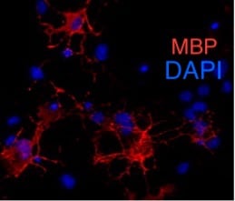
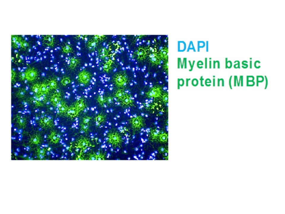
MBP in Rat Cortical Stem Cells.
MBP was detected in immersion fixed rat cortical stem cells differentiated for 7 days to oligodendrocytes using Mouse Anti-Human/Mouse/Rat MBP Monoclonal Antibody (Catalog # MAB42282) at 10 µg/mL for 3 hours at room temperature. Cells were stained using the NorthernLights™ 557-conjugated Anti-Mouse IgG Secondary Antibody (red; Catalog # NL007) and counterstained with DAPI (blue). Specific staining was localized to cell surfaces, cytoplasm, and nuclei. View our protocol for Fluorescent ICC Staining of Stem Cells on Coverslips.
MBP in Human Brain.
MBP was detected in immersion fixed paraffin-embedded sections of human brain (cortex) using Mouse Anti-Human/Mouse/Rat MBP Monoclonal Antibody (Catalog # MAB42282) at 1.7 µg/mL for 1 hour at room temperature followed by incubation with the Anti-Mouse IgG VisUCyte™ HRP Polymer Antibody (Catalog # VC001). Before incubation with the primary antibody, tissue was subjected to heat-induced epitope retrieval using Antigen Retrieval Reagent-Basic (Catalog # CTS013). Tissue was stained using DAB (brown) and counterstained with hematoxylin (blue). Specific staining was localized to neuronal processes. View our protocol for IHC Staining with VisUCyte HRP Polymer Detection Reagents.
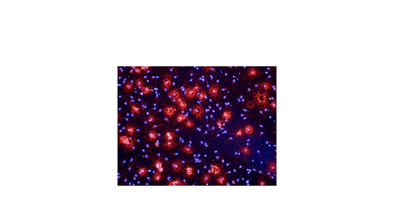
MBP in Mouse Brain.
MBP was detected in perfusion fixed frozen sections of mouse brain using Mouse Anti-Human/Mouse/Rat MBP Monoclonal Antibody (Catalog # MAB42282) at 0.1 µg/mL overnight at 4 °C. Tissue was stained using the NorthernLights™ 557-conjugated Anti-Mouse IgG Secondary Antibody (red; Catalog # NL007) and counterstained with DAPI (blue). Specific staining was localized to myelinated fibers. View our protocol for Fluorescent IHC Staining of Frozen Tissue Sections.
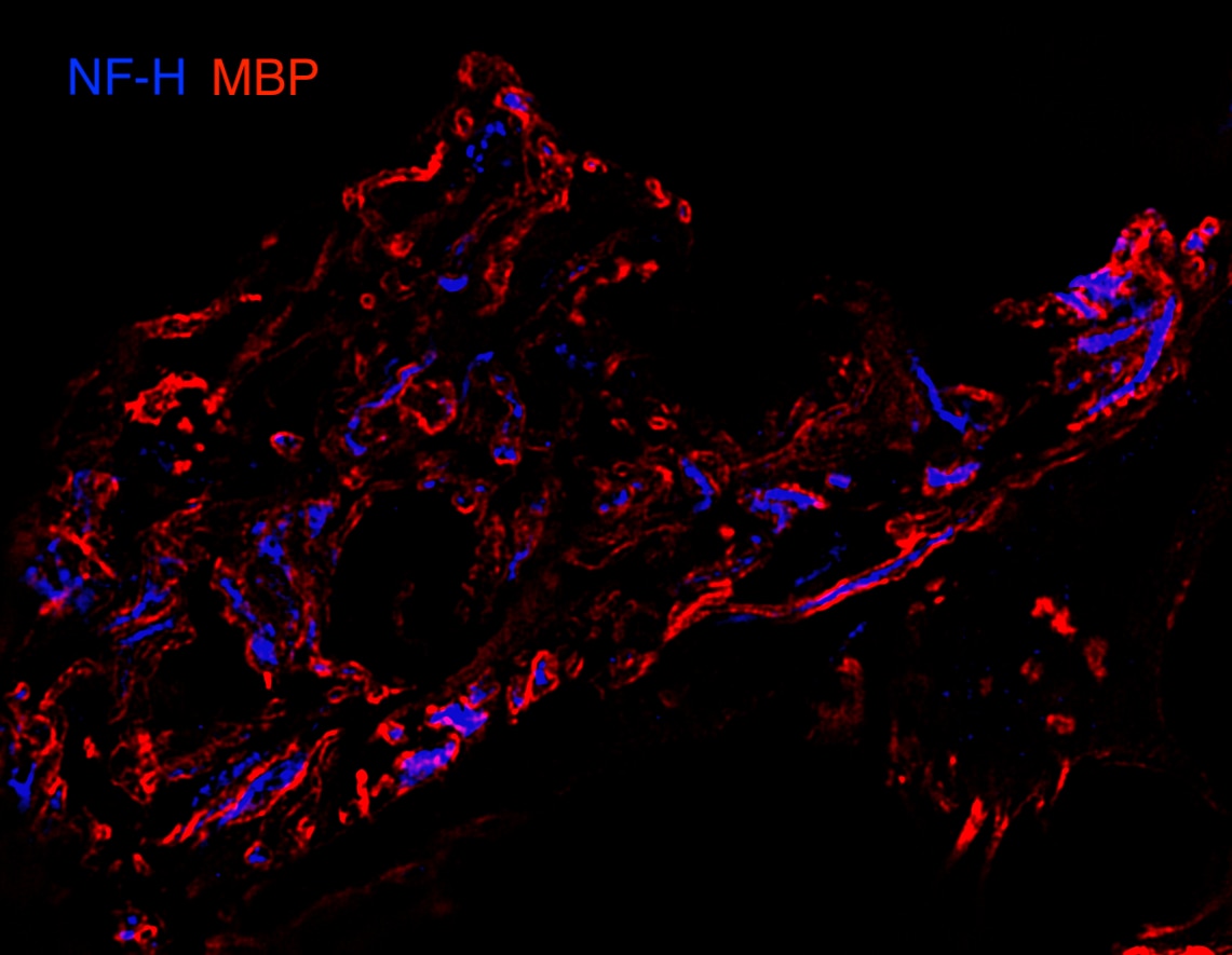
MBP in Rat Brain.
MBP was detected in perfusion fixed frozen sections of rat brain using Mouse Anti-Human/Mouse/Rat MBP Monoclonal Antibody (Catalog # MAB42282) at 0.1 µg/mL overnight at 4 °C. Tissue was stained using the NorthernLights™ 557-conjugated Anti-Mouse IgG Secondary Antibody (red; Catalog # NL007) and counterstained with DAPI (blue). Specific staining was localized to myelinated fibers. View our protocol for Fluorescent IHC Staining of Frozen Tissue Sections.
Detection of Human MBP by Immunohistochemistry
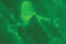
Pronounced myelin loss in FTD-GRN. (A, C) Representative western blots and (B, D) densitometric quantification of PLP, CNP, MBP, beta III-tubulin ( beta III-T) and neurofilament light chain (NF-L) in (A, B) superior frontal white matter, and (C, D) superior parietal white matter from control (n = 11), FTD-C9orf72 (n = 11), and FTD-GRN (n = 6) cases. Protein levels were normalised to beta -actin or GAPDH as a loading control, and are expressed relative to the mean of the control group. (E) Representative images and (F) myelination scores from LFB staining of superior frontal gyrus white matter from control (n = 5), FTD-C9orf72 (n = 4), and FTD-GRN (n = 3) cases from which tissue fixed for < 2 weeks was available. (G) Representative ASPA (red) and MBP (green) staining in superior frontal gyrus white matter, and (H) ASPA-positive cell density. Groups were compared by one-way ANOVA with Tukey’s post-test (B,D,H) or Kruskall-Wallis test with Dunn’s post-test (F): *p < 0.05; **p < 0.01; ***p < 0.001 Image collected and cropped by CiteAb from the following open publication (https://pubmed.ncbi.nlm.nih.gov/36967384), licensed under a CC-BY license. Not internally tested by R&D Systems.
Detection of Human MBP by Immunohistochemistry
Pronounced myelin loss in FTD-GRN. (A, C) Representative western blots and (B, D) densitometric quantification of PLP, CNP, MBP, beta III-tubulin ( beta III-T) and neurofilament light chain (NF-L) in (A, B) superior frontal white matter, and (C, D) superior parietal white matter from control (n = 11), FTD-C9orf72 (n = 11), and FTD-GRN (n = 6) cases. Protein levels were normalised to beta -actin or GAPDH as a loading control, and are expressed relative to the mean of the control group. (E) Representative images and (F) myelination scores from LFB staining of superior frontal gyrus white matter from control (n = 5), FTD-C9orf72 (n = 4), and FTD-GRN (n = 3) cases from which tissue fixed for < 2 weeks was available. (G) Representative ASPA (red) and MBP (green) staining in superior frontal gyrus white matter, and (H) ASPA-positive cell density. Groups were compared by one-way ANOVA with Tukey’s post-test (B,D,H) or Kruskall-Wallis test with Dunn’s post-test (F): *p < 0.05; **p < 0.01; ***p < 0.001 Image collected and cropped by CiteAb from the following open publication (https://pubmed.ncbi.nlm.nih.gov/36967384), licensed under a CC-BY license. Not internally tested by R&D Systems.
Detection of MBP by Western Blot
GCN2 deficiency impacts the length of myelinated segments and myelin levels. Sections (30 μm) of the brain derived from two-month-old male WT (n = 3) and genetically deficient GCN2 (GCN2−/−, n = 3) mice were obtained and processed for immunofluorescence to visualize myelin segments. (A) Representative images showing the myelin basic protein (MBP) (red) staining used as a myelin marker, the nuclei DAPI (blue), and the composed image (merge). An augmented section (white box) of the images are shown at the right (Zoom). Arrows on the zoom image show the type of MBP-positive objects quantified. (B) Images obtained in A were quantified using the Image J software (1.53t version). The plot shows the number and length of myelin segments (upper panel) and the average length (lower panel) of myelin segments found in WT and GCN2−/− mice. (C) Protein extracts were prepared from brains derived from two-month-old WT (n = 4) and GCN2−/− (n = 4) mice. MBP levels were analyzed by Western blot using alpha -Tubulin as a loading control (left). Each lane represents an individual (30 μg/lane). Densitometry analysis of the blot was performed by ImageJ to quantify the intensities of the two MBP bands and then normalized to alpha -Tubulin (right) levels. Scale bar: 100 μm. **** p < 0.00001, * p < 0.05. t-test analysis. Error bars represent the mean ± SEM. The whole Western blot membrane is shown in Supplementary Figure S1. The MBP quantification strategy is described in the “Materials and Methods” section. Image collected and cropped by CiteAb from the following open publication (https://pubmed.ncbi.nlm.nih.gov/40004088), licensed under a CC-BY license. Not internally tested by R&D Systems.Applications for MBP Antibody (932908)
Application
Recommended Usage
Immunocytochemistry
8-25 µg/mL
Sample: Immersion fixed rat cortical stem cells differentiated for 7 days to oligodendrocytes
Sample: Immersion fixed rat cortical stem cells differentiated for 7 days to oligodendrocytes
Immunohistochemistry
0.1-25 µg/mL
Sample: Perfusion fixed frozen sections of human brain, mouse brain, and rat brain
Sample: Perfusion fixed frozen sections of human brain, mouse brain, and rat brain
Western Blot
0.1 µg/mL
Sample: Human brain (cerebellum) tissue, Human brain (motor cortex) tissue, Mouse brain (cerebellum) tissue, and Rat brain (cerebellum) tissue
Sample: Human brain (cerebellum) tissue, Human brain (motor cortex) tissue, Mouse brain (cerebellum) tissue, and Rat brain (cerebellum) tissue
Reviewed Applications
Read 5 reviews rated 5 using MAB42282 in the following applications:
Formulation, Preparation, and Storage
Purification
Protein A or G purified from hybridoma culture supernatant
Reconstitution
Reconstitute at 0.5 mg/mL in sterile PBS. For liquid material, refer to CoA for concentration.
Loading...
Formulation
Lyophilized from a 0.2 μm filtered solution in PBS with Trehalose. *Small pack size (SP) is supplied either lyophilized or as a 0.2 µm filtered solution in PBS.
Shipping
Lyophilized product is shipped at ambient temperature. Liquid small pack size (-SP) is shipped with polar packs. Upon receipt, store immediately at the temperature recommended below.
Stability & Storage
Use a manual defrost freezer and avoid repeated freeze-thaw cycles.
- 12 months from date of receipt, -20 to -70 °C as supplied.
- 1 month, 2 to 8 °C under sterile conditions after reconstitution.
- 6 months, -20 to -70 °C under sterile conditions after reconstitution.
Calculators
Background: MBP
Long Name
Myelin Basic Protein
Alternate Names
Hmbpr, mld, shi
Gene Symbol
MBP
UniProt
Additional MBP Products
Product Documents for MBP Antibody (932908)
Certificate of Analysis
To download a Certificate of Analysis, please enter a lot or batch number in the search box below.
Note: Certificate of Analysis not available for kit components.
Product Specific Notices for MBP Antibody (932908)
For research use only
Related Research Areas
Citations for MBP Antibody (932908)
 Powered by Bioz
Customer Reviews for MBP Antibody (932908) (5)
5 out of 5
5 Customer Ratings
Have you used MBP Antibody (932908)?
Submit a review and receive an Amazon gift card!
$25/€18/£15/$25CAN/¥2500 Yen for a review with an image
$10/€7/£6/$10CAN/¥1110 Yen for a review without an image
Submit a review
Customer Images
Showing
1
-
5 of
5 reviews
Showing All
Filter By:
-
Application: ImmunohistochemistrySample Tested: Spinal cord tissueSpecies: MouseVerified Customer | Posted 11/29/2021

-
Application: Immunocytochemistry/ImmunofluorescenceSample Tested: NHP brainSpecies: MouseVerified Customer | Posted 08/31/2021
-
Application: Immunocytochemistry/ImmunofluorescenceSample Tested: IPS2 induced pluripotent stem cellsSpecies: MouseVerified Customer | Posted 07/27/2018
-
Application: Immunocytochemistry/ImmunofluorescenceSample Tested: Mouse iPSSpecies: MouseVerified Customer | Posted 01/06/2018
-
Application: Immunocytochemistry/ImmunofluorescenceSample Tested: Mouse Pluripotent Stem CellsSpecies: MouseVerified Customer | Posted 10/23/2017
There are no reviews that match your criteria.
Protocols
Find general support by application which include: protocols, troubleshooting, illustrated assays, videos and webinars.
- Antigen Retrieval Protocol (PIER)
- Antigen Retrieval for Frozen Sections Protocol
- Appropriate Fixation of IHC/ICC Samples
- Cellular Response to Hypoxia Protocols
- Chromogenic IHC Staining of Formalin-Fixed Paraffin-Embedded (FFPE) Tissue Protocol
- Chromogenic Immunohistochemistry Staining of Frozen Tissue
- ClariTSA™ Fluorophore Kits
- Detection & Visualization of Antibody Binding
- Fluorescent IHC Staining of Frozen Tissue Protocol
- Graphic Protocol for Heat-induced Epitope Retrieval
- Graphic Protocol for the Preparation and Fluorescent IHC Staining of Frozen Tissue Sections
- Graphic Protocol for the Preparation and Fluorescent IHC Staining of Paraffin-embedded Tissue Sections
- Graphic Protocol for the Preparation of Gelatin-coated Slides for Histological Tissue Sections
- ICC Cell Smear Protocol for Suspension Cells
- ICC Immunocytochemistry Protocol Videos
- ICC for Adherent Cells
- IHC Sample Preparation (Frozen sections vs Paraffin)
- Immunocytochemistry (ICC) Protocol
- Immunocytochemistry Troubleshooting
- Immunofluorescence of Organoids Embedded in Cultrex Basement Membrane Extract
- Immunofluorescent IHC Staining of Formalin-Fixed Paraffin-Embedded (FFPE) Tissue Protocol
- Immunohistochemistry (IHC) and Immunocytochemistry (ICC) Protocols
- Immunohistochemistry Frozen Troubleshooting
- Immunohistochemistry Paraffin Troubleshooting
- Preparing Samples for IHC/ICC Experiments
- Preventing Non-Specific Staining (Non-Specific Binding)
- Primary Antibody Selection & Optimization
- Protocol for Heat-Induced Epitope Retrieval (HIER)
- Protocol for Making a 4% Formaldehyde Solution in PBS
- Protocol for VisUCyte™ HRP Polymer Detection Reagent
- Protocol for the Fluorescent ICC Staining of Cell Smears - Graphic
- Protocol for the Fluorescent ICC Staining of Cultured Cells on Coverslips - Graphic
- Protocol for the Preparation & Fixation of Cells on Coverslips
- Protocol for the Preparation and Chromogenic IHC Staining of Frozen Tissue Sections
- Protocol for the Preparation and Chromogenic IHC Staining of Frozen Tissue Sections - Graphic
- Protocol for the Preparation and Chromogenic IHC Staining of Paraffin-embedded Tissue Sections
- Protocol for the Preparation and Chromogenic IHC Staining of Paraffin-embedded Tissue Sections - Graphic
- Protocol for the Preparation and Fluorescent ICC Staining of Cells on Coverslips
- Protocol for the Preparation and Fluorescent ICC Staining of Non-adherent Cells
- Protocol for the Preparation and Fluorescent ICC Staining of Stem Cells on Coverslips
- Protocol for the Preparation and Fluorescent IHC Staining of Frozen Tissue Sections
- Protocol for the Preparation and Fluorescent IHC Staining of Paraffin-embedded Tissue Sections
- Protocol for the Preparation of Gelatin-coated Slides for Histological Tissue Sections
- Protocol for the Preparation of a Cell Smear for Non-adherent Cell ICC - Graphic
- R&D Systems Quality Control Western Blot Protocol
- TUNEL and Active Caspase-3 Detection by IHC/ICC Protocol
- The Importance of IHC/ICC Controls
- Troubleshooting Guide: Immunohistochemistry
- Troubleshooting Guide: Western Blot Figures
- Western Blot Conditions
- Western Blot Protocol
- Western Blot Protocol for Cell Lysates
- Western Blot Troubleshooting
- Western Blot Troubleshooting Guide
- View all Protocols, Troubleshooting, Illustrated assays and Webinars
Loading...